Targeted echocardiographic screening for latent RHD in Northern Uganda

What’s Known
Echocardiograms use the echoes of sound waves to create “movies” of the beating heart, its valves, and other structures. While rheumatic heart disease (RHD) was prevalent in the United States as late as the 1900s, improved housing conditions and the availability of powerful medicines like antibiotics and penicillin have lowered its incidence to 0.04 to 0.06 cases per 1,000 U.S. children. In regions where streptococcal infections flourish, RHD remains a scourge. Using echocardiographic screening to identify latent RHD— which is apparent on echocardiography before the child has symptoms that can be spotted by clinicians—has the potential to reduce the disease’s global burden.

What’s New
Optimal implementation must account for whom to target, when, in which settings, and how often to screen. The team led by Children’s National Health System researchers and clinicians conducted the first family screening study in Northern Uganda to assess the utility of echocardiographic screening of first-degree relatives of children with latent RHD. They used existing school-based screening data to identify potential participants and invited all first-degree relatives older than 5 years for echocardiography screening. The study recruited 60 RHD-positive schoolchildren and matched them with 67 RHD-negative kids of similar age and gender. Some 1,122 family members were then screened. Children with any RHD were 4.5 times as likely to have a sibling with definite RHD, a risk that increased to 5.6 times if researchers looked solely at index cases with definite RHD. The team, led by Andrea Beaton, MD, a cardiologist at Children’s National, also found that mothers had a 9.3 percent rate of latent RHD—a high rate that was independent of whether their child was RHD-positive.

Questions for Future Research
Q: Many children living in RHD-endemic areas, exposed to the same environmental conditions as RHD-positive kids, are able to fend off disease. Are protective genes to credit for their resilience?
Q: What are the best approaches to train nurses and community workers in how to use lower-cost, handheld echocardiograms to facilitate large-scale screening in countries where healthcare resources are constrained?

Source:  Targeted Echocardiographic Screening for Latent Rheumatic Heart Disease in Northern Uganda: Evaluating Familial Risk Following Identification of an Index Case.” T. Aliku, C. Sable, A. Scheel, A. Tompsett, P. Lwabi, E. Okello, R. McCarter, M. Summar, and A. Beaton. Published online by PLoS June 13, 2016.

Using 3-D MRI for fetal brain imaging during high-risk pregnancies

3DMRI

What’s Known
The placenta plays an essential role in the growth of a healthy fetus and, among other critical tasks, it ferries in oxygen and nutrients. During pregnancies complicated by fetal growth restriction (FGR), the failing placenta cannot support the developing fetus adequately. FGR is a major cause of stillbirth and death, and newborns who do survive face numerous risks for multiple types of ailments throughout their lives. In fact, studies have shown that nutrient depravation during gestation can have lasting consequences that may manifest themselves years or decades later in life. These risks can also cross generations, affecting future pregnancies.

What’s New
A team of researchers applied an advanced imaging technique, three-dimensional (3-D) MRI, to study brain development in these high-risk pregnancies. They are the first to report regional, tissue-specific volume delays for the developing fetal brain in FGR-affected pregnancies. The team compared overall fetal brain volume as well as regional brain volumes for a control group of healthy young pregnant women with a group of young women whose pregnancies were complicated by FGR. While fetuses in both groups grew exponentially as pregnancies progressed, the researchers began to see dramatic differences when they compared the volumes of specific regions of the brain, including the cerebellum, which coordinates balance and smooth movement; the deep gray matter, which also is involved in complex functions, such as memory and emotion; and the white matter, which is made up of millions of nerve fibers that connect to neurons in different regions. Because there are no biomarkers to spot early brain failure, 3-D MRI imaging may fill this knowledge gap.

Questions for Future Research
Q: Certain regions of the brains of FGR-affected infants show accelerated volume. Are these differences regional or global?
Q: Is accelerated brain volume in FGR-affected infants a result of heightened stress that these fetuses experience in the womb?
Q: How do differences in regional brain volume relate to later neurodevelopmental impairment that some FGR-affected infants experience?

Source: “Impaired Global and Tissue-Specific Brain Development in the Growth-Restricted Fetus.N. Andescavage, J. Cruz, M. Metzler, A. du Plessis, and C. Limperopoulos. Presented during the 2016 Pediatric Academic Societies Annual Meeting, Baltimore, MD. May 2, 2016.

Unlocking the ‘black box’ of NICU monitors to protect vulnerable preemies

MiningdatafromNICUmonitors

What’s Known
Around the world, some 15 million infants are born prematurely each year. Babies born prematurely can spend their first weeks to months of life in the neonatal intensive care unit (NICU) tethered to machines that closely monitor vital signs, such as breathing and heart rate.

After discharge, preemies have a very high risk of returning to the NICU, often due to breathing difficulties, such as experiencing excessively long pauses between breaths. Such acute life-threatening events are a major cause of preemies’ hospital readmission and may result in death.

What’s New
During infants’ NICU stays, cardiorespiratory monitors amass a mountain of data about each child. Through the unprecedented collaboration of researchers working in various divisions of Children’s National Health System, the team was able to unlock that black box of information by creating algorithms to extract data and by using retrospective analyses to tease out new insights. This multidisciplinary team has been able to predict with a greater degree of precision which babies are at higher risk of returning to the NICU after discharge. What these most vulnerable preemies have in common is the degree of maturation of their autonomic nervous system, which controls such involuntary actions as heart rate and breathing. The sympathetic nervous system, which the body leverages as it copes with the stress of life-threatening events (ALTE), also plays a role in these infants’ heightened vulnerability. Being able to identify these newborns earlier has the potential to lower readmissions and save lives.

Questions for Future Research
Q: How can further computer-based analyses of NICU monitor data be used to determine how preemies respond to routine activities, such as feeding to predict which infants have compromised cardiorespiratory systems?
Q: How can we develop a test to assess all premature infants for physiologic readiness for safe NICU discharge and, thus, prevent ALTE and sudden death in this vulnerable population?

Source: Vagal Hypersensitivity in Premature Infants and Risk of Hospital Readmission Due to Acute Life-Threatening Events (ALTE).” G. Nino, R. Govindan, T. AlShargabi, M. Metzler, R. Joshi, G. Perez, A.N. Massaro, R. McCarter, and A. du Plessis. Presented during the 2016 Pediatric Academic Societies Annual Meeting, Baltimore, MD. May 2, 2016.

Congenital Zika Viral Infection Linked to Significant Fetal Brain Abnormalities

mosquito

PDF Version

What’s Known
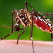
According to the Centers for Disease Control and Prevention, Zika viral transmission is occurring extensively throughout Central and South America. Like other mosquito-borne viruses, Zika virus can be passed by pregnant women to developing fetuses. Unlike these other viruses, Zika has been implicated in a growing number of cases of Brazilian infants born with microcephaly, a condition characterized by undersized heads and severe brain damage. The precise strategy that the Zika virus uses to elude the immune system and the reason why fetal brain cells are particularly vulnerable remain unknown.

What’s New
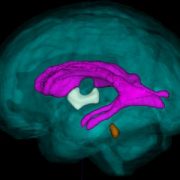
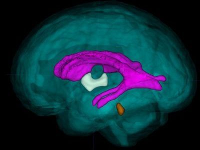
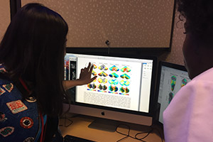
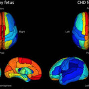
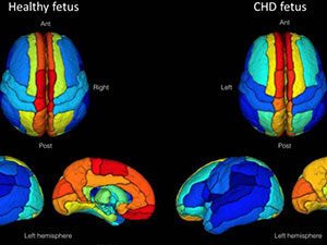
A 33-year-old Finnish woman was 11 weeks pregnant when she and her husband traveled on vacation to Mexico, Guatemala, and Belize in late November 2015. The pair was bitten by mosquitoes during their trip, particularly in Guatemala. One day after returning to their Washington, DC home, the woman got sick, experiencing eye pain, muscle pain, a mild fever, and a rash. A series of early ultrasounds showed no sign of microcephaly or brain calcifications. A fetal ultrasound at the 19th week and a fetal MRI at the 20th week, however, revealed severe brain damage.

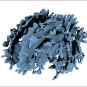
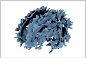
The brain of the 21-week-old aborted fetus weighed only 30 grams. Zika RNA, viral particles, and infectious virus were detected, and Zika virus isolated from the fetal brain remained infectious when tested. The concentration of virus was highest in the fetal brain, umbilical cord, and placenta. The mother remained infected with Zika virus at 21 weeks, some 10 weeks after her initial infection.

Questions for Future Research

  • Could serial measurements and blood tests more accurately detect and, ultimately, predict fetal abnormalities following Zika virus infection?
  • Why does the Zika virus replicate with ease within the womb?
  • At which stage of pregnancy are fetuses most vulnerable?
  • Which specific brain cells does Zika target?

Source:Zika Virus Infection with Prolonged Maternal Viremia and Fetal Brain Abnormalities.” R.W. Driggers, C.Y. Ho, E.M. Korhonen, S. Kuivanen, A.J. Jääskeläinen, T. Smura, D.A. Hill, R. DeBiasi, G. Vezina, J. Timofeev, F.J. Rodriguez, L. Levanov, J. Razak, P. Iyengar, A. Hennenfent, R. Kennedy, R. Lanciotti, A. du Plessis, and O. Vapalahti. The New England Journal of Medicine. June 2, 2016.

Training t-cells, essential players in the immune system, to fight a trio of viruses

Children's is the only U.S. pediatric hospital that manufactures specialized T-cells from native cord blood

What’s Known
Following treatment, patients with leukemia, lymphoma, and other cancers may receive a transplant in order to restore their body’s natural ability to fight infection and, sometimes, such transplants are a component of leukemia treatment. (Leukemia is the second most common blood cancer, after lymphoma, and its incidence rate has increased by 0.2 percent annually from 2002 to 2011.) A stem cell or cord blood transplant restores the body’s ability to produce infection-fighting white blood cells. After such transplants, however, patients can face heightened risk of developing a life-threatening infection with such viruses as adenovirus, cytomegalovirus, or Epstein-Barr virus.

What’s New
A head-to-head comparison of two strategies to thwart such viral infections shows that both approaches leverage the power of multivirus-specific, donor-derived T-cells (mCTL), which are highly skilled at recognizing foreign invaders. The research team, made up of nine scientists and clinicians affiliated with Children’s National Health System, grew personalized T-cells from peripheral blood (PB) of adult donors who were seropositive for CMV and also coaxed T-cells to grow from naïve cord blood (CB). PB-derived cells have long memories of past battles; naïve CB-derived cells need additional training to acquire such skills. From 35 to 384 days after their stem cell or cord blood transplant, 13 patients were infused with PB mCTL and 12 patients were infused with CB mCTL. Within four weeks, patients experienced up to a 160-fold increase in virus-specific T-cells, which coincided with their response to therapy. Overall response rate was 81 percent.

Questions for Future Research
Q: Could T-cells be personalized to attack other viruses that infect patients post-transplant, such as human parainfluenza virus and BK polyomavirus, providing the potential to target five viruses in a single infusion?
Q: Could the proteins that are used to train T-cells to attack certain viruses also be used to create a personalized approach to tumor suppression?

Source: “A Phase 1 Perspective: Multivirus-Specific T Cells From Both Cord Blood and Bone Marrow Transplant Donors.” Hanley, P., M. D. Keller, M. Martin Manso, C. Martinez, K. Leung, C.R. Cruz, C. Barese, S. McCormack, M. Luo, R.A. Krance, D. Jacobsohn, C. Rooney, H. Heslop, E.J. Shpall, and C. Bollard. Presented during the International Society for Cellular Therapy 2016 Annual Meeting, Singapore. May 26, 2016.

The search for precise blood biomarkers of neonatal brain injury

Bloodbiomarkers

PDF Version

What’s Known:
Hypoxic-ischemic encephalopathy (HIE) is characterized by reduced blood and oxygen flow to a baby’s brain around birth and may cause neurologic disability or death. It occurs most commonly after intrauterine asphyxia brought on by such difficulties as circulatory problems, placental abruption, or inflammatory processes. Newborns with HIE may suffer seizures, difficulty feeding, and disturbed control of heart rate and breathing. Cooling therapy, which is the standard of care, offers some protection to the developing brain, but up to 50 percent of HIE-affected infants still have poor outcomes.

What’s New:
Research scientists at Children’s National Health System are involved in a multi-center clinical trial to determine if erythropoietin (EPO), a hormone naturally secreted by the kidneys and commonly used to treat anemia, helps to prevent brain injury in these infants. The trial, called the HEAL Study (High Dose Erythropoietin for Asphyxia and Encephalopathy), is exploring whether EPO, given in addition to hypothermia, further lowers the risk of brain injury in HIE-affected babies. As a part of this study, researchers at Children’s National are leading the investigation to identify biomarkers of brain injury. Biomarkers are telltale chemicals in the blood and are used in tests that evaluate whether patients have suffered a heart attack. While available biomarkers warn when the heart, kidney, or liver is in trouble, there is no blood biomarker that signals ongoing brain injury. Such blood biomarkers could help to determine which infants are responding to treatment as well as to precisely identify which HIE-affected infants are still struggling and require additional treatments, such as EPO, to protect the brain and improve outcomes.

Questions for Future Research: 

  • Does EPO, in tandem with hypothermia, improve long- term neurodevelopmental outcomes in newborns with HIE?
  • Which biomarkers, or panel of biomarkers, best reflect the timing and severity of neonatal brain injury?
  • Can biomarkers direct which types of treatments are best for specific patients and when they should be used?

Source: Plasma Biomarkers of Brain Injury in Neonatal HIE (Hypoxic-Ischemic Encephalopathy).” A.N. Massaro, Y. Wu, T.K. Bammler, A. Mathur, R.C. McKinstry, T. Chang, D.E. Mayock, S. Mulkey, K. Van Meurs, L. Dong, R. Ballard, and S. Juul. Presented during the 2016 Pediatric Academic Societies Annual Meeting, Baltimore, MD. May 3, 2016.

Why subtle cellular changes can result in dramatically different genetic disorders

cellular_changes

PDF Version

What’s Known
One single gene, lamin A/C, is to blame for a multitude of genetic disorders, such as premature aging and problems with nerves, the heart, and muscles. Uncertainties linger in the scientific and medical community about why subtle changes of this gene cause such dramatically different disorders, such as Emery-Dreifuss muscular dystrophy, a condition that can lead to progressive muscle weakness in childhood and heart problems by adulthood.

What’s New
The nuclear envelope is where attached regions are pulled from genetic circulation never to be used again. The process of attachment signals which parts of the genome the cell no longer considers useful. Discarding superfluous DNA keeps the cell focused on what matters more: Its future role. Proper cell differentiation hinges on “the coordinated execution of three key cellular programs,” the study authors write. Pluriopotency programs, which give primitive cells the remarkable ability to generate any cell type in the body, are inactivated. Exit from the cell cycle occurs, and cells stop dividing. Myogenesis is induced, ushering in formation of muscle tissue. Mutations in lamin A/C can disrupt this careful choreography with the cumulative effect of slowing exit from cell cycle, slowing exit from pluripotency programs, and poorly coordinating induction of terminal differentiation programs.

Questions for Future Research
Q: What are the regions of human genome that become attached to the nuclear envelope during the development of tissues other than muscle (such as fat, nerve, and heart)?
Q: Can medicines that influence epigenetic pathways help to reverse the inappropriate DNA-lamin associations in Emery-Dreifuss muscular dystrophy?
Q: Can the new knowledge of DNA-lamin associations during muscle cell differentiation help to inform stem cell therapies?

Source:Laminopathies Disrupt Epigenomic Developmental Programs and Cell Fate.” J. Perovanovic, S. Dell’Orso, V.F. Gnochi, J. K. Jaiswal, V. Sartorelli, C. Vigouroux, K. Mamchaoui, V. Mouly, G. Bonne, and E. P. Hoffman. Science Translational Medicine. April 20, 2016.

Lessons learned from newborn screening for critical congenital heart defects

chd_screening

What’s Known

In 2011, screening for critical congenital heart defects (CCHD) became the second point-of-care newborn screening test added to the Recommended Uniform Screening Panel, and it has since been widely adopted. Heart defects are the primary targets for CCHD screening, which often require evaluation by echocardiogram. An original list of seven conditions represented the most common critical lesions which routinely present with hypoxemia for newborns. Endorsed by the American Academy of Pediatrics and four other professional medical societies, the CCHD screening using pulse oximetry is required by law in all but two states. Remaining challenges include national data collection and outcomes analyses at the population level.

What’s New

An expert panel including Gerard R. Martin, MD, a cardiologist at the Center for Translational Science at Children’s National Health System, reviewed current practices in newborn screening for CCHD and identified opportunities for improvement. The panel’s study expanded the list of core conditions to 12 to emphasize the importance of other potentially critical, yet treatable secondary conditions. Roughly 79 percent of “positive” screens for CCHD identify secondary conditions, such as sepsis and pulmonary diseases. The study found algorithm misinterpretation was common in states collecting outcomes data, emphasizing needs for proper training and quality-assurance feedback mechanisms. Public health surveillance varied dramatically, with nearly one-fifth of states neither actively collecting data nor planning to do so. Additional CCHD screening research in special settings like the NICU, out-of hospital settings, and areas with high altitude may result in adaptations to screening protocol. Future improvements to the current screening algorithm and analyses of the impact on CCHD outcomes will rely on further investment in a national data repository.

Questions for Future Research

Q: What will be the impact on present screening for CCHD on outcomes of non-CCHD secondary conditions?
Q: What is the optimal algorithm for CCHD based on screening and testing ease of use, costs, resource utilization, and sensitivity for different treatment settings?
Q: What will be the impact on present screening for CCHD on outcomes of non-CCHD secondary conditions?

Source: Lessons Learned From Newborn Screening for Critical Congenital Heart Defects.” M.E. Oster, S.W. Aucott, J. Glidewell, J. Hackell, L. Kochilas, G.R. Martin, J. Phillippi, N.M.Pinto, A. Saarinen, M. Sontag, and A.R. Kemper. Published by Pediatrics May 2016.

Some functional brain connectivity altered in fetuses with CHD

chd_fetus

What’s Known
Congenital heart disease (CHD), a structural problem with the heart at birth, is the most common birth defect and impacts 8 of every 1,000 newborns.

While many infants with mild disease require no intervention, others have complex CHD that necessitates specialized treatment shortly after birth. Complex defects change how blood flows through the heart and to other organs—including the brain.

What’s New
Newborns with this diagnosis are at an elevated risk for neurodevelopmental disabilities, underscoring the importance of monitoring fetal brain development and function to identify which newborns need additional surveillance and medical intervention. Neuroimaging research in recent years has shown that resting-state functional magnetic resonance imaging (rs-fMRI) can provide critical insights into how the brain functions, at rest. The research team in the Developing Brain Research Laboratory at Children’s National Health System successfully measured brain function in 90 different brain regions in healthy resting fetuses and pregnancies complicated by CHD. The team reports for the first time that there was robust functional connectivity between hemispheres in both fetuses diagnosed with CHD and controls matched by gestational age. The Children’s researchers and clinicians, however, found that some functional connections were weakened in the association and paralimbic regions of the brain that are involved in attention, emotions, and behaviors.

Questions for Future Research
Q: Does decreased regional connectivity in these association and paralimbic brain regions in CHD-complicated pregnancies influence infants’ neurodevelopment after birth?
Q: Can rs-fMRI be used to identify early disturbances in brain development in CHD-complicated pregnancies, and can the imaging technique lead to improved surveillance and more timely therapeutic intervention?

Source: “Functional Brain Connectivity Is Altered in Fetuses With Congenital Heart Disease.” J. De Asis-Cruz, A. Yarish, M. Donofrio, G. Vezina, A. du Plessis, and C. Limperopoulos. Presented during the 2016 American Society of Neuroradiology Annual Meeting, Washington, DC. May 25 2016.

researcher using ice bucket in lab

Spatial and temporal homogeneity of driver mutations in diffuse intrinsic pontine glioma

PDF Version

What’s Known
Needle biopsies help to guide diagnosis and targeted therapies for diffuse intrinsic pontine gliomas (DIPGs), which make up 10 percent to 15 percent of all pediatric brain tumors but carry a median survival of 9 to 12 months. This dismal survival rate compares with a 70 percent chance of children surviving other central nervous system tumors five years post diagnosis. In DIPG, tumors appear in the pons, an area of the brain that houses cranial nerve nuclei. Surgical options are limited. Spatial and temporal tumor heterogeneity is a major obstacle to accurate diagnosis and successful targeted therapy.

What’s New
The team sought to better define DIPG heterogeneity. They analyzed 134 specimens from nine patients and found that H3K27M mutations were ubiquitous in all 41 samples with oncogenic content, and always were associated with at least one partner driver mutation: TP53, PPM1D, ACVR1 or PIK3R1. These H3K27M mutations are the initial oncogenic event in DIPG, writes the research team led by Children’s National Health System. “Driver” mutations, such as H3K27M, are essential to begin and sustain tumor formation. This main driver partnership is maintained throughout the course of the disease, in all cells across the tumor, and as tumors spread throughout the brain. Because homogeneity for main driver mutations persists for the duration of illness, efforts to cure DIPG should be directed at the oncohistone partnership, the authors write. Based on early tumor spread, efforts to cure DIPG should aim for early systemic tumor control, rather focused exclusively on the pons.

Questions for Future Research
Q: If a larger sample size were analyzed, what would it reveal about the true heterogeneity/homogeneity status of DIPGs?
Q: “Accessory” driver mutations are not absolutely essential but do help to further promote and accelerate tumor growth. What is their precise role?

Source: Spatial and Temporal Homogeneity of Driver Mutations in Diffuse Intrinsic Pontine Glioma.” H. Nikbakht, E. Panditharatna, L.G. Mikael, R. Li, T. Gayden, M. Osmond, C.Y. Ho, M. Kambhampati, E.I. Hwang, D. Faury, A. Siu, S. Papillon-Cavanagh, D. Bechet, K.L. Ligon, B. Ellezam, W.J. Ingram, C. Stinson, A.S. Moore, K.E. Warren, J. Karamchandani, R.J. Packer, N. Jabado, J. Majewski, and J. Nazarian. Published by Nature Communications on April 6, 2016.

The role of NG2 proteoglycan in glioma

A large number of staffers contribute to the Children's National team effort to unravel the mysteries of DIPG. We photograph a few essential players in Dr. Nazarian's lab.

PDF Version

What’s Known
Neuron glia antigen-2 (NG2) is a protein expressed by many central nervous system cells during development and differentiation. NG2-expressing oligodendrocyte progenitor cells have been identified as the cells of origin in gliomas, tumors that arise from the brain’s gluey supportive tissue. What’s more, NG2 expression also has been associated with childhood diffuse intrinsic pontine glioma (DIPG) an aggressive tumor that accounts for 10 percent to 20 percent of pediatric central nervous system (CNS) tumors. Radiation can prolong survival by a few months, but children diagnosed with DIPG typically survive less than one year.

What’s New
Researchers are searching for appropriate targets and effective drugs that offer some chance of benefit. A team of Children’s National Health System researchers investigated whether NG2 – which plays a critical role in proliferation and development of new blood vessels and promotes tumor infiltration – could be a potential target for cancer treatment. Of the various options, antibody-mediated mechanisms of targeting NG2 are feasible, but the size of antibodies limits their ability to cross the blood-brain barrier. “Due to its role in maintaining a pluripotent pool of tumor cells, and its role in tumor migration and infiltration, NG2 provides multiple avenues for developing therapeutics,” the research team concludes. “Moreover, the large extracellular domain of NG2 provides an excellent antigen repertoire for immunotherapeutic interventions. As such, further research is warranted to define the role and expression regulation of NG2 in CNS cancers.”

Questions for Future Research

Q: Because healthy oligodendrocyte progenitor cells are important for the child’s developing brain, how could further characterization of NG2 isoforms help prevent drugs from damaging those beneficial cells?

Q: Could NG2-binding peptides cross the blood-brain barrier to deliver anti-cancer therapies precisely to tumor sites?

Source: The Role of NG2 Proteoglycan in Glioma.” S. Yadavilli, E.I. Hwang, R. J. Packer, and J. Nazarian. Published by Translational Oncology on February 2016.

Clinicopathology of diffuse intrinsic pontine glioma and its redefined genomic and epigenomic landscape

Dr. Nazarian's lab

PDF Version

What’s Known
Fewer than 150 U.S. children per year are diagnosed with diffuse intrinsic pontine glioma (DIPG), one of the most lethal pediatric central nervous system cancers. Despite an increasing number of experimental therapies tested via clinical trials, more than 95 percent of these children die within two years of diagnosis. Molecular studies have yielded additional insight about DIPG, including that mutations in histone-encoding genes are associated with 70 percent of cases. Understanding mutations that drive tumors and the genomic landscape can help to guide development of targeted therapies.

What’s New: Frequently found genetic alterations prevalent in DIPGs

dipg-gene-mutations-and-biological-consequences

Source: Clinicopathology of Diffuse Intrinsic Pontine Glioma and Its Redefined Genomic and Epigenomic Landscape.” E. Panditharatna, K. Yaeger, L.B. Kilburn, R.J. Packer, and J. Nazarian. Published by Cancer Genetics on May 1, 2015.