Tag Archive for: Vezina

Members of the Columbia Zika virus research team

School entry neurodevelopmental outcomes of Zika-exposed Colombian children

Members of the Columbia Zika virus research team

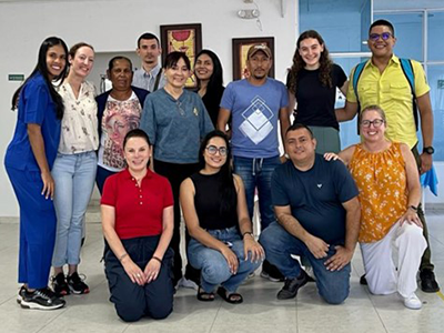
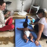
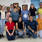
The Children’s National Hospital Zika Research Team and collaborators from Biomelab, in Barranquilla, Colombia take a picture after a study visit in Sabanalarga, Colombia following the neurodevelopmental outcomes of children who had in utero exposure to Zika virus. Pictured from Children’s National Hospital: Dr. Sarah Mulkey, Regan Andringa-Seed, Margarita Arroyave-Wessel, and Dr. Madison Berl.

The long-term neurodevelopmental effects of antenatal Zika virus (ZIKV) exposure in children without congenital Zika syndrome (CZS) remain unclear, as few children have been followed to the age of starting primary school.

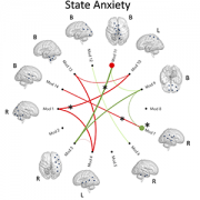
In a new study published in Pathogens, researchers found children with in utero ZIKV exposure appear to have an overall positive developmental trajectory at 4 to 5 years of age but may experience risks to neurodevelopment in areas of emotional regulation and adaptive mobility.

The hold up in the field

Children who were born during the ZIKV epidemic and who had in utero exposure to ZIKV are only now at the age to start school. Child neurodevelopmental outcome data has not been reported at the age of school entry for children with antenatal ZIKV exposure who do not have the severe birth defects of CZS.

“As these children approach the early school-age years, we aim to examine whether there are neurodevelopmental differences in executive function, motor ability, language development or scholastic skills as compared to a group of unexposed control participants from the same communities in Colombia,” says Sarah Mulkey, M.D., Ph.D., prenatal-neonatal neurologist in The Zickler Family Prenatal Pediatrics Institute at Children’s National Hospital and lead author of the study.

Moving the field forward

Building on previous findings, this study presents the longitudinal outcomes of a well-characterized Colombian cohort of ZIKV-exposed children without CZS at ages 4 to 5 years. These children have been seen for neurodevelopmental follow-up as infants and toddlers at approximately 6 months, 18 months and 3 years of age as part of an international collaboration between researchers in Barranquilla, Colombia and at Children’s National beginning in 2016. The objective of this study was to assess the multi-domain neurodevelopmental outcomes in 4 to 5-year-old children with antenatal ZIKV exposure without CZS compared to unexposed controls in Colombia.

Why we’re excited

Many of the children who had antenatal ZIKV exposure are making good progress in multiple areas of their neurodevelopment. However, the researchers found that children with antenatal ZIKV exposure have differences in areas of emotional regulation, executive function, mood and behavior which may relate to virus exposure during their early brain development.

“These areas of brain function are important for future academic achievement, employment, mental health and social relationships,” says Dr. Mulkey. “So, it will be important to continue to follow these children at older ages when they start school.”

Children’s National leads the way

Children’s National is a leader in conducting outcome studies of children born following antenatal ZIKV exposure. This study follows children in Colombia who are now 5 years old who were first studied while they were in the womb. These children have contributed unique longitudinal understanding to early child neurodevelopment following in utero exposure to ZIKV.

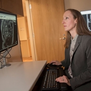
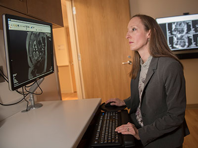
Dr. Mulkey is committed to studying the long-term neurodevelopmental impacts that viruses like Zika and SARS-CoV-2 have on infants born to mothers who were infected during pregnancy through research with the Congenital Infection Program at Children’s National and in collaboration with colleagues in Colombia.

Additional Children’s National authors include Meagan Williams M.S.P.H., C.C.R.C., senior research coordinator; Regan Andringa-Seed, clinical research coordinator, Margarita Arroyave-Wessel, clinical research coordinator; L. Gilbert Vezina, M.D., director, Neuroradiology Program; Dorothy Bulas, M.D., chief, Diagnostic Imaging and Radiology; Robert Podolsky, biostatistician.

Researchers showing paintings of zika virus

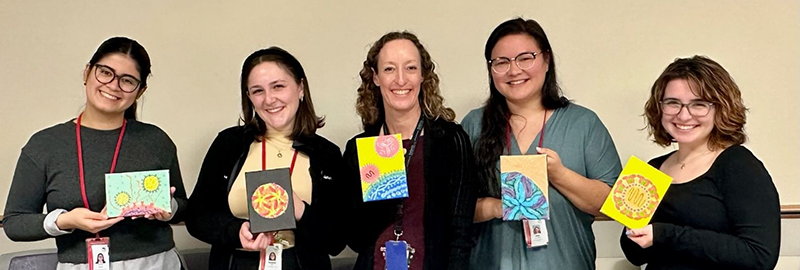
Dr. Sarah Mulkey and Children’s National clinical research coordinators in the Prenatal Pediatrics Institute and the Division of Pediatric Infectious Diseases display their paintings of the Zika virus. Pictured from left to right: Manuela Iglesias, Elizabeth Corn, Dr. Sarah Mulkey, Emily Ansusinha and Meagan Williams.

doctor examining pregnant woman

Low parental socioeconomic status alters brain development in unborn babies

doctor examining pregnant woman

A first-of-its-kind study with 144 pregnant women finds that socioeconomic status (SES) has an impact in the womb, altering several key regions in the developing fetal brain as well as cortical features.

Maternal socioeconomic status impacts babies even before birth, emphasizing the need for policy interventions to support the wellbeing of pregnant women, according to newly published research from Children’s National Hospital.

A first-of-its-kind study with 144 pregnant women finds that socioeconomic status (SES) has an impact in the womb, altering several key regions in the developing fetal brain as well as cortical features. Parental occupation and education levels encompassing populations with lower SES hinder early brain development, potentially affecting neural, social-emotional and cognitive function later in the infant’s life.

Having a clear understanding of early brain development can also help policymakers identify intervention approaches such as educational assistance and occupational training to support and optimize the well-being of people with low SES since they face multiple psychological and physical stressors that can influence childhood brain development, Lu et al. note in the study published in JAMA Network Open.

“While there has been extensive research about the interplay between socioeconomic status and brain development, until now little has been known about the exact time when brain development is altered in people at high-risk for poor developmental outcomes,” said Catherine Limperopoulos, Ph.D., director of the Developing Brain Institute and senior author. “There are many reasons why these children can be vulnerable, including high rates of maternal prenatal depression and anxiety. Later in life, these children may experience conduct disorders and impaired neurocognitive functions needed to acquire knowledge, which is the base to thrive in school, work or life.”

The findings suggest that fetuses carried by women with low socioeconomic backgrounds had decreased regional brain growth and accelerated brain gyrification and surface folding patterns on the brain. This observation in lower SES populations may in part be explained by elevated parental stress and may be associated with neuropsychiatric disorders and mental illness later in life.

In contrast, fetuses carried by women with higher education levels, occupation and SES scores showed an increased white matter, cerebellar and brainstem volume during the prenatal period, and lower gyrification index and sulcal depth in the parietal, temporal and occipital lobes of the brain. These critical prenatal brain growth and development processes lay the foundation for normal brain function, which ready the infant for life outside the womb, enabling them to attain key developmental milestones after birth, including walking, talking, learning and social skills.

There is also a knowledge gap in the association between socioeconomic status and fetal cortical folding — when the brain undergoes structural changes to create sulcal and gyral regions. The study’s findings of accelerated gyrification in low SES adds to the scientific record, helping inform future research, Limperopoulos added.

The Children’s National research team gathered data from 144 healthy women at 24 to 40 weeks gestation with uncomplicated pregnancies. To establish the parameters for socioeconomic status, which included occupation and education in lieu of family income, parents completed a questionnaire at the time of each brain magnetic resonance imaging (MRI) visit. The researchers used MRI to measure fetal brain volumes, including cortical gray matter, white matter, deep gray matter, cerebellum and brain stem. Out of the 144 participants, the scientists scanned 40 brain fetuses twice during the pregnancy, and the rest were scanned once. The 3-dimensional computational brain models among healthy fetuses helped determine fetal brain cortical folding.

Potential proximal risk factors like maternal distress were also measured in the study using a questionnaire accounting for 60% of the participants but, according to the limited data available, there was no significant association with low and high socioeconomic status nor brain volume and cortical features.

Authors in the study from Children’s National include: Yuan-Chiao Lu, Ph.D., Kushal Kapse, M.S., Nicole Andersen, B.A., Jessica Quistorff, M.P.H., Catherine Lopez, M.S., Andrea Fry, B.S., Jenhao Cheng, Ph.D., Nickie Andescavage, M.D., Yao Wu, Ph.D., Kristina Espinosa, Psy.D., Gilbert Vezina, M.D., Adre du Plessis, M.D., and Catherine Limperopoulos, Ph.D.

MRI of the patient's head close-up

Early versus late MRI in newborn brain injury

MRI of the patient's head close-up

A single magnetic resonance imaging (MRI) performed in the first week after birth is adequate to assess brain injury and offer prognostic information in newborn infants with hypoxic ischemic encephalopathy (HIE) treated with therapeutic hypothermia, according to a new study published in The Journal of Pediatrics.

A collaborative team of neonatology, neurology and neuroradiology experts from Children’s National Hospital that included Gilbert Vezina, M.D., Taeun Chang, M.D., and An N. Massaro, M.D., came together to evaluate the agreement in brain injury findings between early and late MRI in newborn infants with hypoxic ischemic encephalopathy (HIE) treated with therapeutic hypothermia. The team then compared the ability of early versus late MRI to predict early neurodevelopmental outcomes.

This was a prospective longitudinal study of 49 patients with HIE who underwent therapeutic hypothermia and had MRI performed at both <7 and ≥7 days of age. MRIs were reviewed by an experienced neuroradiologist and assigned brain injury severity scores according to established systems. Scores for early and late MRIs were assessed for agreement using the kappa statistic. The ability of early and late MRI scores to predict death or developmental delay at 15-30 months of age was assessed by logistic regression analyses.

The results of the study found agreement between the early and late MRI was substantial to near perfect (k>0.75, p<0.001) across MRI scoring systems. In cases of discrepant scoring, early MRI was more likely to identify severe injury when compared with late MRI. Early MRI scores were more consistently predictive of adverse outcomes compared with late MRI.

Read the full study in The Journal of Pediatrics.

Associations Between Resting State Functional Connectivity and Behavior in the Fetal Brain

Maternal anxiety affects the fetal brain

Associations Between Resting State Functional Connectivity and Behavior in the Fetal Brain

Anxiety in gestating mothers appears to affect the course of brain development in their fetuses, changing neural connectivity in the womb, a new study suggests.

Anxiety in gestating mothers appears to affect the course of brain development in their fetuses, changing neural connectivity in the womb, a new study by Children’s National Hospital researchers suggests. The findings, published Dec. 7, 2020, in JAMA Network Open, could help explain longstanding links between maternal anxiety and neurodevelopmental disorders in their children and suggests an urgent need for interventions to diagnose and decrease maternal stress.

Researchers have shown that stress, anxiety or depression in pregnant mothers is associated not only with poor obstetric outcomes but also social, emotional and behavioral problems in their children. Although the care environment after birth complicates the search for causes, postnatal imaging showing significant differences in brain anatomy has suggested that these problems may originate during gestation. However, direct evidence for this phenomenon has been lacking, says Catherine Limperopoulos, Ph.D., director of the Developing Brain Institute at Children’s National.

To help determine where these neurological changes might get their start, Dr. Limperopoulos, along with staff scientist Josepheen De Asis-Cruz, M.D., Ph.D., and their Children’s National colleagues used a technique called resting-state functional magnetic resonance imaging (rs-fMRI) to probe developing neural circuitry in fetuses at different stages of development in the late second and third trimester.

The researchers recruited 50 healthy pregnant volunteers from low-risk prenatal clinics in the Washington, D.C. area who were serving as healthy “control” volunteers in a larger study on fetal brain development in complex congenital heart disease. These study participants, spanning between 24 and 39 weeks in their pregnancies, each filled out widely used and validated questionnaires to screen for stress, anxiety and depression. Then, each underwent brain scans of their fetuses that showed connections between discrete areas that form circuits.

After analyzing rs-fMRI results for their fetuses, the researchers found that those with higher scores for either form of anxiety were more likely to carry fetuses with stronger connections between the brainstem and sensorimotor areas, areas important for arousal and sensorimotor skills, than with lower anxiety scores. At the same time, fetuses of pregnant women with higher anxiety were more likely to have weaker connections between the parieto-frontal and occipital association cortices, areas involved in executive and higher cognitive functions.

“These findings are pretty much in keeping with previous studies that show disturbances in connections reported in the years and decades after birth of children born to women with anxiety,” says Dr. De Asis-Cruz. “That suggests a form of altered fetal programming, where brain networks are changed by this elevated anxiety even before babies are born.”

Whether these effects during gestation themselves linger or are influenced by postnatal care is still unclear, adds Dr. Limperopoulos. Further studies will be necessary to follow children with these fetal differences in neural connectivity to determine whether these variations in neural circuitry development can predict future problems. In addition, it’s unknown whether easing maternal stress and anxiety can avoid or reverse these brain differences. Dr. Limperopoulos and her colleagues are currently studying whether interventions that reduce stress could alter the trajectory of fetal neural development.

In the meantime, she says, these findings emphasize the importance of making sure pregnant women have support for mental health issues, which helps ensure current and future health for both mothers and babies.

“Mental health problems remain taboo, especially in the peripartum period where the expectation is that this is a wonderful time in a woman’s life. Many pregnant mothers aren’t getting the support they need,” Dr. Limperopoulos says. “Changes at the systems level will be necessary to chip away at this critical public health problem and make sure that both mothers and babies thrive in the short and long term.”

Other Children’s National researchers who contributed to this study include Dhineshvikram Krishnamurthy, M.S., software engineer; Li Zhao, Ph.D., research faculty; Kushal Kapse, M.S., staff engineer; Gilbert Vezina, M.D., neuroradiologist; Nickie Andescavage, M.D., neonatologist; Jessica Quistorff, M.P.H., clinical research program lead; and Catherine Lopez, M.S., clinical research program coordinator.

This study was funded by R01 HL116585-01 from the National Heart, Lung, and Blood Institute and U54HD090257 from the Intellectual and Developmental Disabilities Research Center.

Gilbert Vezina

Gilbert Vezina, M.D., recognized with American Society of Pediatric Neuroradiology Gold Medal Award

Gilbert Vezina

Gilbert Vezina, M.D., Director of Neuroradiology in the Division of Diagnostic Imaging and Radiology at Children’s National Hospital, is being recognized at the 2020 American Society of Pediatric Neuroradiology 2nd Annual Meeting with the society’s most distinguished honor, the Gold Medal Award.

The American Society of Pediatric Neuroradiology (ASPNR) Gold Medal is awarded for both professional and personal excellence, honoring individuals who are superb pediatric neuroradiologists, scientists, and/or physicians, and mentors and who also are truly outstanding people. Recipients have consistently extended themselves beyond self-interest to make contributions to the field of pediatric neuroradiology and as such, have elevated the subspecialty. This medal recognizes the exceptional service and achievements of these individuals.

Dr. Vezina completed his undergraduate degree at the Collège Jean-de-Brébeuf, Montréal, Canada and medical school at McGill Medical School, Montréal, Canada. He completed a mixed internship at Montreal General Hospital, Montreal, Canada; residency in Diagnostic Radiology, Massachusetts General Hospital, Boston, Massachusetts followed by a fellowship in Neuroradiology, Boston, Massachusetts.

He began his career at Children’s National Hospital in 1990. He is currently the Director of the Neuroradiology Program at Children’s National Hospital and Professor of Radiology and Pediatrics at George Washington University School of Medicine and Health Sciences, Washington DC. He created the Neuroradiology Fellowship Program in 1993 where he impacted medical students, residents and fellows from around the world. He served as president of ASPNR from 2001-2002 and past President from 2002-2005. He also served as the Interim Chief, Diagnostic Imaging and Radiology at Children’s National for a brief period in 2017.

Congratulations, Dr. Vezina!

Sarah Mulkey

MRI and ultrasound imaging detect the spectrum of Zika’s impact

Sarah Mulkey

“A combination of prenatal MRI and US was able to detect Zika-related brain abnormalities during pregnancy, giving families timely information to prepare for the potential complex care needs of these infants,” says Sarah B. Mulkey, M.D., Ph.D.

Worldwide, thousands of babies have been born to mothers who were infected during pregnancy with Zika, a virus associated with neurological deficits, impaired vision and neurodevelopmental disabilities, among other birth defects. These birth defects are sometimes severe, causing lifelong disability. But they’re also relatively rare compared with the overall rates of infection.

Predicting how many Zika-exposed babies would experience neurological birth defects has been challenging.

However, an international study led by Children’s faculty suggests that ultrasound (US) imaging performed during pregnancy and after childbirth revealed most Zika-related brain abnormalities experienced by infants exposed to the Zika virus during pregnancy, according to a prospective cohort study published online Nov. 26, 2018, in JAMA Pediatrics. Some Zika-exposed infants whose imaging had been normal during pregnancy had mild brain abnormalities detected by US and magnetic resonance imaging (MRI) after they were born.

“A combination of prenatal MRI and US was able to detect Zika-related brain abnormalities during pregnancy, giving families timely information to prepare for the potential complex care needs of these infants,” says Sarah B. Mulkey, M.D., Ph.D., a fetal-neonatal neurologist at Children’s National Health System and the study’s lead author. “In our study, we detected mild brain abnormalities on postnatal neuroimaging for babies whose imaging was normal during pregnancy. Therefore, it is important for clinicians to continue to monitor brain development for Zika-exposed infants after birth.”

As of Nov. 20 2018, nearly 2,500 pregnant women in the U.S. had laboratory confirmed Zika infection, and about 2,400 of them had given birth, according to the Centers for Disease Control and Prevention (CDC). While more than 100 U.S. infants were born with Zika-associated birth defects, the vast majority of Zika-exposed U.S. infants were apparently normal at birth. The sequential neuroimaging study Dr. Mulkey leads seeks to determine the spectrum of brain findings in infants exposed to Zika in the womb using both US and MRI before and after birth.

The international research team enrolled 82 women in the study from June 15, 2016, through June 27, 2017. All of the women had been exposed to Zika during pregnancy; all but one experienced clinical symptoms by a mean gestational age of 8.2 weeks. Eighty of those women lived in or near Barranquilla, Colombia, and were exposed to Zika there. Two U.S. study participants were exposed to the primarily mosquito-borne illness during travel to Zika hot zones.

All women received fetal MRIs and US during the second and/or third trimester of pregnancy. After their infants were born, the children received brain MRI and cranial US. Blood samples from both mothers and babies were tested for Zika using polymerase chain reaction and serology.

Fetal MRI was able to discern Zika-related brain damage as early as 18 weeks gestation and picked up significant fetal brain abnormalities not fully appreciated in US imaging. In one case, the US remained normal while fetal MRI alone detected brain abnormalities. Three fetuses (4 percent) had severe fetal brain abnormalities consistent with Zika infection, including:

Seventy-five infants were born at term. One pregnancy was terminated at 23 weeks gestation due to the gravity of the fetal brain abnormalities. One fetus with normal imaging died during pregnancy. One newborn who was born with significant fetal brain abnormalities died at age 3 days.

Cranial US and brain MRI was performed on the majority of infants whose prenatal imaging had been normal.  Seven of 53 (13 percent) Zika-exposed infants had mild brain abnormalities detected by MRI after birth. In contrast, postnatal cranial US was better at detecting changes of lenticulostriate vasculopathy, cysts within the brain’s choroid plexus (cells that produce cerebrospinal fluid), germinolytic/subependymal cysts and/or calcifications, which were seen in 21 of 57 (37 percent) infants.

“Sequential neuroimaging revealed that the majority of Zika-exposed fetuses had normal brain development. Tragically, in a small number of pregnancies, Zika-related brain abnormalities were quite severe,” Dr. Mulkey adds. “Our data support the CDC’s recommendation that cranial US be performed after Zika-exposed babies are born. In addition, there is clearly a need to follow these babies over time to gauge whether the brain anomalies we see in imaging affects language, motor and social skills.”

Companion editorial: Revealing the effects of Zika

In addition to Dr. Mulkey, study co-authors include Dorothy I. Bulas, M.D.Gilbert Vezina, M.D., Margarita Arroyave-Wessel, MPH,  Stephanie Russo, B.S, Youssef A. Kousa, D.O, Ph.D.Roberta L. DeBiasi, M.D., MS, Senior Author Adré J. du Plessis, M.B.Ch.B., MPH, all of Children’s National; Christopher Swisher, BS, Georgetown University and Caitlin Cristante, BS, Loyola University, both of  whose contributions included research performed at Children’s National; Yamil Fourzali, M.D., Armando Morales, M.D., both of Sabbag Radiologos; Liliana Encinales, M.D., Allied Research Society; Nelly Pacheco, Bacteriologa, Bio-Nep; Robert S. Lanciotti, Ph.D., Arbovirus Diseases Branch, Centers for Disease Control and Prevention; and Carlos Cure, M.D., BIOMELAB.

Research reported in this news release was supported by the IKARIA fund.

Sarah Mulkey Columbia Zika Study

Damage may lurk in “normal” Zika-exposed brains

Sarah Mulkey Columbia Zika Study

An international study that includes Sarah B. Mulkey, M.D., Ph.D., aims to answer one of the most vexing questions about Zika: If babies’ brains appear “normal” at birth, have they survived Zika exposure in the womb with few neurological repercussions? Dr. Mulkey presented preliminary findings at PAS2017.

It has been well established by researchers, including scientists at Children’s National Health System, that the Zika virus is responsible for a slew of birth defects – such as microcephaly, other brain malformations and retinal damage – in babies of infected mothers. But how the virus causes these often devastating effects, and who exactly is affected, has not been explained fully.

Also unknown is whether exposed babies that appear normal at birth are truly unaffected by the virus or have hidden problems that might surface later. The majority of babies born to Zika-infected mothers in the United States appear to have no evidence of Zika-caused birth defects, but that’s no guarantee that the virus has not caused lingering damage.

Recently, Sarah B. Mulkey, M.D., Ph.D., made a trip to Colombia, where Children’s National researchers are collaborating on a clinical study. There, she tested Zika-affected babies’ motor skills as they sat, stood and lay facing upward and downward. The international study aims to answer one of the most vexing questions about Zika: If babies’ brains appear “normal” at birth, have they survived Zika exposure in the womb with few neurological repercussions?

“We don’t know the long-term neurological consequences of having Zika if your brain looks normal,” says Dr. Mulkey, a fetal-neonatal neurologist who is a member of Children’s Congenital Zika Virus Program. “That is what’s so scary, the uncertainty about long-term outcomes.”

According to the Centers for Disease Control and Prevention (CDC), one in 10 pregnancies across the United States with laboratory-confirmed Zika virus infection results in birth defects in the fetus or infant. For the lion’s share of Zika-affected pregnancies, then, babies’ long-term prospects remain a mystery.

“This is a huge number of children to be impacted and the impact, as we understand, has the potential to be pretty significant,” Dr. Mulkey adds.

Dr. Mulkey, the lead author, presented the research group’s preliminary findings during the 2017 annual meeting of the Pediatric Academic Societies (PAS). The presentation was one of several that focused on the Zika virus. Roberta L. DeBiasi, M.D., M.S., chief of the Division of Pediatric Infectious Diseases at Children’s National, organized two invited symposia devoted to the topic of Zika: Clinical perspectives and knowledge gaps; and the science of Zika, including experimental models of disease and vaccines. Dr. DeBiasi’s presentation included an overview of the 68 Zika-exposed or infected women and infants seen thus far by Children’s multidisciplinary Congenital Zika Virus Program.

“As the world’s largest pediatric research meeting, PAS2017 is an ideal setting for panelists to provide comprehensive epidemiologic and clinical updates about the emergence of Congenital Zika Syndrome and to review the pathogenesis of infection as it relates to the fetal brain,” Dr. DeBiasi says. “With temperatures already rising to levels that support spread of the Aedes mosquito, it is imperative for pediatricians around the world to share the latest research findings to identify the most effective interventions.”

As one example, Dr. Mulkey’s research sought to evaluate the utility of using magnetic resonance imaging (MRI) to evaluate fetal brain abnormalities in 48 babies whose mothers had confirmed Zika infection during pregnancy. Forty-six of the women/infant pairs enrolled in the prospective study are Colombian, and two are Washington, D.C. women who were exposed during travel to a Zika hot zone.

The women were infected with Zika during all three trimesters and experienced symptoms at a mean gestational age of 8.4 weeks. The first fetal MRIs were performed as early as 18 weeks’ gestation. Depending upon the gestational age when they were enrolled in the study, the participants had at least one fetal MRI as well as serial ultrasounds. Thirty-six fetuses had a second fetal MRI at about 31.1 gestational weeks. An experienced pediatric neuroradiologist evaluated the images.

Among the 48 study participants, 45 had “normal” fetal MRIs.

Three fetuses exposed to Zika in the first or second trimester had abnormal fetal MRIs:

  • One had heterotopia and an early, abnormal fold on the surface of the brain, indications that neurons did not migrate to their anticipated destination during brain development. This pregnancy was terminated at 23.9 gestational weeks.
  • One had parietal encephalocele, a rare birth defect that results in a sac-like protrusion of the brain through an opening in the skull. According to the CDC, this defect affects one in 12,200 births, or 340 babies, per year. It is not known if this rare finding is related to Zika infection.
  • One had a thin corpus callosum, dysplastic brainstem, heterotopias, significant ventriculomegaly and generalized cerebral/cerebellar atrophy.

“Fetal brain MRI detected early structural brain changes in fetuses exposed to the Zika virus in the first and second trimester,” Dr. Mulkey says. “The vast majority of fetuses exposed to Zika in our study had normal fetal MRI, however. Our ongoing study, underwritten by the Thrasher Research Fund, will evaluate their long-term neurodevelopment.”

Adré J. du Plessis, MB.Ch.B., M.P.H., director of the Fetal Medicine Institute and senior author of the paper, notes that this group “is a very important cohort to follow as long as Dr. Mulkey’s funding permits. We know that microcephaly is among the more devastating side effects caused by Zika exposure in utero. Unanswered questions remain about Zika’s impact on hearing, vision and cognition for a larger group of infants. Definitive answers only will come with long-term follow-up.”

Many of the Colombian families live in Sabanalarga, a relatively rural, impoverished area with frequent rain, leaving pockets of fresh water puddles that the mosquito that spreads Zika prefers, Dr. Mulkey adds. Families rode buses for hours for access to fetal MRI technology, which is not common in Colombia.

“The mothers are worried about their babies. They want to know if their babies are doing OK,” she says.

Catherine Limperopoulous

The brain’s fluid-filled spaces during growth

Catherine Limperopoulous

Catherine Limperopoulous, Ph.D., and her colleagues used volumetric MRIs to assess how the ventricles, cerebrospinal fluid and the rest of the fetal brain normally change over time.

The human brain is not one solid mass. Buried within its gray and white matter are a series of four interconnected chambers, called ventricles, which produce cerebrospinal fluid. These ventricles are readily apparent on the fetal ultrasounds that have become the standard of prenatal care in the United States and most developed countries around the world. Abnormalities in the ventricles’ size or shape – or both – can give doctors an early warning that fetal brain development might be going awry.

But what is abnormal? It is not always clear, says Catherine Limperopoulos, Ph.D., director of the Developing Brain Research Laboratory at Children’s National Health System. Limperopoulos explains that despite having many variations in fetal ventricles, some infants have completely normal neurodevelopmental outcomes later. On the other hand, some extremely subtle variations in shape and size can signal problems.

On top of these complications are the tools clinicians typically use to assess the ventricles. Limperopoulos explains that most early indications of ventricle abnormalities come from ultrasounds, but the finer resolution of magnetic resonance imaging (MRI) can provide a more accurate assessment of fetal brain development. Still, both standard MRI and ultrasound provide only two-dimensional pictures, making it difficult to quantify slight differences in the volume of structures.

To help solve these problems, Limperopoulos and her colleagues recently published a paper in Developmental Neuroscience that takes a different tack. The team performed volumetric MRIs – a technique that provides a precise three-dimensional measure of structural volumes – on the brains of healthy fetuses to assess how the ventricles, cerebrospinal fluid and the rest of the brain normally change over time. Limperopoulos’ team recently performed a similar study to assess normal volumetric development in the brain’s solid tissues.

Previous studies published on comparable topics typically used information gathered from subjects who initially had clinical concerns but eventually were dismissed from these studies for not having worrisome diagnoses in the end. This might not truly reflect a typical population of pregnant women, Limperopoulos says.

Working with 166 pregnant women with healthy pregnancies spanning from 18 to 40 weeks gestation, the researchers performed volumetric MRIs on their singleton fetuses that covered every week of this second half of pregnancy. This technique allowed them to precisely calculate the volumes of structures within the fetal brain and get an idea of how these volumes changed over time within the group.

Their results show that over the second and third trimester:

  • The lateral ventricles, the largest ventricles found in the cerebrum with one for each brain hemisphere, grew about two-fold;
  • The third ventricle, found in the forebrain, grew about 23-fold;
  • The fourth ventricle, found in the hindbrain, grew about eight-fold;
  • And the extra-axial cerebrospinal fluid, found under the lining of the brain, increased about 11-fold.

The total brain volume increased 64-fold over this time, with the parenchyma – the solid brain tissue that encompasses gray and white matter – growing significantly faster than the cerebrospinal fluid-filled spaces.

Limperopoulos points out that the ability to measure the growth of the brain’s fluid-filled spaces relative to the surrounding brain tissue can provide critical information to clinicians caring for developing fetuses. In most cases, knowing what is normal allows doctors to reassure pregnant women that their fetus’ growth is on track. Abnormalities in these ratios can provide some of the first signals to alert doctors to blockages in cerebrospinal fluid flow, abnormal development, or the loss of brain tissue to damage or disease. Although the neurodevelopmental outcomes from each of these conditions can vary significantly, traditional ultrasounds or MRIs might not be able to distinguish these possibilities from each other. Being able to differentiate why cerebrospinal fluid spaces have abnormal shapes or sizes might allow doctors to better counsel parents, predict neurological outcomes, or potentially intervene before or after birth to mitigate brain damage.

“By developing a better understanding of what’s normal,” Limperopoulos says, “we can eventually identify reliable biomarkers of risk and guide interventions to minimize risks for vulnerable fetuses.”

Drs. DeBiasi and du Plessis

Zika virus, one year later

Drs. DeBiasi and du Plessis

A multidisciplinary team at Children’s National has consulted on 66 Zika-affected pregnancies and births since May 2016.

The first pregnant patient with worries about a possible Zika virus infection arrived at the Children’s National Health System Fetal Medicine Institute on Jan. 26, 2016, shortly after returning from international travel.

Sixteen months ago, the world was just beginning to learn how devastating the mosquito-borne illness could be to fetuses developing in utero. As the epidemic spread, a growing number of sun-splashed regions that harbor mosquitoes that efficiently spread the virus experienced a ballooning number of Zika-affected pregnancies and began to record sobering birth defects.

The Washington, D.C. patient’s concerns were well-founded. Exposure to Zika virus early in her pregnancy led to significant fetal brain abnormalities, and Zika virus lingered in the woman’s bloodstream months after the initial exposure — longer than the Centers for Disease Control and Prevention (CDC) then thought was possible.

The research paper describing the woman’s lengthy Zika infection, published by The New England Journal of Medicine, was selected as one of the most impactful research papers written by Children’s National authors in 2016.

In the intervening months, a multidisciplinary team at Children National has consulted on 66 pregnancies and infants with confirmed or suspected Zika exposure. Thirty-five of the Zika-related evaluations were prenatal, and 31 postnatal evaluations assessed the impact of in utero Zika exposure after the babies were born.

The continuum of Zika-related injuries includes tragedies, such as a 28-year-old pregnant woman who was referred to Children’s National after imaging hinted at microcephaly. Follow-up with sharper magnetic resonance imaging (MRI) identified severe diffuse thinning of the cerebral cortical mantle, evidence of parenchymal cysts in the white matter and multiple contractures of upper and lower extremities with muscular atrophy.

According to a registry of Zika-affected pregnancies maintained by the CDC, one in 10 pregnancies across the United States with laboratory-confirmed Zika virus infection has resulted in birth defects in the fetus or infant.

“More surprising than that percentage is the fact that just 25 percent of infants underwent neuroimaging after birth – despite the CDC’s recommendation that all Zika-exposed infants undergo postnatal imaging,” says Roberta L. DeBiasi, M.D., M.S., chief of the Division of Pediatric Infectious Diseases and co-director of the Congenital Zika Virus Program at Children’s National. “Clinicians should follow the CDC’s guidance to the letter, asking women about possible exposure to Zika and providing multidisciplinary care to babies after birth. Imaging is an essential tool to accurately monitor the growing baby’s brain development.”

Adré du Plessis, M.B.Ch.B., M.P.H., director of the Fetal Medicine Institute and Congenital Zika Virus Program co-leader, explains the challenges: ”When it comes to understanding the long-term consequences for fetuses exposed to the Zika virus, we are still on the steepest part of the learning curve. Identifying those children at risk for adverse outcomes will require a sustained and concerted multidisciplinary effort from conception well beyond childhood.”

In addition to counseling families in the greater Washington, D.C. region, the Children’s research team is collaborating with international colleagues to conduct a clinical trial that has been recruiting Zika-infected women and their babies in Colombia. Pediatric Resident Youssef A. Kousa, D.O., Ph.D., M.S., and Neurologist Sarah B. Mulkey, M.D., Ph.D., will present preliminary findings during Research and Education Week 2017.

In Colombia as well as the District of Columbia, a growing challenge continues to be assessing Zika’s more subtle effects on pregnancies, developing fetuses and infants, says Radiologist Dorothy Bulas, M.D., another member of Children’s multidisciplinary Congenital Zika Virus Program.

The most severe cases from Brazil were characterized by interrupted fetal brain development, smaller-than-normal infant head circumference, brain calcifications, enlarged ventricles, seizures and limbs folded at odd angles. In the United States and many other Zika-affected regions, Zika-affected cases with such severe birth defects are outnumbered by infants who were exposed to Zika in utero but have imaging that appears normal.

In a darkened room, Dr. Bulas pores over magnified images of the brains of Zika-infected babies, looking for subtle differences in structure that may portend future problems.

“There are some questions we have answered in the past year, but a number of questions remain unanswered,” Dr. Bulas says. “For neonates, that whole area needs assessment. As the fetal brain is developing, the Zika virus seems to affect the progenitor cells. They’re getting hit quite early on. While we may not detect brain damage during the prenatal period, it may appear in postnatal images. And mild side effects that may not be as obvious early on still have the potential to be devastating.”

Setting a baseline for healthy brain development

Catherine Limperopoulos, Ph.D., and colleagues performed the largest magnetic resonance imaging study of normal fetal brains in the second and third trimesters of pregnancy.

Starting as a speck barely visible to the naked eye and ending the in utero phase of its journey at an average weight of 7.5 pounds, the growth of the human fetus is one of the most amazing events in biology. Of all the organs, the fetal brain undergoes one of the most rapid growth trajectories, expanding over 40 weeks from zero to 100 billion neurons — about as many brain cells as there are stars in the Milky Way Galaxy.

This exponential growth is part of what gives humans our unique abilities to use language or have abstract thoughts, among many other cognitive skills. It also leaves the brain extremely vulnerable should disruptions occur during fetal development. Any veering off the developmental plan can lead to a cascade of results that have long-lasting repercussions. For example, studies have shown that placental insufficiency, or the inability of the placenta to supply the fetus with oxygen and nutrients in utero, is associated with attention deficit hyperactivity disorder, autism, and schizophrenia.

Recent research has identified differences in the brains of people with these disorders compared with those without. Despite the almost certain start of these conditions within the womb, they have remained impossible to diagnose until children begin to show clinical symptoms. If only researchers could spot the beginnings of these problems early in development, says Children’s National Health System researcher Catherine Limperopoulos, Ph.D., they might someday be able to develop interventions that could turn the fetal brain back toward a healthy developmental trajectory.

“Conventional tools like ultrasound and magnetic resonance imaging (MRI) can identify structural brain abnormalities connected to these problems, but by the time these differences become apparent, the damage already has been done,” Limperopoulos says. “Our goal is to be able to pick up very early deviations from normal in the high-risk pregnancy before an injury to the fetus might become permanent.”

Before scientists can recognize abnormal, she adds, they first need to understand what normal looks like.

In a new study published in Cerebral Cortex, Limperopoulos and colleagues begin to tackle this question through the largest MRI study of normal fetal brains in the second and third trimesters of pregnancy. While other studies have attempted to track normal fetal brain growth, that research has not involved nearly as many subjects and typically relied on data collected when fetuses were referred for MRIs for a suspected problem. When the suspected abnormality was ruled out by the scan, these “quasi-controls” were considered “normal” — even though they may be at risk for problems later in life, Limperopoulos explains.

By contrast, the study she led recruited 166 healthy pregnant women from nearby low-risk obstetrics practices. Each woman had an unremarkable singleton pregnancy and ended up having a normal full-term delivery, with no evidence of problems affecting either the mother or fetus over the course of 40 weeks.

At least one time between 18 and 39 gestational weeks, the fetuses carried by these women underwent an MRI scan of their brains. The research team developed complex algorithms to account for movement (since neither the mothers nor their fetuses were sedated during scans) and to convert the two-dimensional images into three dimensions. They used the information from these scans to measure the increasing volumes of the cerebellum, an area of the brain connected to motor control and known to mediate cognitive skills; as well as regions of the cerebrum, the bulk of the brain, that is pivotal for movement, sensory processing, olfaction, language, and learning and memory.

Their results in uncomplicated, full-term pregnancies show that over 21 weeks in the second half of pregnancy, the cerebellum undergoes an astounding 34-fold increase in size. In the cerebrum, the fetal white matter, which connects various brain regions, grows 22-fold. The cortical gray matter, key to many of cerebrum’s functions, grows 21-fold. And the deep subcortical structures (thalamus and basal ganglia), important for relaying sensory information and coordination of movement and behavior, grow 10-fold. Additional examination showed that the left hemisphere has a larger volume than the right hemisphere early in development, but sizes of the left and right brain halves were equal by birth.

By developing similar datasets on high-risk pregnancies or births—for example, those in which fetuses are diagnosed with a problem in utero, mothers experience a significant health problem during pregnancy, babies are born prematurely, or fetuses have a sibling diagnosed with a health problem with genetic risk, such as autism—Limperopoulos says that researchers might be able to spot differences during gestation and post-natal development that lead to conditions such as schizophrenia, attention deficit hyperactivity disorder and autism spectrum disorder.

Eventually, researchers may be able to develop fixes so that babies grow up without life-long developmental issues.

“Understanding ‘normal’ is really opening up opportunities for us to begin to precisely pinpoint when things start to veer off track,” Limperopolous says. “Once we do that, opportunities that have been inaccessible will start to present themselves.”

Analysis of a progressive diffuse intrinsic pontine glioma: a case report

rg_histological-dipg-image

PDF Version

What’s Known
Despite multiple clinical trials testing an assortment of new treatments, the survival rate for diffuse intrinsic pontine glioma (DIPG) remains abysmal, with most children succumbing to the pediatric brainstem tumor within 12 months of diagnosis. Focal radiation therapy, the primary treatment approach, has not improved overall survival. While the majority of DIPG tumors grow within the brainstem, metastases can occur elsewhere in the brain. Due to recent availability of tissue, new data are emerging about the biologic behavior of tumors, details that could be instrumental in constructing optimal treatment strategies.

What’s New
An otherwise healthy 9-year-old girl developed weakness in the left side of her face; magnetic resonance imagining revealed T2/FLAIR hyperintensity centered within and expanding the pons. Despite various treatments, her pontine lesion increased in size and new metastases were noted. The team led by Children’s National Health System researchers is the first to report comprehensive phenotypic analyses comparing multiple sites in primary and distant tumors. All tumor sites displayed positive staining for the H3K27M mutation, a mutation described in more than two-thirds of DIPGs that may portend a worse overall survival. Persistence of mutational status across multiple metastatic sites is particularly important since the effectiveness of some therapeutic approaches relies on this occurring. mRNA analyses, by contrast, identified a small number of genes in the primary tumor that differed from one metastatic tumor. This divergence implies that a single biopsy analysis for mRNA expression has the potential to be misleading.

Questions for Future Research
Q: Because a small cohort of genes in the girl’s primary tumor were different from genes in portions of the metastatic tumor, would genomic and proteomic analyses provide additional details about this genetic evolution?
Q: How do site-specific differences in mRNA expression affect decisions about which therapies to provide and in which order?

Source: “Histological and Molecular Analysis of a Progressive Diffuse Intrinsic Pontine Glioma and Synchronous Metastatic Lesions: A Case Report.” J. Nazarian, G.E. Mason, C.Y. Ho, E. Panditharatna, M. Kambhampati, L.G. Vezina, R.J. Packer, and E.I. Hwang. Published by Oncotarget on June 14, 2016.

Congenital Zika Viral Infection Linked to Significant Fetal Brain Abnormalities

mosquito

PDF Version

What’s Known
According to the Centers for Disease Control and Prevention, Zika viral transmission is occurring extensively throughout Central and South America. Like other mosquito-borne viruses, Zika virus can be passed by pregnant women to developing fetuses. Unlike these other viruses, Zika has been implicated in a growing number of cases of Brazilian infants born with microcephaly, a condition characterized by undersized heads and severe brain damage. The precise strategy that the Zika virus uses to elude the immune system and the reason why fetal brain cells are particularly vulnerable remain unknown.

What’s New
A 33-year-old Finnish woman was 11 weeks pregnant when she and her husband traveled on vacation to Mexico, Guatemala, and Belize in late November 2015. The pair was bitten by mosquitoes during their trip, particularly in Guatemala. One day after returning to their Washington, DC home, the woman got sick, experiencing eye pain, muscle pain, a mild fever, and a rash. A series of early ultrasounds showed no sign of microcephaly or brain calcifications. A fetal ultrasound at the 19th week and a fetal MRI at the 20th week, however, revealed severe brain damage.

The brain of the 21-week-old aborted fetus weighed only 30 grams. Zika RNA, viral particles, and infectious virus were detected, and Zika virus isolated from the fetal brain remained infectious when tested. The concentration of virus was highest in the fetal brain, umbilical cord, and placenta. The mother remained infected with Zika virus at 21 weeks, some 10 weeks after her initial infection.

Questions for Future Research

  • Could serial measurements and blood tests more accurately detect and, ultimately, predict fetal abnormalities following Zika virus infection?
  • Why does the Zika virus replicate with ease within the womb?
  • At which stage of pregnancy are fetuses most vulnerable?
  • Which specific brain cells does Zika target?

Source:Zika Virus Infection with Prolonged Maternal Viremia and Fetal Brain Abnormalities.” R.W. Driggers, C.Y. Ho, E.M. Korhonen, S. Kuivanen, A.J. Jääskeläinen, T. Smura, D.A. Hill, R. DeBiasi, G. Vezina, J. Timofeev, F.J. Rodriguez, L. Levanov, J. Razak, P. Iyengar, A. Hennenfent, R. Kennedy, R. Lanciotti, A. du Plessis, and O. Vapalahti. The New England Journal of Medicine. June 2, 2016.

Drs. DeBiasi and du Plessis

Suspected domestic zika virus infection in Florida underscores the importance of ongoing vigilance

Drs. DeBiasi and du Plessis

Federal health officials continue to investigate the first possible cases of domestic Zika virus transmission in Florida. In light of the growing number of Zika infections, the vast majority of which have been associated with foreign travel, vigilance for additional cases is warranted – particularly as summer heat intensifies and mosquito populations grow. The Centers for Disease Control and Prevention (CDC) now advises that all pregnant women in the continental United States and U.S. territories be evaluated for Zika infection at each prenatal care visit. The CDC also recognizes that Zika-exposed infants will require long-term, multidisciplinary care.

In mid-May, Children’s National Health System Fetal Medicine Institute and Division of Pediatric Infectious Disease announced the formation of a Congenital Zika Virus Program to serve as a dedicated resource for referring clinicians and for pregnant women to receive counseling and science-driven answers about the impact of the Zika virus on pregnancies and newborns. Children’s clinicians have consulted on 30 pregnancies or births with potential Zika virus exposure and/or infection. As of Aug. 31, eight were Zika-positive or probable. One of the pregnancies was the subject of an article published by The New England Journal of Medicine.

”While we’re hopeful there are few local cases, the Congenital Zika Virus Program has been developing emergency response plans in collaboration with local departments of health to prepare for any eventuality,” says Roberta DeBiasi, MD, MS, Chief of the Division of Infectious Disease and Congenital Zika Virus Program co-leader.

Over the years, Children’s National has invested in equipment and highly trained personnel, building world-class expertise in infectious diseases, pediatric neurology, pediatric cardiology, genetics, neurodevelopment, and other specialties. Children’s clinicians are recognized leaders in next-generation imaging techniques, such as fetal MRI, which detects more subtle and earlier indications of impaired brain growth. A variety of divisions work together to offer multidisciplinary support and coordinated care to infants born with special needs. As the nation braces for the possible expansion of Zika virus infection to other states, Children’s National is facilitating the multi-step process of testing blood, urine, and tissue with state health departments, helping to ensure timely and precise information. Children’s National specialists guide Zika-affected pregnancies through the fetal period and are able to oversee and coordinate the care of Zika-affected infants after delivery. Care and clinical support is provided by a multidisciplinary team of pediatric neurologists, ophthalmologists, audiologists, physical and occupational therapists, infectious disease experts, and neurodevelopmental physicians.

The Children’s National multidisciplinary team includes:

  • Adre du Plessis, M.B.Ch.B., Director of the Fetal Medicine Institute, Chief of the Fetal and Transitional Medicine Division, and Congenital Zika Virus Program co-leader;
  • Roberta DeBiasi, M.D., M.S., Chief of the Division of Infectious Disease and Congenital Zika Virus Program co-leader;
  • Cara Biddle, M.D., M.P.H., Medical Director, Children’s Health Center, and a bilingual expert on complex care;
  • Dorothy Bulas, M.D., Radiologist in the Division of Diagnostic Imaging and Radiology;
  • Taeun Chang, M.D., Director, Neonatal Neurology Program in the Division of Neurophysiology, Epilepsy and Critical Care Neurology;
  • Sarah Mulkey, M.D., Ph.D., Fetal-Neonatal Neurologist, Fetal Medicine Institute;
  • Lindsay Pesacreta, M.S., F.N.P.-B.C., Board-Certified Family Nurse Practitioner; and
  • Gilbert Vezina, M.D., attending Radiologist in the Division of Diagnostic Imaging and Radiology and Director of the Neuroradiology Program.

[Updated Sept. 13, 2016]

New program provides science-driven answers about zika virus’s impact on pregnancies

Drs. DeBiasi and du Plessis

Each week, as temperatures rise, the likelihood increases that the United States will experience domestic Zika virus transmission. Indeed, such domestic Zika transmission already is occurring in Puerto Rico and the U.S. Virgin Islands. The Children’s National Health System Fetal Medicine Institute and Division of Pediatric Infectious Disease announced the formation of a Congenital Zika Virus Program to serve as a dedicated resource for referring clinicians and for pregnant women to receive counseling and science-driven answers about the impact of the Zika virus on their pregnancies.

Over years, Children’s National has invested in equipment and highly trained personnel, building expertise in infectious diseases, pediatric neurology, pediatric cardiology, genetics, neurodevelopment, and other specialties. Children’s clinicians are recognized as national leaders in next-generation imaging techniques, such as fetal MRI, and a variety of divisions work together to offer multidisciplinary support and coordinated care to infants born with special needs. As the nation prepares for the Zika virus, Children’s National is facilitating the multi-step process of blood testing, helping to ensure timely and precise information. Children’s National specialists are able to guide Zika-affected pregnancies through the fetal period and can oversee the care of Zika-affected infants after delivery. Care and clinical support is provided by a multidisciplinary team of pediatric neurologists, physical therapists, infectious disease experts, and neurodevelopmental physicians.

Catherine Limperopoulos

Connection between abnormal placenta and impaired growth of fetuses discovered

CLimperopoulous

A team of researchers used 3-D volumetric magnetic resonance imaging (MRI) in an innovative study that reported that when the placenta fails to grow adequately in a fetus with congenital heart disease (CHD), it contributes to impaired fetal growth and premature birth. Fetal CHD involves an abnormality of the heart and is associated with increased risk for neurodevelopmental morbidity.Until now, CHD in the fetus and its relationship to placental function has been unknown. But the advanced fetal imaging study has shown for the first time that abnormal growth in the fetus with CHD relates to impaired placental growth over the third trimester of pregnancy. Catherine Limperopoulos, PhD, Director of Children’s National Developing Brain Research Laboratory in the Division of Diagnostic Imaging and Radiology, is the senior author of the study published in the September 2015 issue of the journal Placenta, “3-D Volumetric MRI Evaluation of the Placenta in Fetuses With Complex Heart Disease.”

Specifically, the decreased 3-D volumetric MRI measurements of pregnant women reported in this study suggest placental insufficiency related to CHD. The placenta nourishes and maintains the fetus, through the delivery of food and oxygen. Its volume and weight can determine fetal growth and birth weight.

Abnormality in placental development may contribute to significant morbidity in this high risk-population. This study shows impaired placental growth in CHD fetuses is associated with the length of the pregnancy and weight at birth. Nearly 1 in every 100 babies is born in the United States with a congenital heart defect.

Developing the capacity to examine the placenta non-invasively using advanced MRI is needed to identify early markers of impaired placental structure and function in the high-risk pregnancy. This is a critical first step towards developing strategies for improved fetal monitoring and management, Dr. Limperopoulos says.

“We are trying to develop the earliest and most reliable indicators of placental health and disease in high-risk pregnancies. Our goal is to bring these early biomarkers into clinical practice and improve our ability to identify placental dysfunction,” Dr. Limperopoulos says. “If we can develop the capacity to reliably identify when things begin to veer off course, we then have a window of opportunity to develop therapies to restore function.”

The study used in-vivo 3-D MRI studies and explored placental development and its relationship to neonatal outcomes by comparing placental volumetric growth in healthy pregnancies and pregnancies complicated by CHD.

While mortality rates continue to decrease steadily in newborns diagnosed with complex CHD, long-term neurodevelopmental impairments are recognized with increasing frequency in surviving infants, Dr. Limperopoulos says.

“Our goal is to better support the developing fetus with CHD. We can best accomplish this if we develop technology that can allow us to safely and effectively monitor the fetal-placental unit as a whole throughout pregnancy,” Dr. Limperopoulos says.

“This is the new frontier, not only to ensure survival but to safeguard the fetus and to ensure the best possible quality of life,” she says.