Tag Archive for: pregnancy

borrelia bacteria

First-of-its-kind study on impacts of Lyme disease in pregnancy and infant development

borrelia bacteria

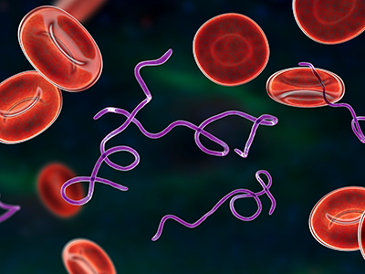
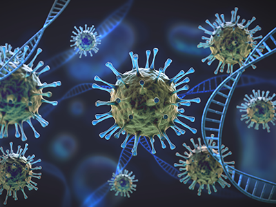
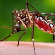
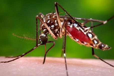
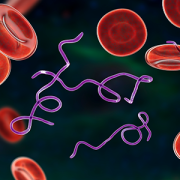
Lyme disease is caused by the bacterium Borrelia burgdorferi.

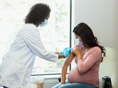
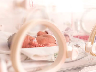
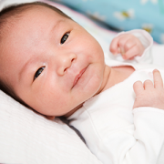
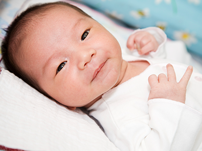
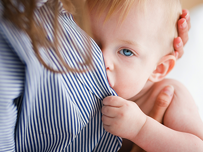
Understanding the effects of Lyme disease on the developing fetal brain is essential to ensure timely prenatal and postnatal treatments to protect the fetus and newborn. In response to this need, Children’s National Hospital is leading a pilot study to establish the groundwork needed for a larger study to determine the effect of in utero exposure to Lyme disease on pregnancy and early childhood neurodevelopmental outcomes.

Why it’s important

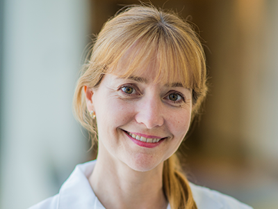
“Insect-borne illnesses have more than doubled during the last twenty years as a result of multiple factors including environmental changes,” says Sarah Mulkey, M.D., Ph.D., principal investigator of the study and prenatal-neonatal neurologist at Children’s National Hospital. Dr. Mulkey is the Co-Director of the Congenital Infection Program at Children’s National Hospital and has led a long-term child outcome study of the mosquito-borne Zika virus infection in pregnancy and impacts on child neurodevelopment. She is now adding Lyme disease to her work on pregnancy and child outcomes. Lyme disease is transmitted to humans via an infected tick and can be acquired throughout much of the United States. “Very little is known about Lyme disease in pregnancy and if and how it may affect the fetus and impact child neurodevelopment,” said Dr. Mulkey.

This pilot study is funded by the Clinical Trials Network for Lyme and other Tickborne Diseases (CTN), supported by the Steven and Alexandra Cohen Foundation. Roberta DeBiasi, M.D., M.S., division chief of infectious diseases at Children’s National, is a CTN Node Principal Investigator. The study team at Children’s National works closely with members of the CTN as well as other Lyme disease-focused organizations and nonprofits to make sure that the pilot study addresses Lyme disease advocates’ research priorities in a scientifically rigorous way. “This is an important opportunity for us to engage with advocacy groups and others who have been stressing the need for this type of research for decades,” says Meagan Williams, M.S.P.H., C.C.R.C., the senior research coordinator for the study. “We know how important it is to maintain a patient-centered and trauma-sensitive lens in our work with families impacted by Lyme disease during pregnancy. We’ve been working hard to build this study in a way that centers our participants’ lived experiences and focuses on the topics they find important. We’re very grateful to have the support of Lyme disease focused organizations and advocates as we embark on this study because they know and have expressed exactly where the gaps are in the literature. It’s our job to do the work to fill those gaps.”

The big picture

This pilot study aims to build upon existing research, case studies and advocacy to assess developmental and other family impacts of Lyme disease exposure during pregnancy.

Especially as tick season begins in Washington, D.C., and surrounding regions, the investigators are determined to make sure that all eligible people diagnosed with Lyme disease during their pregnancy are aware of the study and have the opportunity to participate. Pregnant volunteers can sign up for participation in the study here. Volunteers may be eligible to join if they are currently pregnant, live in the U.S. or Canada and were diagnosed with Lyme disease during pregnancy or have post-treatment Lyme disease syndrome (PTLDS).

Anyone interested in sharing information about the study with their clients or colleagues may email mewilliams@childrensnational.org for access to the team’s recruitment toolkit that includes flyers, FAQs and other resources. “We have resources available for clinicians, public health professionals, friends and family members and anyone else who may be able to help us identify participants and spread the word about this important study,” Williams says.

The study is registered on ClinicalTrials.gov (NCT06026969). If you have questions about the study, please reach out to Meagan at mewilliams@childrensnational.org or email cnhlymestudy@gmail.com.

stressed pregnant mom

Pandemic stress reshapes the placentas of expectant moms

stressed pregnant mom

Elevated maternal stress during the COVID-19 pandemic changed the structure, texture and other qualities of the placenta in pregnant mothers.

Elevated maternal stress during the COVID-19 pandemic changed the structure, texture and other qualities of the placenta in pregnant mothers – a critical connection between mothers and their unborn babies – according to new research from the Developing Brain Institute at Children’s National Hospital.

Published in Scientific Reports, the findings spotlight the underappreciated link between the mental health of pregnant mothers and the health of the placenta – a critical organ that develops during pregnancy to nourish and protect babies. The long-term neurodevelopmental impact on their children is under investigation.

“During the pandemic, mothers were exposed to a litany of negative stressors including social distancing, fear of dying, financial insecurity and more,” said Catherine Limperopoulos, Ph.D., chief and director of the Developing Brain Institute, which led the research. “We now know that this vital organ was changed for many mothers, and it’s essential that we continue to investigate the impact this may have had on children who were born during this global public health crisis.”

The big picture

Dr. Limperopoulos’s team compared magnetic resonance imaging (MRI) of 165 women who were pregnant before March 2020 to 63 women who became pregnant during the pandemic. Those pregnant during the pandemic were not knowingly exposed to COVID-19, and they collectively scored significantly higher on questionnaires measuring stress and depression. They were recruited at Children’s National as part of a clinical trial aimed at reducing pregnant women’s elevated stress levels during the pandemic.

The placenta is a temporary organ that grows during pregnancy to provide oxygen, nutrients and immunological protection to babies, and its health is vital to the well-being of the developing fetus. The data showed key changes in how the placenta grew and developed among women pregnant during the pandemic, especially when compared to placental growth and development among women who were pregnant before the pandemic. Changes in placental development also were associated with the infant’s birth weight at delivery. Importantly, these changes seem to be connected to maternal stress and depression symptoms.

Taken as a whole, the findings suggest that the disturbances measured on placental development in the womb may influence the placenta’s ability to support fetal health and wellness. “We are continuing to follow up on these mother-baby dyads to determine the long-term functional significance of these placental changes in utero,” Dr. Limperopoulos said.

Studies have shown that the placenta adapts to negative changes in the maternal environment and mental health status, and disruptions in placental function impact infant brain development and children’s neurobehavior and temperament.

The patient benefit

Dr. Limperopoulos’s research studying childbirth amid the pandemic builds on her extensive work investigating the impact of maternal stress on unborn children, including its adverse effect on brain structure and biochemistry. She’s also working on treatments and interventions to better support new families. Her program, DC Mother-Baby Wellness, brings together community partners to provide wrap-around care to expectant and new moms with elevated scores for stress, anxiety and depression.

“When identified early, maternal stress is a modifiable risk factor that can be treated with psychotherapy, social support and other personalized, evidence-based interventions,” Dr. Limperopoulos said. “We look forward to continued research in this area to better understand the mechanisms behind these biological changes and the needs of mothers and children who are born during pandemics, natural disasters and other significantly stressful events.”

coronavirus and DNA

Will SARS-CoV-2 during pregnancy impact child’s neurodevelopment?

coronavirus and DNA

Sarah Mulkey, M.D., prenatal-neonatal neurologist at Children’s National, will lead the neurodevelopmental evaluations of the infants born to mothers with SARS-CoV-2 infection during pregnancy to understand any long-term neurological effects in offspring.

Scientists led by the Lieber Institute for Brain Development are studying how a mother’s SARS-CoV-2 infection during pregnancy affects the biology of the placenta and the corresponding trajectory of the child’s brain development, including the risk for neurodevelopmental disorders such as schizophrenia and autism. The work is made possible by a $3 million, five-year grant from the Eunice Kennedy Shriver National Institute of Child Health & Human Development, part of the National Institutes of Health.

The project stems from a collaboration between the Lieber Institute for Brain Development on the Johns Hopkins medical campus in Baltimore, Children’s National Hospital in Washington, D.C., and the Women’s Health Integrated Research Center at Inova Health System in Virginia.

The big picture

The group aims for a clearer picture of how a mother’s SARS-CoV-2 infection during pregnancy affects neurodevelopment in utero, the effects of which may manifest early in a child’s life. The researchers hope to understand how the infection interacts with other factors relevant to brain development, including genomic risk for neurodevelopmental disorders, maternal stress and social determinants of health.

The team will study whether the relationship between maternal SARS-CoV-2 infection and offspring brain development is mediated by changes in the biology of the placenta and the activation of the mother’s immune system. They will also gauge any differences in the effects of SARS-CoV-2 between female and male children and in the offspring of vaccinated and unvaccinated mothers.

Why it matters

Preliminary data show that pregnant people with symptomatic SARS-CoV-2 infections are more likely to have a preterm delivery, abnormalities in the placenta and prenatal and perinatal complications such as preeclampsia and fetal growth restriction. All these complications have been found to increase a child’s risk of neurodevelopmental disorders later in life.

What we hope to discover

Sarah Mulkey, M.D., prenatal-neonatal neurologist at Children’s National, will lead the neurodevelopmental evaluations of the infants born to mothers with SARS-CoV-2 infection during pregnancy to understand any long-term neurological effects in offspring. The researchers will evaluate the children’s neurodevelopment at both 24 and 36 months of age. This work builds upon Dr. Mulkey’s longitudinal neurodevelopmental evaluations in children exposed to Zika virus in utero.

“What we’ve learned is that even when babies don’t have Zika-virus-related birth defects, we still find differences in early child development compared to children who weren’t exposed to Zika virus,” said Dr. Mulkey. “With SARS-CoV-2, there is still so much we don’t know. But by better understanding the long-term impact of COVID exposure during pregnancy, we can ultimately find ways to prevent adverse outcomes.”

pregnant woman getting vaccinated

COVID-19 vaccine may protect pregnant women from SARS-CoV-2 placentitis and stillbirth

pregnant woman getting vaccinated

In a new article published in the American Journal of Obstetrics & Gynecology, researchers conclude that the vaccine not only protects pregnant women but may also be lifesaving for their unborn children.

Stillbirth is a recognized complication of COVID-19 in pregnant women caused by harmful changes to the placenta induced by the virus. Termed SARS-CoV-2 placentitis, it can render the placenta incapable of providing oxygen to the fetus, leading to stillbirth and neonatal death. Researchers now suggest that pregnant women who get the COVID-19 vaccine may be protected from SARS-CoV-2 placentitis and stillbirth. In a new article published in the American Journal of Obstetrics & Gynecology, researchers conclude that the vaccine not only protects pregnant women but may also be lifesaving for their unborn children.

The extensive examination of published literature involved reviewing nearly 100 papers looking at COVID-19’s impacts on pregnant women and the effects on the placenta and pregnancy outcome. Sarah Mulkey, M.D., Ph.D., a prenatal-neonatal neurologist in the Division of Prenatal Pediatrics at Children’s National Hospital and co-author of the article, says the findings make a strong case for vaccination.

“The COVID-19 virus fortunately does not cause birth defects like other viruses such Zika, but it can cause severe injury to the placenta that can result in stillbirth and other pregnancy complications,” says Dr. Mulkey. “I hope patients who are pregnant or planning to become pregnant can learn how the COVID vaccine may help keep them and their baby healthy throughout pregnancy from some of the worst effects of this virus.”

While stillbirths can have many causes, the data analyzed supports that the COVID-19 vaccine is beneficial for pregnancies and for reducing the risk of stillbirth by reducing the risk of the virus impacting the placenta.

“In the multiple reports of SARS-CoV-2 placentitis that have been associated with stillbirths and neonatal deaths, none of the mothers had received COVID-19 vaccinations,” says David Schwartz, M.D., lead author, epidemiologist and perinatal pathologist. “And although not constituting proof, we’re not aware, either personally, via collegial networks, or in the published literature, of any cases of SARS-CoV-2 placentitis causing stillbirths among pregnant women having received the COVID-19 vaccine.”

Earlier in 2022, Dr. Schwartz led a team from 12 countries that found SARS-CoV-2 placentitis destroyed an average of 77.7% of placental tissue, resulting in placental insufficiency and fetal death, all occurring in unvaccinated mothers.

Fortunately, the large majority of pregnancies affected by a COVID-19 infection do not result in stillbirth. The development of SARS-CoV-2 placentitis is complex and likely involves both viral and immunological factors. The characteristics of a SARS-CoV-2 variant may also affect risk.

“Placental pathology is an important component in understanding the pathophysiology of SARS-CoV-2 infection during pregnancy,” says Dr. Mulkey.

As part of the Congenital Infection Program at Children’s National, Dr. Mulkey has been following infants born to mothers who had SARS-CoV-2 infection during pregnancy since the beginning of the pandemic. She will present the results of the early neurodevelopment of these infants at ID Week in Washington, D.C., on Oct. 22, 2022. Dr. Mulkey will also lead the neurodevelopmental follow-up of a large cohort of infants born to mothers with SARS-CoV-2 infection during pregnancy to better understand any long-term neurological effects to offspring.

The study builds upon Dr. Mulkey’s longitudinal studies on Zika virus infection in pregnancy and long-term impacts on the child that is funded by the Thrasher Research Fund.

pregnant hispanic woman

COVID-19’s impact on pregnant women and their babies

pregnant hispanic woman

While pregnant women are at higher risk of severe illness, coronavirus can also trigger inflammatory and vascular responses in the placenta during critical periods of fetal development in symptomatic and asymptomatic cases.

Pregnant women should get vaccinated to minimize the detrimental health effects COVID-19 has on the placenta, the fetus and the newborn, states Roberta L. DeBiasi, M.D., M.S., division chief of Pediatric Infectious Diseases at Children’s National Hospital in an editorial published in The Journal of Infectious Diseases.

The editorial provides a comprehensive review of what is known about the harmful effects of SARS-CoV-2 infection in pregnant women themselves, the effects on their newborns, the negative impact on the placenta and what still is unknown amid the rapidly evolving field. The safety and efficacy of vaccination of pregnant women are also addressed.

While pregnant women are at higher risk of severe illness, the virus can also trigger inflammatory and vascular responses in the placenta during critical periods of fetal development in symptomatic and asymptomatic cases.

In this piece, Dr. DeBiasi comments on two related studies published in the same issue, Guan et al. and Shook et al., demonstrating pathologic findings in women’s placentas who had COVID-19 during pregnancy. Guan et al. published a detailed analysis of a stillbirth resulting from the delta variant infection during the third trimester.

“The authors present a highly plausible mechanism of stillbirth, namely that the virus-induced proinflammatory state ultimately led to placental abruption,” said Dr. DeBiasi.

Shook et al. presented a case series of pregnant women infected with the delta variant associated with stillbirth in two cases and one with severe neonatal illness.

“Taking the studies together, it’s evident that if a pregnant woman gets COVID-19 they’re at an increased risk of severe infection,” says DeBiasi. “They’re also at increased risk of adverse pregnancy outcomes, due to effects on the placenta, which may vary with specific circulating variants.”

Previous studies have documented that the placenta may be detrimentally affected by SARS-CoV-2 infection of the mother. However, maternal comorbidities such as hypertension, preeclampsia and gestational diabetes could also contribute to these findings.

“Despite these previous studies, the precise mechanisms of placental injury are still not clear and require further evaluation,” says Dr. DeBiasi. “Future research should include appropriate controls to better discern nonspecific versus SARS-CoV-2 specific effects and mechanisms of injury.”

Even though these potential risks exist, the vaccination rate among pregnant women is low. Dr. DeBiasi writes that recent publications have demonstrated vaccine efficacy and safety during pregnancy through programs that tracked the use in pregnant women. This data supports that COVID-19 vaccine offers another layer of protection to pregnant women since infants are not yet eligible for vaccination despite the fact that the youngest infants and children are among the most at risk among children for hospitalization.

pregnant woman getting a checkup

Children’s National awarded $4.2 million to lead maternal mental health research programs

pregnant woman getting a checkup

Mothers and their babies often experience stress, depression and anxiety, which impacts the infant’s brain development.

Children’s National Hospital announces a $4.2 million funding award from the Patient-Centered Outcomes Research Institute (PCORI) to support maternal mental health research. The Developing Brain Institute at Children’s National will lead a new program that seeks to advance perinatal mental health and well-being while addressing racial disparities in access to resources that could boost positive health outcomes for women with few opportunities.

Mothers and their babies often experience stress, depression and anxiety, which impacts the infant’s brain development. Maternal psychological distress is more pronounced among low-income mothers — a health disparity that was exacerbated during the COVID-19 pandemic.

“The new fund will support many scientific research portfolios, including our project that will ensure pregnant women in D.C. get the care they need and deserve,” said Catherine Limperopoulos, Ph.D., director of The Developing Brain Institute at Children’s National and co-principal investigator of the project.

“I’m honored to be working alongside Dr. Limperopoulos and our partners. Collectively, our team aims to meet the needs of African American pregnant and postpartum women and their families during this important transition in their lives by providing services to address social determinants of health and prevent and treat maternal distress,” said Huynh-Nhu Le, Ph.D., the co-principal investigator of the project and professor in the Clinical Psychology program, part of the department of Psychological and Brain Sciences at George Washington University.

Cognitive-behavioral intervention, patient navigation and peer support, such tailored strategies developed in the program will provide effective mental health screening and care for 1,000 mothers living in Washington D.C. that is responsive to their cultural, social, environmental, behavioral and medical needs. The participants will access the resources either online or in-person, depending on the type of assistance that fits their lifestyle.

“I am overjoyed that PCORI has provided this essential funding, giving life to our project. The research done here will have a grand effect! Our goals are ambitious: To dissect all aspects of maternal health, beyond just mental health, literally creating a detailed timeline of events a mother can anticipate experiencing from pregnancy, at delivery and postpartum,” said Shanae Bond, one of the women whose firsthand experience giving birth in D.C. informed the study design. “With the maternal health crisis we are currently facing, it’s imperative to gain this type of insight to not only support mothers but to learn how they wish to be supported and how to best improve the care they receive – based on how it impacts, improves (or impairs) their lives,” said Bond.

The multidisciplinary group includes doctors, midwives, psychologists, advisors, community leaders and four prenatal care centers, MedStar Washington Hospital Center, Howard University, The George Washington University and Unity Health Care.

“Our initiative brings together obstetrics, pediatrics, and mental health care in an integrated care model. This collaboration brings early identification and immediate care coordination to its rightful place at the center of care,” said Loral Patchen, Ph.D., CNM, vice chair, Innovation and Community Programs at MedStar Washington Hospital Center. “The prenatal period offers an opportunity for us to support emotional healing, build coping strategies, and offer a safe space for people to prepare for the complex transitions that accompany childbearing. Offering services prior to delivery optimizes opportunity for strong parent-infant attachment and mitigates potential disruptions.”

Kristin L. Atkins, M.D., FACOG, assistant professor in the Obstetrics and Gynecology Department at Howard University College of Medicine, mentioned that Howard University is honored to partner with Children’s National Hospital. “The new program will help discover more about prenatal care interventions related to maternal mental health and how they may impact fetal and pediatric brain development,” said Dr. Atkins. “We are just discovering the impact of long-standing stress on health and well-being, and this starts in utero.”

To Jennifer Keller, M.D., MPH, FACOG, associate professor at The George Washington University School of Medicine & Health Sciences, this project is essential. “The events of the last year have had a profound impact on families in this city,” said Dr. Keller. “This project begins at a time of critical mental health needs for pregnant people in D.C.”

Siobhan Burke, M.D., director of OB/GYN at Unity Health Care, is also thrilled to be part of this partnership. “We all know underlying stressors such as financial difficulties, housing instability and systemic racism can impact health, but it’s important to find out what these things do to the developing fetus and to explore strategies to make lives better,” said Dr. Burke.

In 2020, Children’s National established The Clark Parent & Child Network funded by a $36 million investment from the A. James & Alice B. Clark Foundation. The Clark Network aims to provide families with greater access to mental health care and community resources. New projects like the D.C. mother-infant behavioral wellness program underwritten by PCORI funding will become natural extensions of this essential work.

“This project was selected for PCORI funding not only for its scientific merit and commitment to engaging patients and other stakeholders, but also for its potential to fill an important gap in our health knowledge and give people information to help them weigh the effectiveness of their care options,” said PCORI Executive Director Nakela L. Cook, M.D., M.P.H.. “We look forward to following the study’s progress and working with Children’s National Hospital to share the results.”

This $4.2 million PCORI funding has been approved pending completion of a business and programmatic review by PCORI staff and issuance of a formal award contract.


newborn in incubator

A bronchopulmonary dysplasia primer to guide clinicians and researchers

newborn in incubator

Six months in the writing, the “Bronchopulmonary Dysplasia Primer” published recently by Nature Reviews will be the gold standard review on this topic for years to come.

The term bronchopulmonary dysplasia, or BPD, was first coined in 1967 to describe a chronic lung disease of preterm newborns after treatment with supplemental oxygen via mechanical ventilation in an effort to save their lives. Back then, infants had 50-50 odds of surviving.

In the intervening years, survival has improved and the characteristics of BPD have evolved. Now, BPD is the most common complication of preterm birth for infants born at fewer than 28 weeks’ gestation, as more and more newborns survive premature birth. Hence, the primer.

“The contributing authors are some of the biggest thinkers on this topic,” says Robin H. Steinhorn, M.D., senior vice president, Center for Hospital-Based Specialties, at Children’s National Hospital and author of the section about BPD diagnosis, screening and prevention. “This document will guide clinical education and investigators in the field of BPD. I anticipate this will be the definitive review article on the subject for the next several years.”

Gestational age and low birth weight remain the most potent predictors of BPD. Some 50,000 extremely low gestational age newborns are born each year in the U.S. About 35% will develop some degree of BPD, according to the primer authors.

These newborns are introduced to life outside the womb well before their lungs are ready. Indeed, the pulmonary surfactants needed for normal lung function – a complex mixture of phospholipids that reduce surface tension within the lungs – don’t differentiate until late in pregnancy. Infants who persistently need respiratory support after the 14th day of life are at the highest risk of being diagnosed with BPD at 36 weeks, the coauthors note.

A number of complicating factors can come into play, including maternal diet; fetal exposure to maternal smoking and infection; structural issues such as pre-eclampsia; acute injury from mechanical ventilation and supplemental oxygen; as well as the body’s halting efforts to repair injured, inflamed lung tissue.

“The good news is the number of the smallest and youngest preterm infants who survive extreme preterm birth has steadily increased. Neonatal intensive care units, like our award-winning NICU, now routinely care for babies born at 22 weeks’ gestation,” Dr. Steinhorn says.

Treatment strategies include:

  • Reducing exposure to intubation and ventilation.
  • Leveraging respiratory stimulants, like caffeine.
  • Postnatal steroid therapy.

“Children’s National Hospital is the only center in our immediate region that provides comprehensive care for infants and children with severe BPD,” Dr. Steinhorn adds. “As the population of vulnerable and fragile infants has grown, we have invested in the equipment and the personnel – including at the Hospital for Sick Children Pediatric Center (HSC) – to create a very safe and supportive environment that improves survival and quality of life.”

Some preterm infants spend their first 9 to 10 months of life at Children’s National, and their days are filled with concentrated physical, occupational and speech therapy, as well as music and play therapy to hasten their rehabilitation.

Once their medical condition stabilizes, they transition to HSC to focus more intently on rehabilitation.

“We see HSC as filling a very important role in their care. When our children graduate to HSC, they are going for ongoing care of their lung disease, but also their ongoing rehabilitation. At HSC, they focus on creating the most normal life that we can possibly create and, over time, that is a life free of ventilators and tracheostomy tubes.”

In addition to Dr. Steinhorn, BPD Primer co-authors include Bernard Thébaud, Children’s Hospital of Eastern Ontario; Kara N. Goss, University of Wisconsin-Madison; Matthew Laughon, The University of North Carolina at Chapel Hill; Jeffrey A. Whitsett and Alan H. Jobe, Cincinnati Children’s Hospital Medical Center; Steven H. Abman, Children’s Hospital Colorado;  Judy L. Aschner, Joseph M. Sanzari Children’s Hospital; Peter G. Davis, The Royal Women’s Hospital; Sharon A. McGrath- Morrow, Johns Hopkins University School of Medicine; and Roger F. Soll, University of Vermont.

Financial support for the research described in this post was provided by the National Institutes of Health under grant Nos. U01HL122642, U01HL134745, RO1HL68702, R01HL145679, U01HL12118-01 and K24 HL143283; the Australian National Health and Medical Research Council; the Canadian Institute for Health Research; Stem Cell Network and the Ontario Institute for Regenerative Medicine.

Catherine Limperopoulos

Stressful pregnancies can leave fingerprint on fetal brain

Catherine Limperopoulos

“We were alarmed by the high percentage of pregnant women with a diagnosis of a major fetal heart problem who tested positive for stress, anxiety and depression,” says Catherine Limperopoulos, Ph.D., director of the Center for the Developing Brain at Children’s National and the study’s corresponding author.

When a diagnosis of fetal congenital heart disease causes pregnant moms to test positive for stress, anxiety and depression, powerful imaging can detect impaired development in key fetal brain regions, according to Children’s National Hospital research published online Jan. 13, 2020, in JAMA Pediatrics.

While additional research is needed, the Children’s National study authors say their unprecedented findings underscore the need for universal screening for psychological distress as a routine part of prenatal care and taking other steps to support stressed-out pregnant women and safeguard their newborns’ developing brains.

“We were alarmed by the high percentage of pregnant women with a diagnosis of a major fetal heart problem who tested positive for stress, anxiety and depression,” says Catherine Limperopoulos, Ph.D., director of the Center for the Developing Brain at Children’s National and the study’s corresponding author. “Equally concerning is how prevalent psychological distress is among pregnant women generally. We report for the first time that this challenging prenatal environment impairs regions of the fetal brain that play a major role in learning, memory, coordination, and social and behavioral development, making it all the more important for us to identify these women early during pregnancy to intervene,” Limperopoulos adds.

Congenital heart disease (CHD), structural problems with the heart, is the most common birth defect. Still, it remains unclear how exposure to maternal stress impacts brain development in fetuses with CHD.

The multidisciplinary study team enrolled 48 women whose unborn fetuses had been diagnosed with CHD and 92 healthy women with uncomplicated pregnancies. Using validated screening tools, they found:

  • 65% of pregnant women expecting a baby with CHD tested positive for stress
  • 27% of women with uncomplicated pregnancies tested positive for stress
  • 44% of pregnant women expecting a baby with CHD tested positive for anxiety
  • 26% of women with uncomplicated pregnancies tested positive for anxiety
  • 29% of pregnant women expecting a baby with CHD tested positive for depression and
  • 9% women with uncomplicated pregnancies tested positive for depression

All told, they performed 223 fetal magnetic resonance imaging sessions for these 140 fetuses between 21 and 40 weeks of gestation. They measured brain volume in cubic centimeters for the total brain as well as volumetric measurements for key regions such as the cerebrum, cerebellum, brainstem, and left and right hippocampus.

Maternal stress and anxiety in the second trimester were associated with smaller left hippocampi and smaller cerebellums only in pregnancies affected by fetal CHD. What’s more, specific regions — the hippocampus head and body and the left cerebellar lobe – were more susceptible to stunted growth. The hippocampus is key to memory and learning, while the cerebellum controls motor coordination and plays a role in social and behavioral development.

The hippocampus is a brain structure that is known to be very sensitive to stress. The timing of the CHD diagnosis may have occurred at a particularly vulnerable time for the developing fetal cerebellum, which grows faster than any other brain structure in the second half of gestation, particularly in the third trimester.

“None of these women had been screened for prenatal depression or anxiety. None of them were taking medications. And none of them had received mental health interventions. In the group of women contending with fetal CHD, 81% had attended college and 75% had professional educations, so this does not appear to be an issue of insufficient resources,” Limperopoulos adds. “It’s critical that we routinely to do these screenings and provide pregnant women with access to interventions to lower their stress levels. Working with our community partners, Children’s National is doing just that to help reduce toxic prenatal stress for both the health of the mother and for the future newborns. We hope this becomes standard practice elsewhere.”

Adds Yao Wu, Ph.D., a research associate working with Limperopoulos at Children’s National and the study’s lead author: “Our next goal is exploring effective prenatal cognitive behavioral interventions to reduce psychological distress felt by pregnant women and improve neurodevelopment in babies with CHD.”

In addition to Limperopoulos and Wu , Children’s National study co-authors include Kushal Kapse, MS, staff engineer; Marni Jacobs, Ph.D., biostatistician; Nickie Niforatos-Andescavage, M.D., neonatologist; Mary T. Donofrio, M.D., director, Fetal Heart Program; Anita Krishnan, M.D., associate director, echocardiography; Gilbert Vezina, M.D., director, Neuroradiology Program; David Wessel, M.D., Executive Vice President and Chief Medical Officer; and Adré  J. du Plessis, M.B.Ch.B., director, Fetal Medicine Institute. Jessica Lynn Quistorff, MPH, Catherine Lopez, MS, and Kathryn Lee Bannantine, BSN, assisted with subject recruitment and study coordination.

Financial support for the research described in this post was provided by the National Institutes of Health under grant No. R01 HL116585-01 and the Thrasher Research Fund under Early Career award No. 14764.

doctor checking pregnant woman's belly

Novel approach to detect fetal growth restriction

doctor checking pregnant woman's belly

Morphometric and textural analyses of magnetic resonance imaging can point out subtle architectural deviations associated with fetal growth restriction during the second half of pregnancy, a first-time finding that has the promise to lead to earlier intervention.

Morphometric and textural analyses of magnetic resonance imaging (MRI) can point out subtle architectural deviations that are associated with fetal growth restriction (FGR) during the second half of pregnancy. The first-time finding hints at the potential to spot otherwise hidden placental woes earlier and intervene in a more timely fashion, a research team led by Children’s National Hospital faculty reports in Pediatric Research.

“We found reduced placental size, as expected, but also determined that the textural metrics are accelerated in FGR when factoring in gestational age, suggesting premature placental aging in FGR,” says Nickie Andescavage, M.D., a neonatologist at Children’s National and the study’s lead author. “While morphometric and textural features can discriminate placental differences between FGR cases with and without Doppler abnormalities, the pattern of affected features differs between these sub-groups. Of note, placental insufficiency with abnormal Doppler findings have significant differences in the signal-intensity metrics, perhaps related to differences of water content within the placenta.”

The placenta, an organ shared by the pregnant woman and the developing fetus, delivers oxygen and nutrients to the developing fetus and ferries away waste products. Placental insufficiency is characterized by a placenta that develops poorly or is damaged, impairing blood flow, and can result in still birth or death shortly after birth. Surviving infants may be born preterm or suffer early brain injury; later in life, they may experience cardiovascular, metabolic or neuropsychiatric problems.

Because there are no available tools to help clinicians identify small but critical changes in placental architecture during pregnancy, placental insufficiency often is found after some damage is already done. Typically, it is discovered when FGR is diagnosed, when a fetus weighs less than 9 of 10 fetuses of the same gestational age.

“There is a growing appreciation for the prenatal origin of some neuropsychiatric disorders that manifest years to decades later. Those nine months of gestation very much define the breath of who we later become as adults,” says Catherine Limperopoulos, Ph.D., director of MRI Research of the Developing Brain at Children’s National and the study’s senior author. “By identifying better biomarkers of fetal distress at an earlier stage in pregnancy and refining our imaging toolkit to detect them, we set the stage to be able to intervene earlier and improve children’s overall outcomes.”

The research team studied 32 healthy pregnancies and compared them with 34 pregnancies complicated by FGR. These women underwent up to two MRIs between 20 weeks to 40 weeks gestation. They also had abdominal circumference, fetal head circumference and fetal femur length measured as well as fetal weight estimated.

In pregnancies complicated by FGR, placentas were smaller, thinner and shorter than uncomplicated pregnancies and had decreased placental volume. Ten of 13 textural and morphometric features that differed between the two groups were associated with absolute birth weight.

“Interestingly, when FGR is diagnosed in the second trimester, placental volume, elongation and thickness are significantly reduced compared with healthy pregnancies, whereas the late-onset of FGR only affects placental volume,” Limperopoulos adds. “We believe with early-onset FGR there is a more significant reduction in the developing placental units that is detected by gross measures of size and shape. By the third trimester, the overall shape of the placenta seems to have been well defined so that primarily volume is affected in late-onset FGR.”

In addition to Dr. Andescavage and Limperopoulos, study co-authors include Sonia Dahdouh, Sayali Yewale, Dorothy Bulas, M.D., chief of the Division of Diagnostic Imaging and Radiology, and Biostatistician, Marni Jacobs, Ph.D., MPH, all of Children’s National; Sara Iqbal, of MedStar Washington Hospital Center; and Ahmet Baschat, of Johns Hopkins Center for Fetal Therapy.

Financial support for research described in this post was provided by the National Institutes of Health under award number 1U54HD090257, R01-HL116585, UL1TR000075 and KL2TR000076, and the Clinical-Translational Science Institute-Children’s National.

Andrea Gropman

$5M in federal funding to help patients with urea cycle disorders

Andrea Gropman

Andrea L. Gropman, M.D.: We have collected many years of longitudinal clinical data, but with this new funding now we can answer questions about these diseases that are meaningful on a day-to-day basis for patients with urea cycle disorders.

An international research consortium co-led by Andrea L. Gropman, M.D., at Children’s National Hospital has received $5 million in federal funding as part of an overall effort to better understand rare diseases and accelerate potential treatments to patients.

Urea cycle disorder, one such rare disease, is a hiccup in a series of biochemical reactions that transform nitrogen into a non-toxic compound, urea. The six enzymes and two carrier/transport molecules that accomplish this essential task reside primarily in the liver and, to a lesser degree, in other organs.

The majority of patients have the recessive form of the disorder, meaning it has skipped a generation. These kids inherit one copy of an abnormal gene from each parent, while the parents themselves were not affected, says Dr. Gropman, chief of the Division of Neurodevelopmental Pediatrics and Neurogenetics at Children’s National. Another more common version of the disease is carried on the X chromosome and affects boys more seriously that girls, given that boys have only one X chromosome.

Regardless of the type of urea cycle disorder, when the urea cycle breaks down, nitrogen converts into toxic ammonia that builds up in the body (hyperammonemia), particularly in the brain. As a result, the person may feel lethargic; if the ammonia in the bloodstream reaches the brain in high concentrations, the person can experience seizures, behavior changes and lapse into a coma.

Improvements in clinical care and the advent of effective medicines have transformed this once deadly disease into a more manageable chronic ailment.

“It’s gratifying that patients diagnosed with urea cycle disorder now are surviving, growing up, becoming young adults and starting families themselves. Twenty to 30 years ago, this never would have seemed conceivable,” Dr. Gropman says. “We have collected many years of longitudinal clinical data, but with this new funding now we can answer questions about these diseases that are meaningful on a day-to-day basis for patients with urea cycle disorders.”

In early October 2019, the National Institutes of Health (NIH) awarded the Urea Cycle Disorders Consortium for which Dr. Gropman is co-principal investigator a five-year grant. This is the fourth time that the international Consortium of physicians, scientists, neuropsychologists, nurses, genetic counselors and researchers has received NIH funding to study this group of conditions.

Dr. Gropman says the current urea cycle research program builds on a sturdy foundation built by previous principal investigators Mendel Tuchman, M.D., and Mark Batshaw, M.D., also funded by the NIH. While previous rounds of NIH funding powered research about patients’ long-term survival prospects and cognitive dysfunction, this next phase of research will explore patients’ long-term health.

Among the topics they will study:

Long-term organ damage. Magnetic resonance elastrography (MRE) is a state-of-the-art imaging technique that combines the sharp images from MRI with a visual map that shows body tissue stiffness. The research team will use MRE to look for early changes in the liver – before patients show any symptoms – that could be associated with long-term health impacts. Their aim is spot the earliest signs of potential liver dysfunction in order to intervene before the patient develops liver fibrosis.

Academic achievement. The research team will examine gaps in academic achievement for patients who appear to be underperforming to determine what is triggering the discrepancy between their potential and actual scholastics. If they uncover issues such as learning difficulties or mental health concerns like anxiety, there are opportunities to intervene to boost academic achievement.

“And if we find many of the patients meet the criteria for depression or anxiety disorders, there are potential opportunities to intervene.  It’s tricky: We need to balance their existing medications with any new ones to ensure that we don’t increase their hyperammonemia risk,” Dr. Gropman explains.

Neurologic complications. The researchers will tap continuous, bedside electroencephalogram, which measures the brain’s electrical activity, to detect silent seizures and otherwise undetectable changes in the brain in an effort to stave off epilepsy, a brain disorder that causes seizures.

“This is really the first time we will examine babies’ brains,” she adds. “Our previous imaging studies looked at kids and adults who were 6 years and older. Now, we’re lowering that age range down to infants. By tracking such images over time, the field has described the trajectory of what normal brain development should look like. We can use that as a background and comparison point.”

In the future, newborns may be screened for urea cycle disorder shortly after birth. Because it is not possible to diagnose it in the womb in cases where there is no family history, the team aims to better counsel families contemplating pregnancy about their possible risks.

Research described in this post was underwritten by the NIH through its Rare Diseases Clinical Research Network.

allopregnanolone molecule

Autism spectrum disorder risk linked to insufficient placental steroid

allopregnanolone molecule

A study led by Children’s National Hospital and presented during Neuroscience 2019 finds that loss of allopregnanolone, a key hormone supplied by the placenta, leads to long-term structural alterations of the cerebellum – a brain region essential for smooth motor coordination, balance and social cognition – and increases the risk of developing autism.

An experimental model study suggests that allopregnanolone, one of many hormones produced by the placenta during pregnancy, is so essential to normal fetal brain development that when provision of that hormone decreases – as occurs with premature birth – offspring are more likely to develop autism-like behaviors, a Children’s National Hospital research team reports at the Neuroscience 2019 annual meeting.

“To our knowledge, no other research team has studied how placental allopregnanolone (ALLO) contributes to brain development and long-term behaviors,” says Claire-Marie Vacher, Ph.D., lead author. “Our study finds that targeted loss of ALLO in the womb leads to long-term structural alterations of the cerebellum – a brain region that is essential for motor coordination, balance and social cognition ­– and increases the risk of developing autism,” Vacher says.

According to the Centers for Disease Control and Prevention, about 1 in 10 infants is born preterm, before 37 weeks gestation; and 1 in 59 children has autism spectrum disorder.

In addition to presenting the abstract, on Monday, Oct. 21, Anna Penn, M.D., Ph.D., the abstract’s senior author, will discuss the research with reporters during a Neuroscience 2019 news conference. This Children’s National abstract is among 14,000 abstracts submitted for the meeting, the world’s largest source of emerging news about brain science and health.

ALLO production by the placenta rises in the second trimester of pregnancy, and levels of the neurosteroid peak as fetuses approach full term.

To investigate what happens when ALLO supplies are disrupted, a research team led by Children’s National created a novel transgenic preclinical model in which they deleted a gene essential in ALLO synthesis. When production of ALLO in the placentas of these experimental models declines, offspring had permanent neurodevelopmental changes in a sex- and region-specific manner.

“From a structural perspective, the most pronounced cerebellar abnormalities appeared in the cerebellum’s white matter,” Vacher adds. “We found increased thickness of the myelin, a lipid-rich insulating layer that protects nerve fibers. From a behavioral perspective, male offspring whose ALLO supply was abruptly reduced exhibited increased repetitive behavior and sociability deficits – two hallmarks in humans of autism spectrum disorder.”

On a positive note, providing a single ALLO injection during pregnancy was enough to avert both the cerebellar abnormalities and the aberrant social behaviors.

The research team is now launching a new area of research focus they call “neuroplacentology” to better understand the role of placenta function on fetal and newborn brain development.

“Our team’s data provide exciting new evidence that underscores the importance of placental hormones on shaping and programming the developing fetal brain,” Vacher notes.

  • Neuroscience 2019 presentation
    Sunday, Oct. 20, 9:30 a.m. (CDT)
    “Preterm ASD risk linked to cerebellar white matter changes”
    Claire-Marie Vacher, lead author; Sonia Sebaoui, co-author; Helene Lacaille, co-author; Jackie Salzbank, co-author; Jiaqi O’Reilly, co-author; Diana Bakalar, co-author; Panagiotis Kratimenos, M.D., neonatologist and co-author; and Anna Penn, M.D., clinical neonatologist and developmental neuroscientist and senior author.

Paradoxical outcomes for Zika-exposed tots

In the midst of an unprecedented Zika crisis in Brazil, there were a few flickers of hope: Some babies appeared to be normal at birth, free of devastating birth defects that affected other Brazilian children exposed to the virus in utero.

In the midst of an unprecedented Zika crisis in Brazil, there were a few flickers of hope: Some babies appeared to be normal at birth, free of devastating birth defects that affected other Brazilian children exposed to the virus in utero. But according to a study published online July 8, 2019, in Nature Medicine and an accompanying commentary co-written by a Children’s National clinician-researcher, the reality for Zika-exposed infants is much more complicated.

Study authors led by Karin Nielsen-Saines at David Geffen UCLA School of Medicine followed 216 infants in Rio de Janeiro who had been exposed to the Zika virus during pregnancy, performing neurodevelopmental testing when the babies ranged in age from 7 to 32 months. These infants’ mothers had had Zika-related symptoms themselves, including rash.

Although many children had normal assessments, 29% scored below average in at least one domain of neurological development, including cognitive performance, fine and gross motor skills and expressive language, Sarah B. Mulkey, M.D., Ph.D., and a colleague write in a companion commentary published online by Nature Medicine July 29, 2019.

The study authors found progressively higher risks for developmental, hearing and eye abnormality depending on how early the pregnancy was at the time the infants were exposed. Because Zika virus has an affinity for immature neurons, even babies who were not born with microcephaly remained at continued risk for suffering abnormalities.

Of note, 24 of 49 (49%) infants who had abnormalities at birth went on to have normal test results in the second or third year of life. By contrast, 17 of 68 infants (25%) who had normal assessments at birth had below-average developmental testing or had abnormalities in hearing or vision by age 32 months.

“This work follows babies who were born in 2015 and 2016. It’s heartening that some babies born with abnormalities tested in the normal range later in life, though it’s unclear whether any specific interventions help to deliver these positive findings,” says Dr. Mulkey, a fetalneonatal neurologist in the Division of Fetal and Transitional Medicine at Children’s National in Washington, D.C. “And it’s quite sobering that babies who appeared normal at birth went on to develop abnormalities due to that early Zika exposure.”

It’s unclear how closely the findings apply to the vast majority of U.S. women whose Zika infections were asymptomatic.

“This study adds to the growing body of research that argues in favor of ongoing follow-up for Zika-exposed children, even if their neurologic exams were reassuring at birth,” Dr. Mulkey adds. “As Zika-exposed children approach school age, it’s critical to better characterize the potential implications for the education system and public health.”

In addition to Dr. Mulkey, the perspective’s senior author, William J. Muller, Northwestern University, was the commentary’s lead author.

zika virus

Neuroimaging essential for Zika cases

zika virus

About three years ago, Zika virus emerged as a newly recognized congenital infection, and a growing body of research indicates the damage it causes differs from other infections that occur in utero.

Seventy-one of 110 Brazilian infants at the highest risk for experiencing problems due to exposure to the Zika virus in the womb experienced a wide spectrum of brain abnormalities, including calcifications and malformations in cortical development, according to a study published July 31, 2019 in JAMA Network Open.

The infants were born at the height of Brazil’s Zika epidemic, a few months after the nation declared a national public health emergency. Already, many of the infants had been classified as having the severe form of congenital Zika syndrome, and many had microcephaly, fetal brain disruption sequence, arthrogryposis and abnormal neurologic exams at birth.

These 110 infants “represented a group of ZIKV-exposed infants who would be expected to have a high burden of neuroimaging abnormalities, which is a difference from other reported cohorts,” Sarah B. Mulkey, M.D., Ph.D., writes in an invited commentary published in JAMA Network Open that accompanies the Rio de Janeiro study. “Fortunately, many ZIKV-exposed infants do not have abnormal brain findings or a clinical phenotype associated with congenital Zika syndrome,” adds Dr. Mulkey, a fetalneonatal neurologist in the Division of Fetal and Transitional Medicine at Children’s National in Washington, D.C.

Indeed, a retrospective cohort of 82 women exposed to Zika during their pregnancies led by a research team at Children’s National found only three pregnancies were complicated by severe fetal brain abnormalities. Compared with the 65% abnormal computed tomography (CT) or magnetic resonance imaging (MRI) findings in the new Brazilian study, about 1 in 10 (10%) of babies born to women living in the continental U.S. with confirmed Zika infections during pregnancy had Zika-associated birth defects, according to the Centers for Disease Control and Prevention.

“There appears to be a spectrum of brain imaging abnormalities in ZIKV-exposed infants, including mild, nonspecific changes seen at cranial US [ultrasound], such as lenticulostriate vasculopathy and germinolytic cysts, to more significant brain abnormalities, such as subcortical calcifications, ventriculomegaly and, in its most severe form, thin cortical mantle and fetal brain disruption sequence,” Dr. Mulkey writes.

About three years ago, Zika virus emerged as a newly recognized congenital infection, and a growing body of research indicates the damage it causes differs from other infections that occur in utero. Unlike congenital cytomegalovirus infection, cerebral calcifications associated with Zika are typically subcortical, Dr. Mulkey indicates. What’s more, fetal brain disruption sequence seen in Zika-exposed infants is unusual for other infections that can cause microcephaly.

“Centered on the findings of Pool, et al, and others, early neuroimaging remains one of the most valuable investigations of the Zika-exposed infant,” Dr. Mulkey writes, including infants who are not diagnosed with congenital Zika syndrome.  She recommends:

  • Cranial ultrasound as the first-line imaging option for infants, if available, combined with neurologic and ophthalmologic exams, and brainstem auditory evoked potentials
  • Zika-exposed infants with normal cranial ultrasounds do not need additional imaging unless they experience a developmental disturbance
  • Zika-exposed infants with abnormal cranial ultrasounds should undergo further neuroimaging with low-dose cranial CT or brain MRI.

Autonomic nervous system appears to function well regardless of mode of childbirth

Late in pregnancy, the human body carefully prepares fetuses for the rigors of life outside the protection of the womb. Levels of cortisol, a stress hormone, ramp up and spike during labor. Catecholamines, another stress hormone, also rise at birth, helping to kick start the necessary functions that the baby will need to regulate breathing, heartbeat, blood pressure and energy metabolism levels at delivery. Oxytocin surges, promoting contractions for the mother during labor and stimulating milk production after the infant is born.

These processes also can play a role in preparing the fetal brain during the transition to life outside the womb by readying the autonomic nervous system and adapting its cerebral connections. The autonomic nervous system acts like the body’s autopilot, taking in information it needs to ensure that internal organs run steadily without willful action, such as ensuring the heart beats and eyelids blink at steady intervals. Its yin, the sympathetic division, stimulates body processes while its yang, the parasympathetic division, inhibits them.

Infants born preterm have reduced autonomic function compared with their full-term peers and also face possible serious neurodevelopmental impairment later in life. But is there a difference in autonomic nervous system function for full-term babies after undergoing labor compared with infants delivered via cesarean section (C-section)?

A team from the Children’s National Inova Collaborative Research Program (CNICA) – a research collaboration between Children’s National in Washington, D.C., and Inova Women’s and Children’s Hospital in Virginia – set out to answer that question in a paper published online July 30, 2019, in Scientific Reports.

They enrolled newborns who had experienced normal, full-term pregnancies and recorded their brain function and heart performance when they were about 2 days old. Infants whose conditions were fragile enough to require observation in the neonatal intensive care unit were excluded from the study. Of 167 infants recruited for the prospective cohort study, 118 newborns had sufficiently robust data to include them in the research.  Of these newborns:

  • 62 (52.5%) were born by vaginal delivery
  • 22 (18.6%) started out with vaginal delivery but ultimately switched to C-section based on failure to progress, failed labor induction or fetal intolerance to labor
  • And 34 (28.8%) were born by elective C-section.

The CNICA research team swaddled infants for comfort and slipped electrode nets over their tiny heads to simultaneously measure heart rate variability and electrocortical function through non-invasive techniques. The team hypothesized that infants who had been exposed to labor would have enhanced autonomic tone and higher cortical electroencephalogram (EEG) power than babies born via C-section.

“In a low-risk group of babies born full-term, the autonomic nervous system and cortical systems appear to function well regardless of whether infants were exposed to labor prior to birth,” says Sarah B. Mulkey, M.D., Ph.D., a fetalneonatal neurologist in the Division of Fetal and Transitional Medicine at Children’s National and the study’s lead author.

However, infants born by C-section following a period of labor had significantly increased accelerations in their heart rates. And the infants born by C-section during labor had significantly lower relative gamma frequency EEG at 25.2 hours old compared with the other two groups studied.

“Together these findings point to a possible increased stress response and arousal difference in infants who started with vaginal delivery and finished delivery with C-section,” Dr. Mulkey says. “There is so little published research about the neurologic impacts of the mode of delivery, so our work helps to provide a normal reference point for future studies looking at high-risk infants, including babies born preterm.”

Because the research team saw little differences in autonomic tone or other EEG frequencies when the infants were 1 day old, future research will explore these measures at different points in the newborns’ early life as well as the role of the sleep-wake cycle on heart rate variability.

In addition to Dr. Mulkey, study co-authors include Srinivas Kota, Ph.D., Rathinaswamy B. Govindan, Ph.D., Tareq Al-Shargabi, MSc, Christopher B. Swisher, BS, Laura Hitchings, BScM, Stephanie Russo, BS, Nicole Herrera, MPH, Robert McCarter, ScD, and Senior Author Adré  J. du Plessis, M.B.Ch.B., MPH, all of Children’s National; and Augustine Eze Jr., MS, G. Larry Maxwell, M.D., and Robin Baker, M.D., all of Inova Women’s and Children’s Hospital.

Financial support for research described in this post was provided by the National Institutes of Health National Center for Advancing Translational Sciences under award numbers UL1TR001876 and KL2TR001877.

Dr. Anitha John, third from right, director of the Washington Adult Congenital Heart Program, hosts the eighth-annual “Adult Congenital Heart Disease in the 21st Century” conference

CME spotlight: Treating adult congenital heart disease

Dr. Anitha John, third from right, director of the Washington Adult Congenital Heart Program, hosts the eighth-annual “Adult Congenital Heart Disease in the 21st Century” conference

Dr. Anitha John, third from right, director of the Washington Adult Congenital Heart Program, hosts the eighth-annual “Adult Congenital Heart Disease in the 21st Century” conference, which takes place Oct. 4-5, 2019.

A two-day continuing medical education (CME) conference for physicians and clinicians treating patients with adult congenital heart disease (ACHD) takes place Oct. 4-5, 2019, at the Bethesda Marriott in Bethesda, Maryland.

The eighth-annual conference, “Adult Congenital Heart Disease in the 21st Century,” hosted by Children’s National Health System and MedStar Washington Hospital Center provides a comprehensive review of the evaluation, diagnosis and management of ACHD, including guidelines to help ACHD patients manage a healthy pregnancy and clinical guidance about the progression of congenital heart disease (CHD) treatment from adolescence through adulthood.

Two tracks accommodate these themes, with the first focusing on a multidisciplinary approach clinicians can use to help ACHD patients assess risks for pregnancy complications, while planning and managing a healthy pregnancy, with input from cardiologists, anesthesiologists and maternal fetal medicine specialists. The second focuses on cardiac defects, starting with anatomical cardiac lessons with 3D heart models, then moves to imaging review, examining echocardiograms and MRI’s, and ends with clinical management review.

“This conference brings the best science and the most innovative approaches to treatment with questions doctors receive in the exam room,” says Anitha John, M.D., Ph.D., the conference organizer and director of the Washington Adult Congenital Heart program at Children’s National. “We’re inviting patients to join the afternoon of the second day of the CME conference again this year to support shared knowledge of these concepts, which supports lifelong treatment and education.”

Dr. John planned this year’s conference with the November 6 ACHD board exams in mind, integrating topics that will appear on the third ACHD certification exam issued by the American Board of Internal Medicine.

At this year’s CME conference, more than a dozen faculty members, including several physicians and nurses from Children’s National, will guide lectures to help attendees meet 13 objectives, from understanding the prevalence of congenital heart disease and its complications to learning about when surgical interventions and referrals to specialists are necessary.

Attendees will review new and innovative PAH therapies, mechanical support therapies, catheter-based interventional procedures and appraise the use of pacemaker and defibrillator therapy among adults with CHD.

Patients and families attending the patient sessions, held from 12:30 to 3:45 p.m. on Saturday, October 5, have a chance to participate in three sessions that support the medical and social needs of ACHD patients. Topics range from workshops that address the neurodevelopment and psychosocial factors of living with a congenital heart defect to sessions that focus on reproductive options for patients and personalized lifestyle recommendations, including fitness and exercise guidelines.

“To support cardiovascular health throughout the lifespan, it helps to educate patients about their heart’s structure and unique needs,” notes Dr. John. “We want to spark a dialogue now and have future conversations with patients, especially while they are young.”

The American College of Cardiology/American Heart Association Task Force on Clinical Practice Guidelines updated ACHD treatment recommendations in August 2018, the first time in 10 years, and many of these guidelines manifest as panel discussions and interactive lectures presented at the 2019 Adult Congenital Heart Disease in the 21st Century conference.

Attendees can receive up to 12.5 credits from the Accreditation Council for Continuing Medical Education, the Accreditation Council for Pharmacy Education, the American Nurses Credentialing Center and the American Academy of PAs.

Those interested in starting their own ACHD program can attend an evening symposium, entitled “ACHD Program Building 101,” hosted by representatives from the Mid-Atlantic ACHD Regional Group. Topics in the six-session panel range from managing ACHD patients in a pediatric hospital setting to the role of clinical nurse coordinators in ACHD care.

To learn more about or to register for the conference, visit CE.MedStarHealth.org/ACHD. You can also listen to an interview with Dr. Anitha John about the upcoming Adult Congenital Heart Disease (ACHD) conference.

Claire Marie Vacher

Placental function linked to brain injuries associated with autism

Claire Marie Vacher

“We saw long-term cerebellar white matter alterations in male experimental models, and behavioral testing revealed social impairments and increased repetitive behaviors, two hallmark features of ASD,” says Claire-Marie Vacher, Ph.D., lead study author.

Allopregnanolone (ALLO), a hormone made by the placenta late in pregnancy, is such a potent neurosteroid that disrupting its steady supply to the developing fetus can leave it vulnerable to brain injuries associated with autism spectrum disorder (ASD), according to Children’s research presented during the Pediatric Academic Societies 2019 Annual Meeting.

In order to more effectively treat vulnerable babies, the Children’s research team first had to tease out what goes wrong in the careful choreography that is pregnancy. According to the Centers for Disease Control and Prevention, about 1 in 10 babies is born preterm, before 37 weeks of gestation. Premature birth is a major risk factor for ASD.

The placenta is an essential and understudied organ that is shared by the developing fetus and the pregnant mother, delivering oxygen, glucose and nutrients and ferrying out waste products. The placenta also delivers ALLO, a progesterone derivative, needed to ready the developing fetal brain for life outside the womb.

ALLO ramps up late in gestation. When babies are born prematurely, their supply of ALLO stops abruptly. That occurs at the same time the cerebellum – a brain region essential for motor coordination, posture, balance and social cognition– typically undergoes a dramatic growth spurt.

“Our experimental model demonstrates that losing placental ALLO alters cerebellar development, including white matter development,” says Anna Penn, M.D., Ph.D., a neonatologist in the divisions of Neonatology and Fetal Medicine, and a developmental neuroscientist at Children’s National. “Cerebellar white matter development occurs primarily after babies are born, so connecting a change in placental function during pregnancy with lingering impacts on later brain development is a particularly striking result.”

The research team created a novel experimental model in which the gene encoding the enzyme responsible for producing ALLO is deleted in the placenta. They compared these preclinical models with a control group and performed whole brain imaging and RNAseq gene expression analyses for both groups.

“We saw long-term cerebellar white matter alterations in male experimental models, and behavioral testing revealed social impairments and increased repetitive behaviors, two hallmark features of ASD,” says Claire-Marie Vacher, Ph.D., lead study author. “These male-specific outcomes parallel the increased risk of brain injury and ASD we see in human babies born prematurely.”

ALLO binds to specific GABA receptors, which control most inhibitory signaling in the nervous system.

“Our findings provide a new way to frame poor placental function: Subtle but significant changes in utero may set in motion neurodevelopmental disorders that children experience later in life,” adds Dr. Penn, the study’s senior author. “Future directions for our research could include identifying new targets in the placenta or brain that could be amenable to hormone supplementation, opening the potential for earlier treatment for high-risk fetuses.”

Pediatric Academic Societies 2019 Annual Meeting presentation

  • “Placental allopregnanolone loss alters postnatal cerebellar development and function.”
    • Sunday, April 28, 2019, 5:15 p.m. to 5:30 p.m. (EST)

Claire-Marie Vacher, Ph.D., lead author; Jackie Salzbank, co-author; Helene Lacaille, co-author; Dana Bakalar, co-author; Jiaqi O’Reilly, co-author; and Anna Penn, M.D., Ph.D., a neonatologist in the divisions of Neonatology and Fetal Medicine, developmental neuroscientist and senior study author.

DNA Molecule

Decoding cellular signals linked to hypospadias

DNA Molecule

“By advancing our understanding of the genetic causes and the anatomic differences among patients, the real goal of this research is to generate knowledge that will allow us to take better care of children with hypospadias,” Daniel Casella, M.D. says.

Daniel Casella, M.D., a urologist at Children’s National, was honored with an AUA Mid-Atlantic Section William D. Steers, M.D. Award, which provides two years of dedicated research funding that he will use to better understand the genetic causes for hypospadias.

With over 7,000 new cases a year in the U.S., hypospadias is a common birth defect that occurs when the urethra, the tube that transports urine out of the body, does not form completely in males.

Dr. Casella has identified a unique subset of cells in the developing urethra that have stopped dividing but remain metabolically active and are thought to represent a novel signaling center. He likens them to doing the work of a construction foreman. “If you’re constructing a building, you need to make sure that everyone follows the blueprints.  We believe that these developmentally senescent cells are sending important signals that define how the urethra is formed,” he says.

His project also will help to standardize the characterization of hypospadias. Hypospadias is classically associated with a downward bend to the penis, a urethra that does not extend to the head of the penis and incomplete formation of the foreskin. Still, there is significant variability among patients’ anatomy and to date, no standardized method for documenting hypospadias anatomy.

“Some surgeons take measurements in the operating room, but without a standardized classification system, there is no definitive way to compare measurements among providers or standardize diagnoses from measurements that every surgeon makes,” he adds. “What one surgeon may call ‘distal’ may be called ‘midshaft’ by another.” (With distal hypospadias, the urethra opening is near the penis head; with midshaft hypospadias, the urethra opening occurs along the penis shaft.)

“By advancing our understanding of the genetic causes and the anatomic differences among patients, the real goal of this research is to generate knowledge that will allow us to take better care of children with hypospadias,” he says.

Parents worry about lingering social stigma, since some boys with hypospadias are unable to urinate while standing, and in older children the condition can be associated with difficulties having sex. Surgical correction of hypospadias traditionally is performed when children are between 6 months to 1 year old.

When reviewing treatment options with family, “discussing the surgery and postoperative care is straight forward. The hard part of our discussion is not having good answers to questions about long-term outcomes,” he says.

Dr. Casella’s study hopes to build the framework to enable that basic research to be done.

“Say we wanted to do a study to see how patients are doing 15-20 years after their surgery.  If we go to their charts now, often we can’t accurately describe their anatomy prior to surgery.  By establishing uniform measurement baselines, we can accurately track long-term outcomes since we’ll know what condition that child started with and where they ended up,” he says.

Dr. Casella’s research project will be conducted at Children’s National under the mentorship of Eric Vilain, M.D., Ph.D., an international expert in sex and genitalia development; Dolores J. Lamb, Ph.D., HCLD, an established leader in urology based at Weill Cornell Medicine; and Marius George Linguraru, DPhil, MA, MSc, an expert in image processing and artificial intelligence.

toddler nursing

Newborns with suspected food allergies breastfed significantly longer

toddler nursing

Mothers whose newborns had suspected food allergies reported breastfeeding them significantly longer than women whose infants had no adverse reactions after food exposure, according to preliminary research led by Karen A. Robbins, M.D., and presented during the American Academy of Allergy, Asthma & Immunology 2019 Annual Meeting.

According to the Centers for Disease Control and Prevention (CDC), food allergies affect 4 to 6 percent of U.S. children, making such allergies a growing public health concern. Researchers are attempting to learn more about the interplay between food allergies and what, when and how children eat to inform allergy-prevention efforts. Little is known about the association between perceived food allergies, intolerance or hypersensitivity among babies eating their first bites of solid food and how long they’re breastfed.

Dr. Robbins and colleagues analyzed data gathered through a longitudinal study led by the Food and Drug Administration (FDA) and the CDC from 2005 to 2007. The Infant Feeding Practices Study II tracked diet and feeding practices of about 2,000 women late in their pregnancies and followed their babies’ diets through the first year of life.

Some 2,586 breastfeeding mothers in the study completed surveys when their infants were 4, 9 and 12 months old. The women were asked whether there were problems caused by food, such as an allergic reaction, sensitivity or intolerance. The majority of these infants (84.6 percent) had no suspected allergic reaction to either food they ate on their own or to food they were exposed to via breastmilk. The mothers reported that nearly 11 percent of infants reacted to something they ate; 2.4 percent reacted to food products they were exposed to via breastmilk; and 2.4 percent reacted to both food they consumed directly or were exposed to via breastfeeding. They also found:

  • Infants with suspected food allergies after exposure to food their mothers ate were breastfed a mean of 45.8 weeks.
  • Infants with food intolerance after both exposure to food their mother consumed and food they ate themselves were breastfed a mean of 40.2 weeks.

That contrasts with infants with no concern for food reactions, who were breastfed a mean of 32 weeks.

“Breastfeeding a newborn for the first few months of life helps their developing immune system become more robust, may affect the microbiome, and could influence or prevent development of allergy later in life,” says Dr. Robbins, an allergist at Children’s National Health System and lead author of the research. “However, mothers’ perceptions of their newborns’ adverse reactions to food appears to factor into how long they breastfeed.”

One potential concern is that extended breastfeeding can impact solid food introduction practices.

“Gradually transitioning to solid food gives infants an opportunity to sample an array of foods, nibble by nibble, including food allergens like peanut and eggs. We know from previously published research that introducing high-risk babies to a food allergen like peanuts early in life appropriately primes their immune system and dramatically decreases how often these children actually develop peanut allergies,” Dr. Robbins adds. “The relationship between breastfeeding and allergy development is complex, so understanding mothers’ practices is important. We also do not know how often these early reactions result in true food allergy, compared with transient food intolerance.”

American Academy of Allergy, Asthma & Immunology 2019 Annual Meeting presentation

  • “Perceived food allergy, sensitivity or intolerance and its impact on breastfeeding practices.”

Monday, Feb. 25, 2019, 9:45-10:45 a.m. (PST)

Karen A. Robbins M.D., lead author; Marni Jacobs, Ph.D., co-author; Ashley Ramos Ph.D., co-author; Daniel V. DiGiacomo, M.D., co-author; Katherine M. Balas BS, co-author; and Linda Herbert, Ph.D., director of Children’s Division of Allergy and Immunology’s psychosocial clinical program and senior author.

Breastfeeding Mom

Exclusive breastfeeding lowers odds of some schoolchildren having eczema

Breastfeeding Mom

Children exclusively breastfed for the first three months of life had significantly lower odds of having eczema at age 6 compared with peers who were not breastfed or were breastfed for less time, according to preliminary research presented during the American Academy of Allergy, Asthma & Immunology 2019 Annual Meeting.

Eczema is a chronic condition characterized by extremely itchy skin that, when scratched, becomes inflamed and covered with blisters that crack easily. While genes and the environment are implicated in this inflammatory disease, many questions remain unanswered, such as how best to prevent it. According to the Centers for Disease Control and Prevention (CDC), breastfed infants have reduced risks for developing many chronic conditions, including asthma and obesity.

“The evidence that being exclusively breastfed protects children from developing eczema later in life remains mixed,” says Katherine M. Balas, BS, BA, a clinical research assistant at Children’s National and the study’s lead author. “Our research team is trying to help fill that data gap.”

Balas and colleagues tapped data collected in Infant Feeding Practices Study II, a longitudinal study co-led by the CDC and the Food and Drug Administration (FDA) from 2005 to 2007, as well as the agencies’ 2012 follow-up examination of that study cohort. This study first tracked the diets of about 2,000 pregnant women from their third trimester and examined feeding practices through their babies’ first year of life. Their follow-up inquiry looked at the health, development and dietary patterns for 1,520 of these children at 6 years of age.

About 300 of the children had been diagnosed with eczema at some point in their lives, and 58.5 percent of the 6-year-olds had eczema at the time of the CDC/FDA Year Six Follow-Up. Children with higher socioeconomic status or a family history of food allergies had higher odds of being diagnosed with eczema.

“Children who were exclusively breastfed for three months or longer were significantly less likely (adjusted odds ratio: 0.477) to have continued eczema at age 6, compared with peers who were never breastfed or who were breastfed for less than three months,” Balas adds. “While exclusive breastfeeding may not prevent kids from getting eczema, it may protect them from experiencing extended flare-ups.”

American Academy of Allergy, Asthma & Immunology 2019 Annual Meeting presentation

  • “Exclusive breastfeeding in infancy and eczema diagnosis at 6 years of age.”

Sunday, Feb. 24, 2019, 9:45 a.m. (PST)

Katherine M. Balas BS, BA, lead author; Karen A. Robbins M.D., co-author; Marni Jacobs, Ph.D., co-author; Ashley Ramos Ph.D., co-author; Daniel V. DiGiacomo, M.D., co-author; and Linda Herbert, Ph.D., director of Children’s Division of Allergy and Immunology’s psychosocial clinical program and senior author.

pregnant woman holding eggs

How does diet during pregnancy impact allergies in offspring?

pregnant woman holding eggs

A small percentage of women said they consumed fewer allergens during pregnancy to stave off food allergies in their newborns, according to preliminary research Karen Robbins, M.D., presented during the American College of Asthma Allergy and Immunology 2018 Annual Scientific Meeting.

Pregnant women routinely swear off alcohol and tobacco to boost their chances of having a healthy baby. What about common food allergens like nuts and milk?

There are scant data that describe how often pregnant women deliberately stop eating a specific food item in order to prevent future food allergies in their newborns. As a first step toward addressing this data gap, a research team led by Karen Robbins, M.D., an allergist at Children’s National Health System, pored through a longitudinal study conducted by the Food and Drug Administration (FDA) and the Centers for Disease Control and Prevention.

About 4,900 pregnant women completed the Infant Feeding Practices Study II prenatal questionnaire from May 2005 to June 2007. The study tracked 2,000 pregnant women from the third trimester of pregnancy and their infants through the first year of life. A small percentage of women said they had consumed fewer allergens during pregnancy to stave off food allergies in their newborns, according to a poster Dr. Robbins presented during the American College of Asthma Allergy and Immunology 2018 Annual Scientific Meeting. While their numbers were small, most of these women reported giving up major allergens like nuts, milk or eggs during pregnancy, including:

  • 144 (2.9 percent) reported restricting their diet in some way to prevent future food allergies in their offspring
  • 84 women (1.7 percent) ate fewer nuts
  • 15 women (.3 percent) ate fewer eggs and
  • 2 women (.04 percent) ate/drank consumed less dairy/milk.

“At the time the survey was conducted, few pregnant women in this large data set said they gave up certain foods with the express aim of avoiding a food allergy in their babies,” Dr. Robbins says. “However, mothers who had an older child with a food allergy or who had food allergies themselves had significantly higher odds of trying this food avoidance strategy.”

Despite the diet changes, infants born to these expectant mothers were twice as likely to experience problems with food at age 4 months – though not at age 9 months or 12 months. And these infants were no more likely to be diagnosed with a food allergy.

According to the FDA, millions of Americans suffer a food allergy each year. Reactions can range from mild to life-threatening and can begin soon after eating a problematic food item or an ingredient from that food. Among the most common allergenic foods are milk, eggs, fish, shellfish, tree nuts, peanuts, wheat and soybeans.

“We really need to know more about how often targeted food avoidance occurs among U.S. pregnant women who have a family history of food allergies,” Dr. Robbins adds. “We hope to learn what factors into these women’s decision-making as well as why many of them settled on food avoidance as a potential strategy to try to prevent food allergy in their infants.”

American College of Asthma Allergy and Immunology 2018 Annual Scientific Meeting presentation

  • “Prenatal food allergen avoidance practices for food allergy prevention.”

Karen Robbins M.D., lead author; Ashley Ramos Ph.D., co-author; Marni Jacobs, Ph.D., co-author; Kate Balas BS, co-author; and Linda Herbert, Ph.D., director of Children’s Division of Allergy and Immunology’s psychosocial clinical program, and senior author.