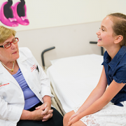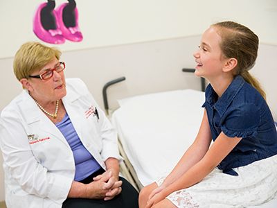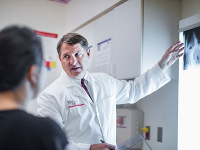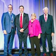
Laura Tosi, M.D., celebrated as a 2024 POSNA Hall of Fame inductee
 Laura Tosi, M.D., was one of four inductees nominated into the Pediatric Orthopaedic Society of North America (POSNA) Hall of Fame, which honors members who have displayed dedication to teaching and mentoring, studying musculoskeletal conditions in children and caring for children with musculoskeletal conditions.
“I was particularly pleased to be recognized because my contributions have not been typical,” said Dr. Tosi. “I have never developed a new surgical technique or made a scientific breakthrough. Rather, I have come to recognize the importance of maintaining good bone health to support the quality of life of our patient population.”
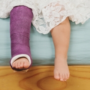
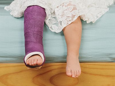
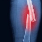
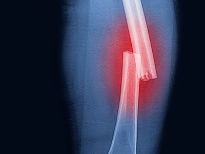
Dr. Tosi began her career at Children’s National Hospital in 1984, focused on the care of children with disabilities such as spina bifida, cerebral palsy and arthrogryposis. Over the course of her tenure, she noted that children with these conditions were surviving at increasingly higher rates, but their quality of life was often derailed by disuse osteoporosis and pathologic fractures.
Her work on secondary fragility fracture prevention ultimately led her to develop the Children’s National Bone Health Program, which launched in 2003. She has since leveraged this program to expand interest in rare bone disease, thanks in part, to her collaborative work with the National Institutes of Health and Osteogenesis Imperfecta Foundation.
“We are all so proud of Dr. Tosi for this amazing recognition,” said Matthew Oetgen, M.D., M.B.A., chief of orthopaedic surgery and sports medicine at Children’s National. The POSNA Hall of Fame is a BIG DEAL! There really is no more powerful recognition than that of colleagues, and this award shows how much of an impact Dr. Tosi has had worldwide. She is a true leader in pediatric orthopaedic surgery and pediatric bone health, and this award is well deserved. Her dedication to Children’s National, the children of the region and us, her partners, has been unwavering, and we thank her for it.”