Painful NICU procedures change neurological development in preterm babies
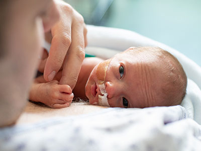
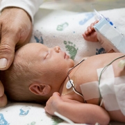
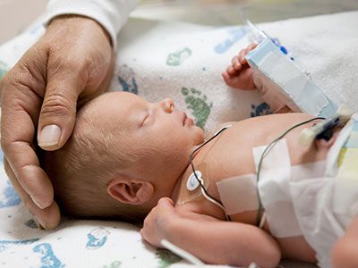
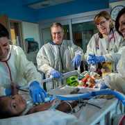
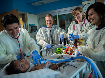
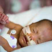
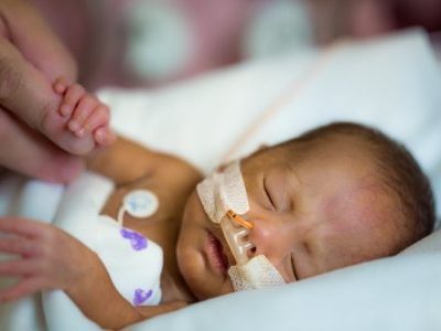
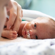
Premature infants exposed to pain while in the Neonatal Intensive Care Unit (NICU) are at greater risk for motor delays, language deficits and autism, even in the absence of structural brain injuries, according to findings from the new Center for Prenatal, Neonatal & Maternal Health Research at Children’s National Hospital.
Premature infants exposed to pain while in the Neonatal Intensive Care Unit (NICU) are at greater risk for motor delays, language deficits and autism, even in the absence of structural brain injuries, according to findings from the new Center for Prenatal, Neonatal & Maternal Health Research at Children’s National Hospital.
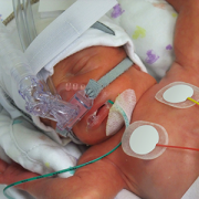
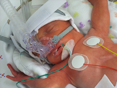
The research sheds light on the potential outcomes of routine medical interventions – such as heel pricks, venipunctures and IV placements – and correlates these skin breaks to changes in neurological connectivity in the preterm infants’ brains. Published in BMC Medicine, the work provides valuable insights about the far-reaching impact of early medical care.
“We know that premature babies are often exposed to repeated medical interventions, light, sound and other stimuli that they would not experience in utero, and we wanted to better understand the long-term effect,” said Kevin Cook, Ph.D., research faculty at the new center and an expert in fetal and neonatal neurology. “Through this study, we can see that early and repeated exposure to pain appears to alter brain development and put children at risk for poor neurodevelopmental outcomes.”
The big picture
Globally, nearly 1 in 10 babies is born preterm, and the Children’s National team was particularly interested in the experience of those born “very” and “extremely” preterm, which is considered any delivery earlier than 32 and 28 weeks of gestation, respectively. While rates of prematurity have been relatively stable, survival rates of these babies have increased remarkably in recent decades, thanks to improved interventions and therapies for preterm infants. Yet neurodevelopmental challenges among these children persist, with noteworthy risks of autism and other neurological deficits.
At Children’s National, researchers are working to understand the mechanism behind those challenges. Given that the late second trimester and the third trimester are critical periods for brain development, the team wanted to study the effects of exposing babies to the world outside the womb early.
The fine print
Dr. Cook and his colleagues collected resting-state functional MRI (fMRI) scans from 148 infants born at least four weeks prematurely, along with 99 infants born full term. The fMRI scans, uniquely suited for studying the resting state of the brain in non-responsive infants, revealed significant hyperconnectivity within the cerebellum, which coordinates muscle activity, and the limbic and paralimbic regions, which govern emotions, motivation and cognitive functions.
Notably, the hyperconnectivity correlated with the number of skin break procedures, including heel pricks, venipunctures and IV placements. When the children returned for developmental evaluations at 18 months, the skin breaks were strongly associated with an increased risk of autism and lower motor and language scores. The toddlers identified at risk for autism had an average of 118 skin breaks, which is significantly more than the average of 65 skin breaks in those who were not at risk.
What’s ahead
Catherine Limperopoulos, Ph.D., director of the Center for Prenatal, Neonatal & Maternal Health Research, said the findings have important implications for understanding how painful NICU procedures can impact long-term outcomes and how physicians conceptualize the risks of care given to preterm babies. She and her team at the center recommend further research into managing pain in premature babies, especially given the limits of current options and the known risk of opioids.
“With this foundational study, we should consider ways to improve pain management for preterm infants and methods to better weigh the interventions used on these incredibly vulnerable patients,” Dr. Limperopoulos said. “Saving their lives is certainly the priority, and the quality of that life should also be forefront of our minds.”