Tag Archive for: magnetic resonance imaging

Sarah Mulkey

Fetal MRI plus ultrasound assess Zika-related brain changes

Sarah Mulkey

Magnetic resonance imaging and ultrasound provide complementary data needed to assess ongoing changes to the brains of fetuses exposed to Zika in utero, says Sarah B. Mulkey, M.D., Ph.D.

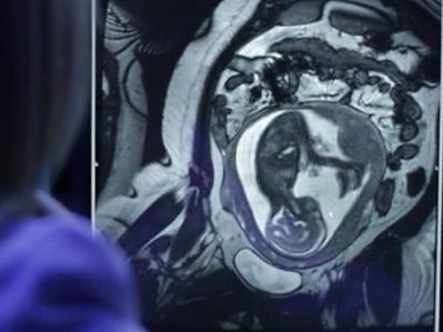
For Zika-affected pregnancies, fetal magnetic resonance imaging (MRI) used in addition to standard ultrasound (US) imaging can better assess potential brain abnormalities in utero, according to research presented by Children’s National Health System during IDWeek 2017. In cases of abnormal brain structure, fetal MRI can reveal more extensive areas of damage to the developing brain than is seen with US.

“MRI and US provide complementary data needed to assess ongoing changes to the brains of fetuses exposed to Zika in utero,” says Sarah B. Mulkey, M.D., Ph.D., a fetal/neonatal neurologist at Children’s National Health System and lead author of the research paper. “In addition, our study found that relying on ultrasound alone would have given one mother the false assurance that her fetus’ brain was developing normally while the sharper MRI clearly pointed to brain abnormalities.”

As of Sept. 13, the Centers for Disease Control and Prevention (CDC) reported that 1,901 U.S. women were exposed to Zika at some point during their pregnancies but their infants appeared normal at birth. Another 98 U.S. women, however, gave birth to infants with Zika-related birth defects.  And eight more women had pregnancy losses with Zika-related birth defects, according to CDC registries.

The longitudinal neuroimaging study led by Children’s National enrolled 48 pregnant women exposed to the Zika virus in the first or second trimester whose infection was confirmed by reverse transcription polymerase chain reaction, which detects Zika viral fragments shortly after exposure, and/or Immunoglobulin M testing, which reveals antibodies the body produces to neutralize the virus. Forty-six of the study volunteers live in Barranquilla, Colombia, where Zika infection is endemic. Two women live in the Washington region and were exposed to Zika during travel elsewhere.

All of the women underwent at least one diagnostic imaging session while pregnant, receiving an initial MRI or US at 25.1 weeks’ gestational age. Thirty-six women underwent a second MRI/US imaging pair at roughly 31 weeks’ gestation. Children’s National radiologists read every image.

Three of 48 pregnancies, or 6 percent, were marked by abnormal fetal MRIs:

  • One fetus had heterotopias (clumps of grey matter located at the wrong place) and abnormal cortical indent (a deformation at the outer layer of the cerebrum, a brain region involved in consciousness). The US taken at the same gestational age for this fetus showed its brain was developing normally.
  • Another fetus had parietal encephalocele (an uncommon skull defect) and Chiari malformation Type II (a life-threatening structural defect at the base of the skull and the cerebellum, the part of the brain that controls balance). The US for this fetus also detected these brain abnormalities.
  • The third fetus had a thin corpus callosum (bundle of nerves that connects the brain’s left and right hemispheres), an abnormally developed brain stem, temporal cysts, subependymal heterotopias and general cerebral/cerebellar atrophy. This fetal US showed significant ventriculomegaly (fluid-filled structures in the brain that are too large) and a fetal head circumference that decreased sharply from the 32nd to 36th gestational week, a hallmark of microcephaly.

After they were born, infants underwent a follow-up MRI without sedation and US. For nine infants, these ultrasounds revealed cysts in the choroid plexus (cells that produce cerebrospinal fluid) or germinal matrix (the source for neurons and glial cells that migrate during brain development). And one infant’s US after birth showed lenticulostriate vasculopathy (brain lesions).

“Because a number of factors can trigger brain abnormalities, further studies are needed to determine whether the cystic changes to these infants’ brains are attributable to Zika exposure in the womb or whether some other insult caused these troubling results,” Dr. Mulkey says.

Spectral data shine light on placenta

preemie baby

A research project led by Subechhya Pradhan, Ph.D., aims to shed light on metabolism of the placenta, a poorly understood organ, and characterize early biomarkers of fetal congenital heart disease.

The placenta serves as an essential intermediary between a pregnant mother and her developing fetus, transporting in life-sustaining oxygen and nutrients, ferrying out waste and serving as interim lungs, kidneys and liver as those vital organs develop in utero.

While the placenta plays a vital role in supporting normal pregnancies, it remains largely a black box to science. A research project led by Subechhya Pradhan, Ph.D., and partially funded by a Clinical and Translational Science Institute Research Award aims to shed light on placenta metabolism and characterize possible early biomarkers of impaired placental function in fetal congenital heart disease (CHD), the most common type of birth defect.

“There is a huge information void,” says Pradhan, a research faculty member of the Developing Brain Research Laboratory at Children’s National Health System. “Right now, we do not have very much information about placenta metabolism in vivo. This would be one of the first steps to understand what is actually going on in the placenta at a biochemical level as pregnancies progress.”

The project Pradhan leads will look at the placentas of 30 women in the second and third trimesters of healthy, uncomplicated pregnancies and will compare them with placentas of 30 pregnant women whose fetuses have been diagnosed with CHD. As volunteers for a different study, the women are already undergoing magnetic resonance imaging, which takes detailed images of the placenta’s structure and architecture. The magnetic resonance spectroscopy scans that Pradhan will review show the unique chemical fingerprints of key metabolites: Choline, lipids and lactate.

Choline, a nutrient the body needs to preserve cellular structural integrity, is a marker of cell membrane turnover. Fetuses with CHD have higher concentrations of lactate in the brain, a telltale sign of a shortage of oxygen. Pradhan’s working hypothesis is that there may be differing lipid profiles and lactate levels in the placenta in pregnancies complicated by CHD.  The research team will extract those metabolite concentrations from the spectral scans to describe how they evolve in both groups of pregnant women.

“While babies born with CHD can undergo surgery as early as the first few days (or sometimes hours) of life to correct their hearts, unfortunately, we still see a high prevalence of neurodevelopmental impairments in infants with CHD. This suggests that neurological dysfunctional may have its origin in fetal life,” Pradhan says.

Having an earlier idea of which fetuses with CHD are most vulnerable has the potential to pinpoint which pregnancies need more oversight and earlier intervention.

Placenta spectral data traditionally have been difficult to acquire because the pregnant mother moves as does the fetus, she adds. During the three-minute scans, the research team will try to limit excess movement using a technique called respiratory gating, which tells the machine to synchronize image acquisition so it occurs in rhythm with the women’s breathing.

Catherine Limperopoulous

The brain’s fluid-filled spaces during growth

Catherine Limperopoulous

Catherine Limperopoulous, Ph.D., and her colleagues used volumetric MRIs to assess how the ventricles, cerebrospinal fluid and the rest of the fetal brain normally change over time.

The human brain is not one solid mass. Buried within its gray and white matter are a series of four interconnected chambers, called ventricles, which produce cerebrospinal fluid. These ventricles are readily apparent on the fetal ultrasounds that have become the standard of prenatal care in the United States and most developed countries around the world. Abnormalities in the ventricles’ size or shape – or both – can give doctors an early warning that fetal brain development might be going awry.

But what is abnormal? It is not always clear, says Catherine Limperopoulos, Ph.D., director of the Developing Brain Research Laboratory at Children’s National Health System. Limperopoulos explains that despite having many variations in fetal ventricles, some infants have completely normal neurodevelopmental outcomes later. On the other hand, some extremely subtle variations in shape and size can signal problems.

On top of these complications are the tools clinicians typically use to assess the ventricles. Limperopoulos explains that most early indications of ventricle abnormalities come from ultrasounds, but the finer resolution of magnetic resonance imaging (MRI) can provide a more accurate assessment of fetal brain development. Still, both standard MRI and ultrasound provide only two-dimensional pictures, making it difficult to quantify slight differences in the volume of structures.

To help solve these problems, Limperopoulos and her colleagues recently published a paper in Developmental Neuroscience that takes a different tack. The team performed volumetric MRIs – a technique that provides a precise three-dimensional measure of structural volumes – on the brains of healthy fetuses to assess how the ventricles, cerebrospinal fluid and the rest of the brain normally change over time. Limperopoulos’ team recently performed a similar study to assess normal volumetric development in the brain’s solid tissues.

Previous studies published on comparable topics typically used information gathered from subjects who initially had clinical concerns but eventually were dismissed from these studies for not having worrisome diagnoses in the end. This might not truly reflect a typical population of pregnant women, Limperopoulos says.

Working with 166 pregnant women with healthy pregnancies spanning from 18 to 40 weeks gestation, the researchers performed volumetric MRIs on their singleton fetuses that covered every week of this second half of pregnancy. This technique allowed them to precisely calculate the volumes of structures within the fetal brain and get an idea of how these volumes changed over time within the group.

Their results show that over the second and third trimester:

  • The lateral ventricles, the largest ventricles found in the cerebrum with one for each brain hemisphere, grew about two-fold;
  • The third ventricle, found in the forebrain, grew about 23-fold;
  • The fourth ventricle, found in the hindbrain, grew about eight-fold;
  • And the extra-axial cerebrospinal fluid, found under the lining of the brain, increased about 11-fold.

The total brain volume increased 64-fold over this time, with the parenchyma – the solid brain tissue that encompasses gray and white matter – growing significantly faster than the cerebrospinal fluid-filled spaces.

Limperopoulos points out that the ability to measure the growth of the brain’s fluid-filled spaces relative to the surrounding brain tissue can provide critical information to clinicians caring for developing fetuses. In most cases, knowing what is normal allows doctors to reassure pregnant women that their fetus’ growth is on track. Abnormalities in these ratios can provide some of the first signals to alert doctors to blockages in cerebrospinal fluid flow, abnormal development, or the loss of brain tissue to damage or disease. Although the neurodevelopmental outcomes from each of these conditions can vary significantly, traditional ultrasounds or MRIs might not be able to distinguish these possibilities from each other. Being able to differentiate why cerebrospinal fluid spaces have abnormal shapes or sizes might allow doctors to better counsel parents, predict neurological outcomes, or potentially intervene before or after birth to mitigate brain damage.

“By developing a better understanding of what’s normal,” Limperopoulos says, “we can eventually identify reliable biomarkers of risk and guide interventions to minimize risks for vulnerable fetuses.”

Setting a baseline for healthy brain development

Catherine Limperopoulos, Ph.D., and colleagues performed the largest magnetic resonance imaging study of normal fetal brains in the second and third trimesters of pregnancy.

Starting as a speck barely visible to the naked eye and ending the in utero phase of its journey at an average weight of 7.5 pounds, the growth of the human fetus is one of the most amazing events in biology. Of all the organs, the fetal brain undergoes one of the most rapid growth trajectories, expanding over 40 weeks from zero to 100 billion neurons — about as many brain cells as there are stars in the Milky Way Galaxy.

This exponential growth is part of what gives humans our unique abilities to use language or have abstract thoughts, among many other cognitive skills. It also leaves the brain extremely vulnerable should disruptions occur during fetal development. Any veering off the developmental plan can lead to a cascade of results that have long-lasting repercussions. For example, studies have shown that placental insufficiency, or the inability of the placenta to supply the fetus with oxygen and nutrients in utero, is associated with attention deficit hyperactivity disorder, autism, and schizophrenia.

Recent research has identified differences in the brains of people with these disorders compared with those without. Despite the almost certain start of these conditions within the womb, they have remained impossible to diagnose until children begin to show clinical symptoms. If only researchers could spot the beginnings of these problems early in development, says Children’s National Health System researcher Catherine Limperopoulos, Ph.D., they might someday be able to develop interventions that could turn the fetal brain back toward a healthy developmental trajectory.

“Conventional tools like ultrasound and magnetic resonance imaging (MRI) can identify structural brain abnormalities connected to these problems, but by the time these differences become apparent, the damage already has been done,” Limperopoulos says. “Our goal is to be able to pick up very early deviations from normal in the high-risk pregnancy before an injury to the fetus might become permanent.”

Before scientists can recognize abnormal, she adds, they first need to understand what normal looks like.

In a new study published in Cerebral Cortex, Limperopoulos and colleagues begin to tackle this question through the largest MRI study of normal fetal brains in the second and third trimesters of pregnancy. While other studies have attempted to track normal fetal brain growth, that research has not involved nearly as many subjects and typically relied on data collected when fetuses were referred for MRIs for a suspected problem. When the suspected abnormality was ruled out by the scan, these “quasi-controls” were considered “normal” — even though they may be at risk for problems later in life, Limperopoulos explains.

By contrast, the study she led recruited 166 healthy pregnant women from nearby low-risk obstetrics practices. Each woman had an unremarkable singleton pregnancy and ended up having a normal full-term delivery, with no evidence of problems affecting either the mother or fetus over the course of 40 weeks.

At least one time between 18 and 39 gestational weeks, the fetuses carried by these women underwent an MRI scan of their brains. The research team developed complex algorithms to account for movement (since neither the mothers nor their fetuses were sedated during scans) and to convert the two-dimensional images into three dimensions. They used the information from these scans to measure the increasing volumes of the cerebellum, an area of the brain connected to motor control and known to mediate cognitive skills; as well as regions of the cerebrum, the bulk of the brain, that is pivotal for movement, sensory processing, olfaction, language, and learning and memory.

Their results in uncomplicated, full-term pregnancies show that over 21 weeks in the second half of pregnancy, the cerebellum undergoes an astounding 34-fold increase in size. In the cerebrum, the fetal white matter, which connects various brain regions, grows 22-fold. The cortical gray matter, key to many of cerebrum’s functions, grows 21-fold. And the deep subcortical structures (thalamus and basal ganglia), important for relaying sensory information and coordination of movement and behavior, grow 10-fold. Additional examination showed that the left hemisphere has a larger volume than the right hemisphere early in development, but sizes of the left and right brain halves were equal by birth.

By developing similar datasets on high-risk pregnancies or births—for example, those in which fetuses are diagnosed with a problem in utero, mothers experience a significant health problem during pregnancy, babies are born prematurely, or fetuses have a sibling diagnosed with a health problem with genetic risk, such as autism—Limperopoulos says that researchers might be able to spot differences during gestation and post-natal development that lead to conditions such as schizophrenia, attention deficit hyperactivity disorder and autism spectrum disorder.

Eventually, researchers may be able to develop fixes so that babies grow up without life-long developmental issues.

“Understanding ‘normal’ is really opening up opportunities for us to begin to precisely pinpoint when things start to veer off track,” Limperopolous says. “Once we do that, opportunities that have been inaccessible will start to present themselves.”