Tag Archive for: Ishibashi

Children’s National Hospital at American Heart Association Scientific Sessions 2024
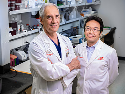
American Heart Association Scientific Sessions logoExperts from Children’s National Heart Center presented and shared their latest research findings at this year’s American Heart Association (AHA) Scientific Sessions, held in Chicago, Illinois, in mid-November.

The annual AHA Scientific Sessions are attended by scientists, clinicians, researchers and other health care professionals from around the globe who have an interest in cardiovascular disease. Children’s National Hospital experts highlighted work focused on caring for the full spectrum of people who live with congenital heart disease — from tiny neonates through adulthood.

Presentations

  • Transvenous cardiac re-synchronization: When is it effective in CHD? Charles Berul, M.D.
  • Leveling the Playing Field: Creating Equity within Pediatric Cardiology Leadership and Salary, Wayne Franklin, M.D.
  • Debate: Patients with small coronary artery aneurysms SHOULD be discharged – US experience, Ashraf Harahsheh, M.D.
  • Science of Engagement: Inclusion of Adults with Congenital Heart Disease Living with Neurodevelopmental Disability in PCOR, Anitha John, M.D., Ph.D.
  • 100 Years of AHA Leading Global Health, Craig Sable, M.D.
  • Best Oral Abstract: Safety of Discontinuing Secondary Antibiotic Prophylaxis After Echocardiographic Normalization in Early Rheumatic Heart Disease, GOAL-Post Study, Craig Sable, M.D., co-author
  • Su2032│CMR can discriminate need for biopsy and rejection therapy in children post heart transplant, Ravi Vamsee Vegulla, M.D.

Posters and poster presentations

  • Minimally-Invasive Intrapericardial Injections under Direct Visualization via Thoracic Cavity Access in Infant and Pediatric-sized Pre-clinical Model, Charles Berul, M.D., Ryan O’Hara, Ph.D.
  • Early total cfDNA, but not donor fraction, predicts late events after heart transplantation, Shriprasad Deshpande, M.D.
  • Impact of Angiotensin Receptor Neprilysin Inhibitor on Chronic Heart Failure with Reduced Ejection, Shriprasad Deshpande, M.D.
  • Fraction in Adult Congenital Heart Disease Patients: A Systematic Review and Meta-analysis, Shriprasad Deshpande, M.D.
  • Trough Level Prediction of Major Adverse Transplant Events: A Report from the TEAMMATE Trial, Shriprasad Deshpande, M.D.
  • Thrombocytosis is Prevalent and Associated with Greater Inflammation and Coronary Artery Involvement in Both Kawasaki Disease and Multisystem Inflammatory Syndrome in Children Associated with COVID-19, Ashraf Harahsheh, M.D.
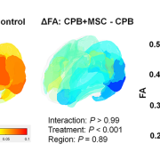
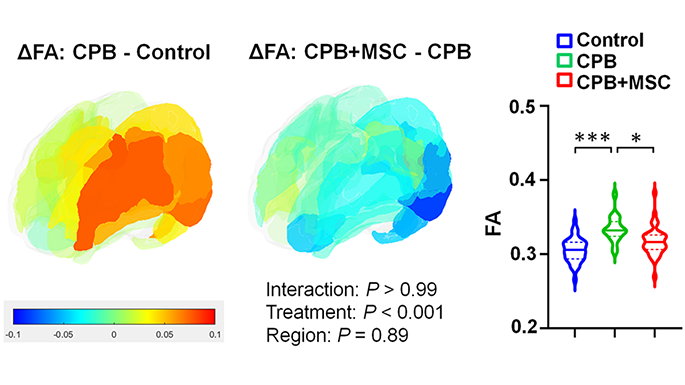
  • Mesenchymal Stromal Cell Delivery through Cardiopulmonary Bypass in Pediatric Cardiac Surgery – MeDCaP Phase I Trial, Nobuyuki Ishibashi, M.D., Shriprasad Deshpande, M.D., et. al.
  • Social Determinants of Health: Impact on Mortality and Care Status for Adults with CHD, Jamie Jackson, Ph.D.; Anitha John, M.D., Ph.D., co-author
  • Loss to Follow-Up Among Adults with Congenital Heart Defects: A Report from Congenital Heart Disease Project to Understand Lifelong Survivor Experience (CHD PULSE), Anitha John, M.D., Ph.D., co-author
  • The Burden of Adult Congenital Heart Disease in the United States, Vasupradha Suresh Kumar, M.D.
  • Determining the Physiologic Effect of the Cavopulmonary Connection on Caval Flows Using 4D Flow MRI , Vasupradha Suresh Kumar, M.D.
  • Shape Variations in Right Ventricular 3D Geometry are associated with adverse outcomes in Hypoplastic Left Heart Syndrome Patients: A Fontan Outcomes Registry using CMR Examination (FORCE) Study, Yue-Hin Loke, M.D.
  • Matrix Metalloproteinases and Tissue Inhibitors of Metalloproteinases as Biomarkers in Duchenne Muscular Dystrophy Cardiomyopathy, Christopher Spurney, M.D., co-author
  • Duchenne Muscular Dystrophy Boys Have Diastolic Dysfunction Based on Cardiac Magnetic Resonance, Christopher Spurney, M.D., co-author

Read more about these presentations and posters on the AHA’s meeting website: Programming – Scientific Sessions 2024.

Cell therapy mitigates neurological impacts of cardiac surgery in pre-clinical model

Differences of cortical fractional anisotropy between cardiopulmonary bypass and control (left), cardiopulmonary bypass + mesenchymal stromal cells and cardiopulmonary bypass (center), and 3 groups (right).

A pre-clinical study in the journal JACC: Basic to Translational Science shows that infusing bone marrow-derived mesenchymal stromal cells (BM-MSCs) during cardiac surgery provides both cellular-level neuroprotection for the developing brain and improvements in behavior alterations after (or resulting from) surgery.

What this means

According to lead author Nobuyuki Ishibashi, M.D., Oxidative and inflammatory stresses that are thought to be related to cardiopulmonary bypass cause prolonged microglia activation and cortical dysmaturation in the neonatal and infant brain. These issues are a known contributor to neurodevelopmental impairments in children with congenital heart disease.

This study found that, in a pre-clinical model, the innovative use of cardiopulmonary bypass to deliver these mesenchymal stromal cells minimizes microglial activation and neuronal apoptosis (cell death), with subsequent improvement of cortical dysmaturation and behavioral alteration after neonatal cardiac surgery.

Additionally, the authors note that further transcriptomic analyses provided a possible mechanism for the success: Exosome-derived miRNAs such as miR-21-5p, which may be key drivers of the suppressed apoptosis and STAT3-mediated microglial activation observed following BM-MSC infusion.

Why it matters

Significant neurological delay is emerging as one of the most important current challenges for children with congenital heart disease, yet few treatment options are currently available.

Applications of BM-MSC treatment will provide a new therapeutic paradigm for potential MSC-based therapies as a form of neuroprotection in children with congenital heart disease.

Children’s National Hospital leads the way

The Ishibashi lab is the first research team to demonstrate the safety, efficacy and utility of using cardiopulmonary bypass to deliver BM-MSCs with the goal of improving neurological impairments in children undergoing surgery for congenital heart disease. In addition to this pre-clinical research, a phase 1 clinical trial, MeDCaP, is underway at Children’s National.

Recent additional funding from the NIH will allow the team to identify molecular signatures of BM-MSC treatment and mine specific BM-MSC exosomes for unique cardiopulmonary bypass pathology to further increase understanding of precisely how and why this cell-based treatment shows success.

Cell therapy research for neuroprotection in congenital heart disease receives another $3.3 million from NIH

x-ray of child with congenital heart disease

Significant neurological delay is emerging as one of the most important current challenges for children with congenital heart disease, yet few treatment options are currently available.

The research lab of Nobuyuki Ishibashi, M.D., at Children’s National Hospital, recently received $3.3 million in additional funding for research into cell therapy for neuroprotection in children with congenital heart disease. The new support comes from the National Heart, Lung and Blood Institute (NHLBI) of the National Institutes of Health.

The research goal

The overarching goal of the award is to establish detailed molecular signatures from critical cell populations for tissue repair and regeneration at single cell resolution after bone marrow-derived mesenchymal stromal cell (BM-MSC) delivery. The team has shown cellular, structural and behavioral improvements in pre-clinical models after delivery of BM-MSCs through cardiopulmonary bypass for children with congenital heart disease. However, the mechanisms underlying the therapeutic action of BM-MSCs still remain largely unknown. This R01 renewal will address the key knowledge gap.

Why it matters

Significant neurological delay is emerging as one of the most important current challenges for children with congenital heart disease, yet few treatment options are currently available.

The Ishibashi lab has demonstrated the efficacy and utility of using cardiopulmonary bypass to deliver BM-MSCs  to improve neurological impairments in children undergoing surgery for congenital heart disease. Most notably, this included development of a phase 1 clinical trial, MeDCaP, at Children’s National.

The big picture

Together with the ongoing clinical trial established from the previous award, identifying molecular signatures of BM-MSC treatment and mining specific BM-MSC exosomes for unique cardiopulmonary bypass pathology will significantly improve understanding of this cell-based treatment. This work will also provide a new therapeutic paradigm for potential cell-free MSC-based therapies for neuroprotection in children with congenital heart disease.

Caspases may link brain cell degeneration and cardiac surgery

caspase molecule

The review summarizes both the known physiological roles of caspases as well as some of the well-characterized neurotoxic effects of anesthetics in pre-clinical models.

A review article in the journal Cell Press: Trends in Neuroscience outlines the wide variety of cellular signaling roles for caspase proteins — a type of cellular enzyme best known for its documented role in the natural process of cell death (apoptosis). The authors, including Nemanja Saric, Ph.D., Kazue Hashimoto-Torii, Ph.D., and Nobuyuki Ishibashi, M.D., all from Children’s National Research Institute, pay particular attention to what the scientific literature shows about caspases’ non-apoptotic roles in the neurons specifically. They also highlight research showing how, when activated during a cardiac surgery with anesthesia and cardiopulmonary bypass, these enzymes may contribute to the degeneration of brain cells seen in young children who undergo heart surgery for critical congenital heart defects (CHDs).

Why it matters

The review summarizes both the known physiological roles of caspases as well as some of the well-characterized neurotoxic effects of anesthetics in pre-clinical models.

The authors propose that these non-apoptotic activities of caspases may be behind some of the adverse effects on the developing brain related to cardiac surgery and anesthesia. Those adverse effects are known to increase risk of behavioral impairments in children with congenital heart disease who underwent cardiac surgery with both anesthesia and cardiopulmonary bypass at a very young age.

This work is the first to propose a possible link between developmental anesthesia neurotoxicity and caspase-dependent cellular responses.

The patient benefit

Better understanding of the time and dose-dependent effects of general anesthetics on the developing brain, particularly in children who have genetic predispositions to conditions such as CHDs, will help researchers understand their role (if any) in behavioral problems often encountered by these patients after surgery.

If found to be a contributing factor, perhaps new therapies to mitigate this caspase activity might be explored to alleviate some of these adverse effects on the developing brain.

What’s next?

The authors hope to stimulate more in-depth research into caspase signaling events, particularly related to how these signaling events change when an anesthetic is introduced. Deeper understanding of how anesthetics impact caspase activation in the developing brain will allow for better assessments of the risk for children who need major surgery early in life.

Children’s National leads the way

Children’s National Hospital leads studies funded by the U.S. Department of Defense to better understand how these other roles of caspases, which until now have not been well-documented, may contribute to brain cell degeneration when activated by prolonged anesthesia and cardiopulmonary bypass during cardiac surgery for congenital heart disease.

Cardiopulmonary bypass may cause significant changes to developing brain and nerve cells

brain network illustration

Cardiopulmonary bypass, more commonly known as heart-and-lung bypass, has some unique impacts on the creation and growth of brain cells in the area of a child’s brain called the subventricular zone (SVZ), according to a study in the Annals of Neurology. The SVZ is a critical area for the growth and migration of neurons and nerve cells called neuroblasts, both of which ultimately contribute to the proper development of key brain structures and functions during the early years of life.

The findings, from a study conducted in the Cardiac Surgery Research Laboratory at Children’s National Hospital, provide new insight into the cellular impacts of the cardiopulmonary bypass machine on brain growth and development for newborn infants with congenital heart disease. They will have an important role in the refinement of strategies to help protect the fragile brains of children who require lifesaving cardiac surgery with cardiopulmonary bypass immediately after birth.

Specifically, the research team found that during cardiopulmonary bypass:

  • Creation of neurons (neurogenesis) in the neonatal and infant subventricular zone is altered.
  • Migration of nerve cells, called neuroblasts, to the frontal lobe is potentially disrupted.
  • Changes to the growth and movement of neurons in the SVZ are prolonged.
  • Cortical development and expansion is impaired.
  • Specific types of neurons found only in the brain and spinal cord, called interneurons, are also affected.

The study uses an innovative pre-clinical model of the developing brain that is more anatomically and physiologically similar to human neonates and infants than those used in prior studies and in most neurological laboratory-based research.

Cardiopulmonary bypass is one of several key factors thought to cause children with congenital heart disease to sometimes demonstrate delays in the development of cognitive and motor skills. These disabilities often persist into adolescence and adulthood and can ultimately represent long-term neurocognitive disabilities. It is also believed that genetic factors, abnormal blood flow to the brain while in utero or low cardiac output after surgical procedures on the heart may contribute to these challenges.

“Unraveling cellular and molecular events during surgery using this preclinical model will allow us to design therapeutic approaches that can be restorative or reparative to the neurogenic potential of the neuronal stem precursor cells found in the subventricular zone of the neonatal or infant brain,” says Nobuyuki Ishibashi. M.D., Foglia-Hills Professor of Pediatric Cardiac Research, director of the Cardiac Surgery Research Laboratory at Children’s National and senior author on the study. “In particular, previous studies in our laboratory have shown improvement in the neurogenic activities of these precursor cells when they are treated with mesenchymal stromal cells (MSCs).”

The findings from this study further support the work already underway in the NIH-funded MeDCaP clinical trial for neonates and infants undergoing cardiac surgery using the cardiopulmonary bypass machine. That trial uses the heart and lung machine itself to deliver MSCs directly into the main arteries that carry blood to the brain.

Study suggests chronic hypoxia delays cardiac maturation in CHD

newborn baby

Every year, nearly 40,000 babies are born with a congenital heart defect (CHD) — the leading cause of birth defect-associated infant illness and death.

Every year, nearly 40,000 babies are born with a congenital heart defect (CHD) — the leading cause of birth defect-associated infant illness and death. An event that may contribute to cyanotic CHD is the lack of oxygen, known as hypoxia, before and after birth, impacting gene expression and cardiac function that delay postnatal cardiac maturation, according to a new pre-clinical model led by researchers at Children’s National Hospital.

Single ventricle, transposition of the great arteries, truncus arteriosus and severe forms of tetralogy of Fallot, such cyanotic congenital heart diseases have lower circulating blood oxygen levels. The lack of oxygen in the blood begins prenatally and continues after birth until definitive repair, suggesting a delay on cardiac maturation.

There is little research on the underpinnings that explain the lack of oxygen’s effects on the developing heart, which could help inform adequate therapies in the pediatric population to promote cardiovascular health across the lifetime. The researchers developed the first pre-clinical model that explores the effects of chronic hypoxia in perinatal and postnatal stages on the developing heart under conditions seen in cyanotic CHD.

“To the best of our knowledge, ours is the first study to perform complete gene expression arrays on animals after perinatal hypoxia,” said Jennifer Romanowicz, senior noninvasive imaging fellow at Boston Children’s Hospital and lead author of the study. “Not only did these studies allow us to determine the effects of hypoxia on heart development, but the detailed results of our study will be available to other researchers to independently address other questions about perinatal hypoxia and heart development.”

The study published in the American Journal of Physiology: Heart and Circulatory Physiology suggests that chronic lack of oxygen alters the electrical properties of heart tissue, called the electrophysiological substrate, and the contractile apparatus, a muscle composed of proteins that control cardiac contraction. Multiple genes involved with the contractile apparatus were expressed differently in the non-human subjects.

“What was remarkable was that most abnormalities normalized after the animals recovered in normal oxygen levels,” said Romanowicz. “This is an optimistic sign that early repair of cyanotic congenital heart disease may allow the heart to finish development.”

The researchers placed pregnant non-human subjects in hypoxic chambers starting on embryonic day 16, mimicking the second trimester in humans. The same subjects gave birth in the hypoxic chambers, and the newborns were kept there until postnatal day eight when the heart muscle maturation is nearly complete. To understand how human infants recover with normalized oxygen levels after surgical repair of cyanotic CHD, the researchers moved hypoxic subjects to normal oxygen conditions for recovery and tested again at postnatal day 30.

“Next steps include using a pre-clinical model of cyanotic congenital heart disease that more accurately represents human neonatal physiology,” said Devon Guerrelli, Ph.D. candidate at Children’s National. We plan to work with the cardiac surgery team at Children’s National to investigate changes in the myocardium due to hypoxia in pediatric patients who are undergoing surgical repair.”

Nikki Posnack, Ph.D., principal investigator at Sheikh Zayed Institute for Pediatric Surgical Innovation and Nobuyuki Ishibashi, M.D., director of Cardiac Surgery Research Laboratory at Children’s National, led and guided the team of researchers involved in the study.

Innovative phase 1 trial to protect brains of infants with CHD during and after surgery

A novel phase 1 trial looking at how best to optimize brain development of babies with congenital heart disease (CHD) is currently underway at Children’s National Hospital.

Children with CHD sometimes demonstrate delay in the development of cognitive and motor skills. This can be a result of multiple factors including altered prenatal oxygen delivery, brain blood flow and genetic factors associated with surgery including exposure to cardiopulmonary bypass, also known as the heart lung machine.

This phase 1 trial is the first to deliver mesenchymal stromal cells from bone marrow manufactured in a lab (BM-MSC) into infants already undergoing cardiac surgery via cardiopulmonary bypass. The hypothesis is that by directly infusing the MSCs into the blood flow to the brain, more MSCs quickly and efficiently reach the subventricular zone and other areas of the brain that are prone to inflammation. The trial is open to eligible patients ages newborn to six months of age.


Learn more in this overview video.

The trial is part of a $2.5 million, three-year grant from the National Institutes of Health (NIH) led by Richard Jonas, M.D., Catherine Bollard, M.B.Ch.B., M.D., and Nobuyuki Ishibashi, M.D.. The project involves collaboration between the Prenatal Cardiology program of Children’s National Heart Institute, the Center for Cancer and Immunology Research, the Center for Neuroscience Research and the Sheikh Zayed Institute for Pediatric Surgical Innovation.

“NIH supported studies in our laboratory have shown that MSC therapy may be extremely helpful in improving brain development in animal models after cardiac surgery,” says Dr. Ishibashi. “MSC infusion can help reduce inflammation including prolonged microglia activation that can occur during surgery that involves the heart lung machine.”

Staff from the Cellular Therapy Laboratory, led by director Patrick Hanley, Ph.D., manufactured the BM-MSC at the Center for Cancer and Immunology Research, led by Dr. Bollard.

The phase 1 safety study will set the stage for a phase 2 effectiveness trial of this highly innovative MSC treatment aimed at reducing brain damage, minimizing neurodevelopmental disabilities and improving the postoperative course in children with CHD. The resulting improvement in developmental outcome and lessened behavioral impairment will be of enormous benefit to individuals with CHD.

For more information about this new treatment, contact the clinical research team: Gil Wernovsky, M.D., Shriprasad Deshpande, M.D., Maria Fortiz.

Primary cilia safeguard cortical neurons from environmental stress-induced dendritic degeneration

neuron on teal background

Fetus and neonates are under the risk of exposure to various external agents, such as alcohol and anesthetics taken by the mother. However, primary cilia can protect neurons by activating cilia-localized molecular signaling that inhibits degeneration of neuronal processes, according to the study’s findings.

A new study led by Kazue Hashimoto-Torii, Ph.D. and Masaaki Torii, Ph.D., both principal investigators for the Center for Neuroscience Research at Children’s National Hospital, found that primary cilia – tiny hair-like protrusions from the body of neuronal cells – protect neurons in the developing brain from adverse impacts of prenatal exposure.

Fetus and neonates are under the risk of exposure to various external agents, such as alcohol and anesthetics taken by the mother. However, primary cilia can protect neurons by activating cilia-localized molecular signaling that inhibits degeneration of neuronal processes, according to the study’s findings.

“Remarkably, the developing brain is equipped with intrinsic cell protection that helps to minimize the adverse impacts of to various external agents,” said Dr. Hashimoto-Torii. “However, the mechanisms of such protection have been unclear. Our study provides the first evidence that the tiny hair-like organelle protects neurons in the perinatal brain from adverse impacts of such external agents taken by the mother.”

The findings suggest that subtle alterations in primary cilia due to genetic conditions may lead to various neurodevelopmental disorders if combined with exposure to external agents from the environment. The findings also suggest that ciliopathy patients who have abnormal ciliary function due to genetic causes may have increased risk of abnormal brain development upon exposure to external agents.

“Clarifying diverse roles of cilia provides essential information for clinicians and patients with potential deficits in primary cilia to take extra precautions to avoid the risks for long-term negative impacts of external factors,” Dr. Torii explained. “We hope that further studies will define the whole picture of cilia-mediated neuroprotection and help us to advance our understanding of its importance in the pathogenesis of neurodevelopmental disorders.

This may ultimately lead to the development of treatment for various neurodevelopmental disorders,” he added.

The uniqueness of the study stems from the investigation of the role of cilia in brain development at the risk of exposure to various external factors that occur in the real world. Little is known about how the normal and abnormal brain development progresses in an environment where many external factors interact with intrinsic cellular mechanisms.

The study is a collaboration with researchers at Yale University and Keio University, Japan. Other Children’s National researchers who contributed to this study include Seiji Ishii, Ph.D.; Nobuyuki Ishibashi, M.D.; Toru Sasaki, M.D., Ph.D.; Shahid Mohammad, Ph.D.; Hye Hwang; Edwin Tomy; and Fahad Somaa.

U.S. DoD awards $2M for study to protect neurological function after cardiac surgery

doctors operating

A collaboration between clinical and basic science researchers including Drs. Ishibashi, Hashimoto-Torii, Jonas, and Deutsch, seeks to to understand how caspase enzyme activation plays a role in the development of fine and gross motor skills in children who underwent cardiac surgery for CHD repair.

The U.S. Department of Defense has awarded $2 million to Children’s National Hospital to study how a family of protease enzymes known as caspases may contribute to brain cell degeneration when activated by prolonged anesthesia and cardiopulmonary bypass during cardiac surgery for congenital heart disease.

This U.S. Army Medical Research Acquisition Activity Award, Anesthesia Neurotoxicity in Congenital Heart Disease, is led by principal investigator Nobuyuki Ishibashi, M.D., with both clinical and basic science co-investigators including Kazue Hashimoto-Torii, Ph.D., (Neuroscience), Richard Jonas, M.D., (Cardiovascular Surgery) and Nina Deutsch, M.D., (Anesthesiology).

While the specific cellular and molecular mechanisms of how anesthesia and cardiac surgery impact cortical development are poorly understood, both seem to impact brain growth and development in young children. The most common neurologic deficit seen in children after CHD surgical repair is the impairment of fine and gross motor skills.

Both anesthetic agents and inflammation like that seen as a result of cardiopulmonary bypass have also been shown to contribute to the activation of a specific group of enzymes that play an essential role in the routine (programmed) death of cells: caspases. However, recent pre-clinical research shows that these enzymes may also contribute to other alterations to cells beyond cell death, including making changes to other cell structures. In pre-clinical models, these changes cause impairments to fine and gross motor skills – the same neurological deficits seen in children with CHD who have undergone procedures requiring prolonged anesthesia and cardiopulmonary bypass.

The research team hypothesizes that caspases are extensively activated as a result of cardiac surgery and while that activation is rarely causing reduced numbers of neurons, the changes that caspase enzymes trigger in neurons are contributing to neurological deficits seen in children with CHD after surgery.

While the study focuses specifically on the impacts of cardiac surgery for correction of a heart defect, the findings could have major implications for any pediatric surgical procedure requiring prolonged anesthesia and/or cardiopulmonary bypass.

R01 grant funds white matter protection study for congenital heart disease

Nobuyuki Ishibashi

Nobuyuki Ishibashi, M.D., is the principal investigator on a $3.2 million NIH R01 to study white matter growth and repair in utero for fetal brains affected by congenital heart disease.

Many of the neurological deficits seen in children with congenital heart disease (CHD) are related to abnormal white matter development early in life caused by reduced oxygen supply to the brain while in utero. Children with immature white matter at birth also commonly sustain additional white matter injuries following cardiac surgery.

The NIH recently awarded a prestigious R01 grant totaling more than $3.2 million to a collaborative project led by the Center for Neuroscience Research, the Sheikh Zayed Institute for Pediatric Surgical Innovation and the Children’s National Heart Institute at Children’s National Hospital as well as MedStar Washington Hospital Center.

The research, titled “White matter protection in the fetus with congenital heart disease,” looks specifically at whether providing a supplemental amount of the naturally occurring tetrahydrobiopterin (BH4) for pregnant women could rescue white matter development of fetuses with congenital heart disease whose brains aren’t receiving enough oxygen – or suffering from hypoxic-ischemic events.

Previous preclinical studies have shown that this lack of oxygen depletes the brain’s natural BH4 level, and the researchers hypothesize that BH4 levels play a critical role in the growth and development of white matter in the fetal brain by triggering key cellular/molecular processes. Specifically, the study will focus on three aims:

  1. Establish in a preclinical model the optimal protective regiment for women pregnant with a fetus who has CHD to receive BH4.
  2. Determine the appropriate approach to deliver BH4 to this population
  3. Leverage genetic tools and biochemical techniques in the laboratory to better understand where and how BH4 levels play a role in the growth (or lack thereof) of oligodendrocytes—the primary cells of white matter.

This laboratory-based work is the first step to determining if the neurodevelopment of babies born with CHD can be preserved or recovered by addressing key brain development that occurs before the baby is even born. Findings related to congenital heart disease may also translate to other populations where white matter development is affected by hypoxia-ischemia, including premature infants.

The project is led by principal investigator Nobuyuki Ishibashi, M.D., with co-investigators Vittorio Gallo, Ph.D., Joseph Scafidi, D.O., and Mary Donofrio, M.D. as well as colleagues at MedStar Washington Hospital Center.

Can cells collected from bone marrow stimulate generation of new neurons in babies with CHD?

Newborn baby laying in crib

The goal of the study will be to optimize brain development in babies with congenital heart disease (CHD) who sometimes demonstrate delay in the development of cognitive and motor skills.

An upcoming clinical trial at Children’s National Hospital will harness cardiopulmonary bypass as a delivery mechanism for a novel intervention designed to stimulate brain growth and repair in children who undergo cardiac surgery for congenital heart disease (CHD).

The NIH has awarded Children’s National $2.5 million to test the hypothesis that mesenchymal stromal cells (MSCs), which have been shown to possess regenerative properties and the ability to modulate immune responses in a variety of diseases, collected from allogeneic bone marrow, may promote regeneration of damaged neuronal and glial cells in the early postnatal brain. If successful, the trial will determine the safety of the proposed treatment in humans and set the stage for a Phase 2 efficacy trial of what could potentially be the first treatment for delays in brain development that happen before birth as a consequence of congenital heart disease. The study is a single-center collaboration between three Children’s National physician-researchers: Richard Jonas, M.D., Catherine Bollard, M.B.Ch.B., M.D. and Nobuyuki Ishibashi, M.D.

Dr. Jonas, chief of cardiac surgery at Children’s National, will outline the trial and its aims on Monday, November 18, 2019, at the American Heart Association’s Scientific Sessions 2019. Dr. Jonas was recently recognized by the Cardiac Neurodevelopmental Outcome Collaborative for his lifelong research of how cardiac surgery impacts brain growth and development in children with CHD.

Read more about the study: Researchers receive $2.5M grant to optimize brain development in babies with CHD.

###

Regenerative Cell Therapy in Congenital Heart Disease – Protecting the Immature Brain
Presented by Richard Jonas, M.D.
AHA Scientific Sessions
Session CH.CVS.608 Congenital Heart Disease and Pediatric Cardiology Seminar: A Personalized Approach to Heart Disease in Children
9:50 a.m. to 10:05 a.m.
November 18, 2019

Cardiac surgery chief recognized for studies of surgery’s impacts on neurodevelopment

Dr. Jonas and research collaborator Nobuyuki Ishibashi in the laboratory.

Dr. Jonas and research collaborator Nobuyuki Ishibashi in the laboratory.

Richard Jonas, M.D., is this year’s recipient of the Newburger-Bellinger Cardiac Neurodevelopmental Award in recognition of his lifelong research into understanding the impact of cardiac surgery on the growth and development of the brain. The award was established in 2013 by the Cardiac Neurodevelopmental Outcome Collaborative (CNOC) to honor Jane Newburger and David Bellinger, pioneers in research designed to understand and improve neurodevelopmental outcomes for children with heart disease.

At Children’s National, Dr. Jonas’ laboratory studies of neuroprotection have been conducted in conjunction with Dr. Vittorio Gallo, director of neuroscience research at Children’s National, and Dr. Nobuyuki Ishibashi, director of the cardiac surgery research laboratory. Their NIH-supported studies have investigated the impact of congenital heart disease and cardiopulmonary bypass on the development of the brain, with particular focus on impacts to white matter, in people with congenital heart disease.

Dr. Jonas’s focus on neurodevelopment after cardiac surgery has spanned his entire career in medicine, starting with early studies in the Harvard psychology department where he developed models of ischemic brain injury. He subsequently undertook a series of highly productive pre-clinical cardiopulmonary bypass studies at the National Magnet Laboratory at MIT. These studies suggested that some of the bypass techniques used at the time were suboptimal. The findings helped spur a series of retrospective clinical studies and subsequently several prospective randomized clinical trials at Boston Children’s Hospital examining the neurodevelopmental consequences of various bypass techniques. These studies were conducted by Dr. Jonas and others, in collaboration with Dr. Jane Newburger and Dr. David Bellinger, for whom this award is named.

Dr. Jonas has been the chief of cardiac surgery and co-director of the Children’s National Heart Institute since 2004. He previously spent 20 years on staff at Children’s Hospital Boston including 10 years as department chief and as the William E. Ladd Chair of Surgery at Harvard Medical School.

As the recipient of the 2019 award, Dr. Jonas will deliver a keynote address at the 8th Annual Scientific Sessions of the Cardiac Neurodevelopmental Outcome Collaborative in Toronto, Ontario, October 11-13, 2019.

Researchers receive $2.5M grant to optimize brain development in babies with CHD

baby cardioilogy patient

Children’s National Health System researchers Richard Jonas, M.D., Catherine Bollard, M.B.Ch.B., M.D., and Nobuyuki Ishibashi, M.D., have been awarded a $2.5 million, three-year grant from the National Institutes of Health (NIH) to conduct a single-center clinical trial at Children’s National. The study will involve collaboration between the Children’s National Heart Institute, the Center for Cancer and Immunology Research, the Center for Neuroscience Research and the Sheikh Zayed Institute for Pediatric Surgical Innovation.

The goal of the study will be to optimize brain development in babies with congenital heart disease (CHD) who sometimes demonstrate delay in the development of cognitive and motor skills. This can be a result of multiple factors including altered prenatal oxygen delivery, brain blood flow and genetic factors associated with surgery including exposure to the heart lung machine.

The award will be used to complete three specific aims of a Phase 1 safety study as described in the NIH grant:

  • Aim 1: To determine the safety and feasibility of delivering allogeneic bone marrow derived mesenchymal stromal cell (BM-MSC) during heart surgery in young infants less than 3 months of age using the heart lung machine. The optimal safe dose will be determined.
  • Aim 2: To determine the impact of MSC infusion on brain structure using advanced neuroimaging and neurodevelopmental outcomes.
  • Aim 3: To determine differences in postoperative inflammatory and patho-physiological variables after MSC delivery in the infant with CHD.

“NIH supported studies in our laboratory have shown that MSC therapy may be extremely helpful in improving brain development in animal models after cardiac surgery,” says Dr. Ishibashi. “MSC infusion can help reduce inflammation including prolonged microglia activation that can occur during surgery that involves the heart lung machine.”

In addition the researchers’ studies have demonstrated that cell-based intervention can promote white matter regeneration through progenitor cells, restoring the neurogenic potential of the brain’s own stem cells that are highly important in early brain development.

The Phase 1 clinical trial is being implemented in two stages beginning with planning, regulatory documentation, training and product development. During the execution phase, the trial will focus on patient enrollment. Staff from the Cellular Therapy Laboratory, led by director Patrick Hanley, Ph.D., manufactured the BM-MSC at the Center for Cancer and Immunology Research, led by Dr. Bollard. The Advanced Pediatric Brain Imaging Laboratory, led by Catherine Limperopoulos, Ph.D., will perform MR imaging.

The phase 1 safety study will set the stage for a phase 2 effectiveness trial of this highly innovative MSC treatment aimed at reducing brain damage, minimizing neurodevelopmental disabilities and improving the postoperative course in children with CHD. The resulting improvement in developmental outcome and lessened behavioral impairment will be of enormous benefit to individuals with CHD.

Cortical dysmaturation in congenital heart disease

Nobuyuki Ishibashi

On Jan. 4, 2019, Nobuyuki Ishibashi, M.D., the director of the Cardiac Surgery Research Laboratory and an investigator with the Center for Neuroscience Research at Children’s National Health System, published a review in Trends in Neurosciences about the mechanisms of cortical dysmaturation, or disturbances in cortical development, that can occur in children born with congenital heart disease (CHD). By understanding the early-life impact and relationship between cardiac abnormalities and cortical neuronal development, Dr. Ishibashi and the study authors hope to influence strategies for neonatal neuroprotection, mitigating the risk for developmental delays among CHD patients.

Dr. Ishibashi answers questions about this review and CHD-neurodevelopmental research:

  1. Tell us more about your research. Why did you choose to study these interactions in this patient population?

My research focuses on studying how CHD and neonatal cardiac surgery affect the rapidly-developing brain. Many children with CHD, particularly the most complex anomalies, suffer from important behavioral anomalies and neurodevelopmental delays after cardiac surgery. As a surgeon scientist, I want to optimize treatment strategy and develop a new standard of care that will reduce neurodevelopmental impairment in our patients.

  1. How does this study fit into your larger body of work? What are a few take-home messages from this paper?

Our team and other laboratories have recently identified a persistent perinatal neurogenesis that targets the frontal cortex – the brain area responsible for higher-order cognitive functions. The main message from this article is that further understanding of the cellular and molecular mechanisms underlying cortical development and dysmaturation will likely help to identify novel strategies to treat and improve outcomes in our patients suffering from intellectual and behavioral disabilities.

  1. What do you want pediatricians and researchers to know about this study? Why is it important right now?

Although the hospital mortality risk is greatly reduced, children with complex CHD frequently display subsequent neurological disabilities affecting intellectual function, memory, executive function, speech and language, gross and fine motor skills and visuospatial functions. In addition to the impact of the neurological morbidity on the patients themselves, the toll on families and society is immense. Therefore it is crucial to determine the causes of altered brain maturation in CHD.

  1. How do you envision this research influencing future studies and pediatric health outcomes? As a researcher, how will you proceed?

In this article we placed special emphasis on the need for well-designed preclinical studies to define disturbances in cortical neurogenesis due to perinatal brain injury. I believe that further study of the impact of hypoxemia on brain development is of broad relevance — not just for children with congenital heart disease, but for other populations where intellectual and behavioral dysfunctions are a source of chronic morbidity, such as survivors of premature birth.

  1. What discoveries do you envision being at the forefront of this field?

One of the important questions is: During which developmental period, prenatal or postnatal, is the brain most sensitive to developmental and behavioral disabilities associated with hypoxemia? Future experimental models will help us study key effects of congenital cortical development anomalies on brain development in children with CHD.

  1. What impact could this research make? What’s the most striking finding and how do you think it will influence the field?

Although cortical neurogenesis at fetal and adult stages has been widely studied, the development of the human frontal cortex during the perinatal period has only recently received greater attention as a result of new identification of ongoing postnatal neurogenesis in the region responsible for important intellectual and behavioral functions. Children’s National is very excited with the discoveries because it has opened new opportunities that may lead to regeneration and repair of the dysmature cortex. If researchers identify ways to restore endogenous neurogenic abilities after birth, the risk of neurodevelopment disabilities and limitations could be greatly reduced.

  1. Is there anything else you would like to add that we didn’t ask you about? What excites you about this research?

In this article we highlight an urgent need to create a truly translational area of research in CHD-induced brain injury through further exploration and integration of preclinical models. I’m very excited about the highly productive partnerships we developed within the Center for Neuroscience Research at Children’s National, led by an internationally-renowned developmental neuroscientist, Vittorio Gallo, Ph.D., who is a co-senior author of this article. Because of our collaboration, my team has successfully utilized sophisticated and cutting-edge neuroscience techniques to study brain development in children born with CHD. To determine the causes of altered brain maturation in congenital heart disease and ultimately improve neurological function, we believe that a strong unity between cardiovascular and neuroscience research must be established.

Additional study authors include Camille Leonetti, Ph.D., a postdoctoral research fellow with the Center for Neuroscience Research and Children’s National Heart Institute, and Stephen Back, M.D., Ph.D., a professor of pediatrics at Oregon Health and Science University.

The research was supported by multiple grants and awards from the National Institutes of Health, inclusive of the National Heart Lung and Blood Institute (RO1HL139712), the National Institute of Neurological Disorders and Stroke (1RO1NS054044, R37NS045737, R37NS109478), the National Institute on Aging (1RO1AG031892-01) and the National Institute of Child Health and Human Development (U54HD090257).

Additional support for this review was awarded by the American Heart Association (17GRNT33370058) and the District of Columbia Intellectual and Developmental Disabilities Research Center, which is supported through the Eunice Kennedy Shriver National Institute of Child Health and Human Development program grant 1U54HD090257.

Children’s receives NIH grant to study use of stem cells in healing CHD brain damage

Nobuyuki Ishibashi

“Bone marrow stem cells are used widely for stroke patients, for heart attack patients and for those with developmental diseases,” explains Nobuyuki Ishibashi, M.D. “But they’ve never been used to treat the brains of infants with congenital heart disease. That’s why we are trying to understand how well this system might work for our patient population.”

The National Institutes of Health (NIH) awarded researchers at Children’s National Health System $2.6 million to expand their studies into whether human stem cells could someday treat and even reverse neurological damage in infants born with congenital heart disease (CHD).

Researchers estimate that 1.3 million infants are born each year with CHD, making it the most common major birth defect. Over the past 30 years, advances in medical technology and surgical practices have dramatically decreased the percentage of infants who die from CHD – from a staggering rate of nearly 100 percent just a few decades ago to the current mortality rate of less than 10 percent.

The increased survival rate comes with new challenges: Children with complex CHD are increasingly diagnosed with significant neurodevelopmental delay or impairment. Clinical studies demonstrate that CHD can reduce oxygen delivery to the brain, a condition known as hypoxia, which can severely impair brain development in fetuses and newborns whose brains are developing rapidly.

Nobuyuki Ishibashi, M.D., the study’s lead investigator with the Center for Neuroscience Research and director of the Cardiac Surgery Research Laboratory at Children’s National, proposes transfusing human stem cells in experimental models through the cardio-pulmonary bypass machine used during cardiac surgery.

“These cells can then identify the injury sites,” says Dr. Ishibashi. “Once these cells arrive at the injury site, they communicate with endogenous tissues, taking on the abilities of the damaged neurons or glia cells they are replacing.”

“Bone marrow stem cells are used widely for stroke patients, for heart attack patients and for those with developmental diseases,” adds Dr. Ishibashi. “But they’ve never been used to treat the brains of infants with congenital heart disease. That’s why we are trying to understand how well this system might work for our patient population.”

Dr. Ishibashi says the research team will focus on three areas during their four-year study – whether the stem cells:

  • Reduce neurological inflammation,
  • Reverse or halt injury to the brain’s white matter and
  • Help promote neurogenesis in the subventricular zone, the largest niche in the brain for creating the neural stem/progenitor cells leading to cortical growth in the developing brain.

At the conclusion of the research study, Dr. Ishibashi says the hope is to develop robust data so that someday an effective treatment will be available and lasting neurological damage in infants with congenital heart disease will become a thing of the past.

The effects of cardiopulmonary bypass on white matter development

 cardiopulmonary bypass

Nobuyuki Ishibashi, M.D., and a team of researchers looked the effects of cardiopulmonary bypass surgery on the white matter of an animal model.

Mortality rates for infants born with congenital heart disease (CHD) have dramatically decreased over the past two decades, with more and more children reaching adulthood. However, many survivors are at risk for neurodevelopmental abnormalities  associated with cardiopulmonary bypass surgery (CPB), including long-term injuries to the brain’s white matter and neural connectivity impairments that can lead to neurological dysfunction.

“Clinical studies have found a connection between abnormal neurological outcomes and surgery, but we don’t know what’s happening at the cellular level,” explains Nobuyuki Ishibashi, M.D., Director of the Cardiac Surgery Research Laboratory at Children’s National. To help shed light on this matter, Ishibashi and a team of researchers looked at the effects of CPB on the white matter of an animal model.

The research team randomly assigned models to receive one of three CPB-induced insults: a sham surgery (control group); full-flow bypass for 60 minutes; and 25°C circulatory arrest for 60 minutes. The team then used fractional anisotropy — a technique that measures the directionality of axon mylenation — to determine white matter organization in the models’ brains. They also used immunohistology techniques to assess the integrity of white matter oligodendrocytes, astrocytes and microglia.

The results, published in the Journal of the American Heart Association, show that white matter experiences region-specific vulnerability to insults associated with CPB, with fibers within the frontal cortex appearing the most susceptible. The team also found that fractional anisotropy changes after CPB were insult dependent and that regions most resilient to CPB-induced fractional anisotropy reduction were those that maintained mature oligodendrocytes.

From these findings, Ishibashi and his co-authors conclude that reducing alterations of oligodendrocyte development in the frontal cortex can be both a metric and a goal to improve neurodevelopmental impairment in the congenital heart disease population. “Because we are seeing cellular damage in these regions, we can target them for future therapies,” explains Ishibashi.

The study also demonstrates the dynamic relationship between fractional anisotropy and cellular events after pediatric cardiac surgery, and indicates that the technique is a clinically relevant biomarker in white matter injury after cardiac surgery.

Congenital heart disease and the brain

Nobuyuki Ishibashi

In a recent review article published in Circulation Research, Nobuyuki Ishibashi, M.D., and his colleagues at Children’s National Health System summarized what is currently known about how congenital heart disease affects brain maturation.

What’s known

Among all known birth defects, congenital heart disease (CHD) is the leading cause of death in infants. Fortunately, advances in surgical techniques and treatments are improving the outlook for these children, and more and more are reaching adulthood. However, because of this increased longevity, it has become increasingly clear that children born with CHD are at risk of developing life-long neurological deficits. Several risk factors for these neurodevelopmental abnormalities have been identified, but direct links between specific factors and neurological defects have yet to be established.

What’s new

In a recent review article published in Circulation Research, a team from Children’s National Health System summarized what is currently known about how CHD affects brain maturation. Drawing from studies conducted at Children’s National as well as other research institutions, Paul D. Morton, Ph.D., Nobuyuki Ishibashi, M.D., and Richard A. Jonas, M.D., write that clinical findings in patients, improvements in imaging analysis, advances in neuromonitoring techniques and the development of animal models have greatly contributed to our understanding of the neurodevelopmental changes that occur with CHD.

Findings from Children’s National include:

  • An assessment of the intraoperative effects of cardiopulmonary bypass surgery on white matter using neonatal piglets.
  • An arterial spin labeling MRI study that showed newborns with complex CHD have a significant reduction in global cerebral blood flow.
  • A rodent study that modeled diffuse white matter brain injury in premature birth and identified the cellular and molecular mechanisms underlying lineage-specific vulnerabilities of oligodendrocytes and their regenerative response after chronic neonatal hypoxia.

The authors conclude that although there is ample clinical evidence of neurological damage associated with CHD, there is limited knowledge of the cellular events associated with these abnormalities. They offer perspectives about what can be done to improve our understanding of neurological deficits in CHD, and emphasize that ultimately, a multidisciplinary approach combining multiple fields and myriad technology will be essential to improve or prevent adverse neurodevelopmental outcomes in individuals with CHD.

Questions for future research

Q: What are the cellular events associated with each factor involved in neurodevelopmental delays?
Q: How does the neurodevelopmental status of a patient with CHD change as they age?
Q: How do the genes involved in structural congenital cardiac anomalies affect brain development and function?

Source: Norton, P.D., Ishibashi, N., Jonas, R.A. Neurodevelopmental Abnormalities and Congenital Heart Disease: Insights Into Altered Brain Maturation,” Circulation Research (2017) 120:960-977.

Congenital heart disease and cortical growth

The cover of  Science Translational Medicine features a new study of the cellular-level changes in the brain induced by congenital heart disease. Reprinted with permission from AAAS. Not for download

Disruptions in cerebral oxygen supply caused by congenital heart disease have significant impact on cortical growth, according to a research led by Children’s National Health System. The findings of the research team, which include co-authors from the National Institutes of Health, Boston Children’s Hospital and Johns Hopkins School of Medicine, appear on the cover of Science Translational Medicine. The subventricular zone (SVZ) in normal newborns’ brains is home to the largest stockpile of neural stem/progenitor cells, with newly generated neurons migrating from this zone to specific regions of the frontal cortex and differentiating into interneurons. When newborns experience disruptions in cerebral oxygen supply due to congenital heart disease, essential cellular processes go awry and this contributes to reduced cortical growth.

The preliminary findings point to the importance of restoring these cells’ neurogenic potential, possibly through therapeutics, to lessen children’s long-­term neurological deficits.

“We know that congenital heart disease (CHD) reduces cerebral oxygen at a time when the developing fetal brain most needs oxygen. Now, we are beginning to understand the mechanisms of CHD-­induced brain injuries at a cellular level, and we have identified a robust supply of cells that have the ability to travel directly to the site of injury and potentially provide help by replacing lost or damaged neurons,” says Nobuyuki Ishibashi, M.D., Director of the Cardiac Surgery Research Laboratory at Children’s National, and co­-senior study author.

The third trimester of pregnancy is a time of dramatic growth for the fetal brain, which expands in volume and develops complex structures and network connections that growing children rely on throughout adulthood. According to the National Heart, Lung, and Blood Institute, congenital heart defects are the most common major birth defect, affecting 8 in 1,000 newborns. Infants born with CHD can experience myriad neurological deficits, including behavioral, cognitive, social, motor and attention disorders, the research team adds.

Cardiologists have tapped non­invasive imaging to monitor fetal hearts during gestation in high-­risk pregnancies and can then perform corrective surgery in the first weeks of life to fix damaged hearts. Long­ term neurological deficits due to immature cortical development also have emerged as major challenges in pregnancies complicated by CHD.

“I think this is an enormously important paper for surgeons and for children and families who are affected by CHD. Surgeons have been worried for years that the things we do during corrective heart surgery have the potential to affect the development of the brain. And we’ve learned to improve how we do heart surgery so that the procedure causes minimal damage to the brain. But we still see some kids who have behavioral problems and learning delays,” says Richard A. Jonas, M.D., Chief of the Division of Cardiac Surgery at Children’s National, and co-­senior study author. “We’re beginning to understand that there are things about CHD that affect the development of the brain before a baby is even born. What this paper shows is that the low oxygen level that sometimes results from a congenital heart problem might contribute to that and can slow down the growth of the brain. The good news is that it should be possible to reverse that problem using the cells that continue to develop in the neonate’s brain after birth.”

Among preclinical models, the spatiotemporal progression of brain growth in this particular model most closely parallels that of humans. Likewise, the SVZ cytoarchitecture of the neonatal preclinical model exposed to hypoxia mimics that of humans in utero and shortly after birth. The research team leveraged CellTracker Green to follow the path traveled by SVZ­ derived cells and to illuminate their fate, with postnatal SVZ supplying the developing cortex with newly generated neurons. SVZ­ derived cells were primarily neuroblasts. Superparamagnetic iron oxide nanoparticles supplied answers about long­ term SVZ migration, with SVZ ­derived cells making their way to the prefrontal cortex and the somatosensory cortex of the brain.

“We demonstrated that in the postnatal period, newly generated neurons migrate from the SVZ to specific cortices, with the majority migrating to the prefrontal cortex,” says Vittorio Gallo, Ph.D., Director of the Center for Neuroscience Research at Children’s National, and co­-senior study author. “Of note, we revealed that the anterior SVZ is a critical source of newborn neurons destined to populate the upper layers of the cortex. We challenged this process through chronic hypoxia exposure, which severely impaired neurogenesis within the SVZ, depleting this critical source of interneurons.”

In the preclinical model of hypoxia as well as in humans, brains were smaller, weighed significantly less and had a significant reduction in cortical gray matter volume. In the prefrontal cortex, there was a significant reduction in white matter neuroblasts. Taken as a whole, according to the study authors, the findings suggest that impaired neurogenesis within the SVZ represents a cellular mechanism underlying hypoxia ­induced, region ­specific reduction in immature neurons in the cortex. The prefrontal cortex, the region of the brain that enables such functions as judgment, decision­ making and problem solving, is most impacted. Impairments in higher ­order cognitive functions involving the prefrontal cortex are common in patients with CHD.

Congenital heart disease and white matter injury

This is the consequential malfunction of the brain during congenital heart defects.

Although recent advances have greatly improved the survival of children with congenital heart disease, up to 55 percent will be left with injury to their brain’s white matter – an area that is critical for aiding connection and communication between various regions in the brain.

What’s known

Eight of every 1,000 children born each year have congenital heart disease (CHD). Although recent advances have greatly improved the survival of these children, up to 55 percent will be left with injury to their brain’s white matter – an area that is critical for aiding connection and communication between various regions in the brain. The resulting spectrum of neurological deficits can have significant costs for the individual, their family and society. Although studies have demonstrated that white matter injuries due to CHD have many contributing factors, including abnormal blood flow to the fetal brain, many questions remain about the mechanisms that cause these injuries and the best interventions.

What’s new

A Children’s National Health System research team combed existing literature, reviewing studies from Children’s as well as other research groups, to develop an article detailing the current state of knowledge on CHD and white matter injury. The scientists write that advances in neuroimaging – including magnetic resonance imaging, magnetic resonance spectroscopy, Doppler ultrasound and diffusion tensor imaging – have provided a wealth of knowledge about brain development in patients who have CHD. Unfortunately, these techniques alone are unable to provide pivotal insights into how CHD affects cells and molecules in the brain. However, by integrating animal models with findings in human subjects and in postmortem human tissue, the scientists believe that it will be possible to find novel therapeutic targets and new standards of care to prevent developmental delay associated with cardiac abnormalities.

For example, using a porcine model, the Children’s team was able to define a strategy for white matter protection in congenital heart surgery through cellular and developmental analysis of different white matter regions. Another study from Children’s combined rodent hypoxia with a brain slice model to replicate the unique brain conditions in neonates with severe and complex congenital heart disease. This innovative animal model provided novel insights into the possible additive effect of preoperative hypoxia on brain insults due to cardiopulmonary bypass and deep hypothermic circulatory arrest.

The Children’s research team also recently published an additional review article describing the key windows of development during which the immature brain is most vulnerable to CHD-related injury.

Questions for future research

Q: Can we create an animal model that recapitulates the morphogenic and developmental aspects of CHD without directly affecting other organs or developmental processes?
Q: What are the prenatal and neonatal cellular responses to CHD in the developing brain?
Q: What are the molecular mechanisms underlying white matter immaturity and vulnerability to CHD, and how can we intervene?
Q: How can we accurately assess the dynamic neurological outcomes of CHD and/or corrective surgery in animal models?
Q: Prenatal or postnatal insults to the developing brain: which is most devastating in regards to developmental and behavioral disabilities?
Q: How can we best extrapolate from, and integrate, neuroimaging findings/correlations in human patients with cellular/molecular approaches in animal models?

Source: Reprinted from Trends in Neurosciences, Vol. 38/Ed. 6, Paul D. Morton, Nobuyuki Ishibashi, Richard A. Jonas and Vittorio Gallo, “Congenital cardiac anomalies and white matter injury,” pp. 353-363, Copyright 2015, with permission from Elsevier.