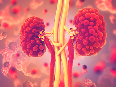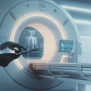
AI’s transformative potential in radiology
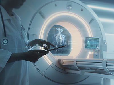
The adoption of artificial intelligence (AI) has the potential to enhance radiological imaging, improve diagnostic capabilities and reduce burnout in the field.
The adoption of artificial intelligence (AI) has the potential to enhance radiological imaging, improve diagnostic capabilities and reduce burnout in the field, provided that physicians and scientists work together to ensure its careful integration into the practice of medicine, according to a special report in Radiology: Artificial Intelligence, a journal of the Radiological Society of North America (RSNA).
Assembled by experts in radiology, medical imaging and machine learning, the special report lays out the clinical, cultural, computation and regulatory considerations that are being introduced, particularly as generative AI models become part of the field.
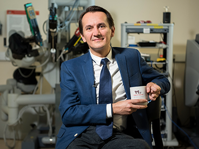
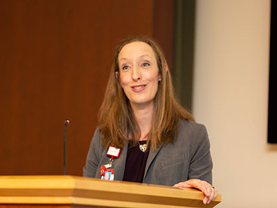
“AI tools can play a key role in radiology, but radiologists must be able to trust in the systems’ design and receive adequate training. As the physicians most familiar with these tools, radiologists should establish clear guidelines regarding clinical accountability,” said Marius George Linguraru, D.Phil., M.A., M.Sc., the Connor Family Professor in Research and Innovation and principal investigator in the Sheikh Zayed Institute for Pediatric Surgical Innovation.
Moving the field forward
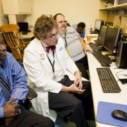
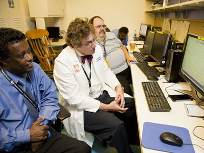
Dr. Linguraru and his peers assembled the report based on a series of seminars hosted by RSNA and the Medical Image Computing and Computer Assisted Intervention (MICCAI) Society. They collected input from multidisciplinary experts to outline the current clinical uses of AI and its future potential.
The experts agreed that collaboration between radiologists and AI scientists will be essential to successfully integrate AI into the discipline of radiology. This partnership should focus on establishing a unified agenda, shared language and clear expectations of the tools developed. By working together, they can ensure that AI tools are designed and implemented to meet the practical needs of radiology, particularly with the incorporation of language and vision models.
What’s next
Among the challenges ahead, clinical institutions must align their staffing, data management and computational resources to deploy and monitor AI systems effectively. This alignment includes ensuring that personnel are adequately trained to use AI tools, that data is managed and processed efficiently and that sufficient computational power is available to support AI operations. Cloud computing may be vital to hospitals that don’t have hardware and technical maintenance resources.
“The successful integration of AI in radiology depends on trust in AI design, collaborative efforts between radiologists and AI scientists, and the alignment of clinical resources to support AI deployment,” Dr. Linguraru said. “With these factors in place, AI can play a transformative role in improving radiological practices and outcomes.”
Read the special report “Clinical, Cultural, Computational, and Regulatory Considerations to Deploy AI in Radiology: Perspectives of RSNA and MICCAI Experts” in Radiology: Artificial Intelligence.