Tag Archive for: Gustavo Nino

computer circuit board

Integrating clinical parameters with lung imaging to predict respiratory outcomes in premature babies

computer circuit board

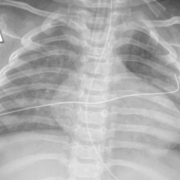
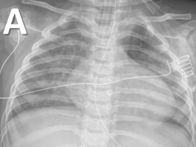
The team will develop an objective framework to predict the risk and assess the severity of respiratory disease in premature babies using non-invasive low-radiation X-ray imaging biomarkers and clinical parameters from the patient bedside.

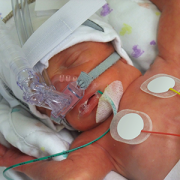
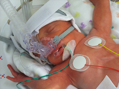
Children’s National Hospital received a $1.7M award from the National Institutes of Health (NIH) National Heart, Lung, and Blood Institute (NHLBI) to develop computational tools that integrate continuous clinical parameters with lung imaging to predict respiratory outcomes for babies born severely premature in newborn intensive care unit (NICU) settings.

The multi-disciplinary team of internationally recognized experts in quantitative imaging, machine learning and neonatal respiratory research believes they can improve clinical practice. To get there, they will develop an objective framework to predict the risk and assess the severity of respiratory disease in premature babies using non-invasive low-radiation X-ray imaging biomarkers and clinical parameters from the patient bedside.

“This computational tool will assist clinicians in making critical decisions about the course of therapy and other necessary follow-ups,” said Gustavo Nino, M.D., M.S.H.S., D’A.B.S.M., principal investigator in the Center for Genetic Medicine at Children’s National. “An objective informed decision about the severity of lung disease in prematurity will result in fewer rehospitalizations, better long-term outcomes and life-saving benefits.”

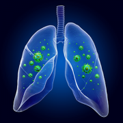
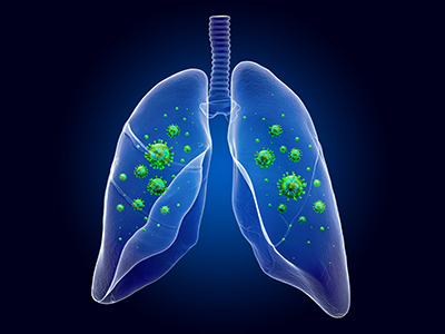
Prematurity is the largest single cause of death in children under five in the world. Lower respiratory tract infections (LRTI) are the top cause of hospitalization and mortality in premature infants. Clinical tools to predict the risk and assess the severity of LRTI in premature babies are needed to allow early interventions that can decrease the high morbidity and mortality in this patient group.

“Our new technology will provide clinicians an accurate, fast and comprehensive summary of the respiratory status of premature babies,” said Dr. Nino. “The data analysis along with the software technology will help determine if a premature baby seen in the NICU can be safely discharged or will require further monitoring and treatment.”

Predictive analytics could help in many ways. For example, there are instances where newborns in the NICU are on the right path with no risks in the future, but there are babies who will come back with severe infections.

“In the first scenario, if we can predict earlier that they’re fine, this could reduce the number of chest X-rays and extra tests, so we assess that this child can be safely sent home,” said Marius George Linguraru, D.Phil., M.A., M.Sc., principal investigator in the Sheikh Zayed Institute for Pediatric Surgical Innovation at Children’s National. “On the other hand, for kids that may come back to the hospital in the near future, we could predict earlier that they are not that well by looking at images and other continuous measurements such as supplemental oxygen.”

This approach, in essence, is a collection of continuous data from the NICU, which is very complex itself because it needs to be collected every day and fed into a machine learning model that digests the data to identify risk patterns for the health of the lung.

“If we find that there is still a risk, it does not necessarily mean that the child has to stay in the NICU any longer, but they might continue treatment, and we will have to define how this integrates into the clinical management of these patients,” said Linguraru. “If there is something in the data that we can put our finger on, we will know which kids require timely attention, hopefully reducing future adverse situations with potential comorbidities and financial burdens.”

smiling baby sleeping

Link between early lower respiratory tract infections and obstructive sleep apnea

smiling baby sleeping

For the first time, researchers at Children’s National Hospital have identified the association between early LRTI and the development of OSA in children.

Several birth cohorts have defined the pivotal role of early lower respiratory tract infections (LRTI) in the inception of pediatric respiratory conditions. However, the association between early LRTI and the development of obstructive sleep apnea (OSA) in children had not previously been made.

Now, for the first time, researchers at Children’s National Hospital have identified the association between early LRTI and the development of OSA in children, according to a study published in the journal SLEEP.

“These results suggest that respiratory syncytial virus LRTI may contribute to the pathophysiology of OSA in children,” said Gustavo Nino, M.D., director of sleep medicine at Children’s National.

The study also demonstrated that children with a history of severe respiratory syncytial virus (RSV) bronchiolitis during early infancy had more than double the odds of developing OSA during the first five years of life independently of other risk factors.

“The results suggest that RSV LRTI may contribute to the pathophysiology of OSA in children, raising concern for the possibility that primary prevention strategies can hinder the initial establishment of OSA following early viral LRTIs,” said Dr. Nino. “Primary prevention of OSA in children would have a dramatic effect in reducing the increasing incidence of this condition and in preventing its detrimental effects on childhood health and beyond.”

The novel findings also raise the possibility that anticipatory strategies and interventions can be developed to identify and prevent the initial establishment of OSA following viral respiratory infections during early infancy. This could provide a dramatic effect in reducing the increasing incidence of this condition and its multiple detrimental effects on childhood health and beyond.

“Our study offers a new paradigm for investigating mechanisms implicated in the early pathogenesis of OSA in the pediatric population,” said Dr. Nino.

Marishka Brown, Ph.D., director of the National Center on Sleep Disorders Research at the National Heart, Lung, and Blood Institute (NHLBI), part of the National Institutes of Health (NIH), agreed.

“The findings from this study suggest that viral lower respiratory tract infections could predispose to the development of sleep-disordered breathing in later childhood,” Brown said. “More research to determine how these infections affect airway function could lead to a better understanding of how sleep apnea develops in pediatric patients.”

This study includes funding support from the NIH, including the NHLBI.

The Pulmonary Division at Children’s National has been ranked as one of the top ten programs in the nation by U.S. News & World Report.

x-ray of child's chest with COVID

Chest X-rays help distinguish COVID-19 from other types of viral respiratory infections

x-ray of child's chest with COVID

COVID-19 in a 9-month infant demonstrating a GGO/consolidation pattern.

Increased infections of COVID-19 and other respiratory viruses in kids are filling up children’s hospitals, pushing them to critical occupancy nationwide. As schools open, the community spread of viral infections has become more common, and the rapid differentiation of pediatric COVID-19 from other viruses is — more than ever — relevant to pediatric clinicians.

“Pediatric cases have increased exponentially and currently represent over 15% of the total cases, and about 26% of the new infections in the U.S. Chest imaging is a powerful tool for determining their status.” said Marius George Linguraru, D.Phil., M.A., M.Sc., principal investigator in the Sheikh Zayed Institute for Pediatric Surgical Innovation at Children’s National Hospital.

In a new peer-reviewed study, researchers from Children’s National found novel and clinically relevant data regarding the specific lung imaging patterns of pediatric COVID-19 on chest radiographs (CXR), their relationship to clinical outcomes and the possible differences from infections caused by other respiratory viruses in children.

“While most studies have focused on clinical manifestations and lung imaging of COVID-19 in adults, this study is the first to define specific patterns of clinical disease and imaging signatures in CXR in different age groups of children infected with COVID-19,” said Gustavo Nino, M.D., director of sleep medicine at Children’s National.

Lung imaging has become critically important for the early identification and treatment of pediatric patients affected by COVID-19 and may play an important role in distinguishing COVID-19 infection from viral bronchiolitis.

“The old perception that COVID-19 only affects older patients is no longer true,” said Dr. Nino. “Pediatric intensive care units and emergency departments are overwhelmed with COVID-19 cases, and now hospitals are admitting more children with COVID-19 than ever.”

As next steps, Nino et al. will develop pediatric-centered technology for early identification, risk stratification, and outcome prediction of COVID-19 in children, similar to what the scientific community has done for adults.

RSV infected infant cells

$2.13M grant accelerates treatments for kids with Down syndrome experiencing respiratory viruses

RSV infected infant cells

Children’s National Hospital received a combined $2.13 million award from the National Institutes of Health’s (NIH) National Heart, Lung and Blood Institute to better understand the mechanisms of severe viral respiratory infections in patients with Down syndrome and to develop new diagnostic tools and innovative precision medicine approaches for this vulnerable population.

“We have a unique opportunity to discover novel targets that can treat severe viral respiratory infections, including SARS-CoV-2,” said Gustavo Nino, M.D., M.S.H.S., D’A.B.S.M., principal investigator in the Center for Genetic Medicine at Children’s National. “Part of the award will help us accelerate the development of these novel approaches to prevent severe respiratory infections caused by SARS-CoV-2 and other viruses like respiratory syncytial virus infection (RSV) in children and adults with Down syndrome.”

Lower respiratory tract infections are a leading cause of hospitalization and death in children with Down syndrome. Those children have a nine times higher risk for hospitalization and mortality due to respiratory viruses that cause lower respiratory tract infections.

Chromosome 21, which is an extra chromosome copy found in patients with Down syndrome, encodes four of the six known interferon receptors, leading to hyperactivation of interferon response in Down syndrome. With the central role of interferons focused on antiviral defense, it remains puzzling how interferon hyperactivation contributes to severe viral lower respiratory tract infections in children with Down syndrome. This is an area that the researchers will explore to better manage and treat viral lower respiratory tract infections in these patients, with the support of NIH’s INCLUDE initiative. INCLUDE provides institutions with grants to help clinical research and therapeutics to understand and diminish risk factors that influence the overall health, longevity, and quality of life for people with Down syndrome related to respiratory viruses.

“While many of the other studies focus on intellectual and other disabilities, we are exploring a novel viral respiratory infectious disease mechanism and are doing so by working directly with patients and patient-derived samples,” said Jyoti Jaiswal, M.Sc., Ph.D., senior investigator in the Center for Genetic Medicine Research at Children’s National.

Children with Down syndrome have historically been excluded in research related to airway antiviral immunity, which is a focus of this human-based transformative study to improve the health and survival of patients with Down syndrome. There is a critical need for studies that define targetable molecular and cellular mechanisms to address dysregulated antiviral responses in this patient population.

“The clinical expertise at Children’s National in studying Down syndrome and the work of our team in caring for these patients with respiratory and sleep disorders positions us well to pursue this work,” said Jaiswal. “This is further supplemented by our initial studies that have identified a novel mechanism of impaired airway antiviral responses in these patients.”

Congresswoman Eleanor Holmes Norton (D-DC) also celebrated Children’s National and its NIH research funding benefitting people with Down syndrome.

“I am pleased to congratulate Dr. Nino and staff on being the recipients of the National Heart, Lung, & Blood Institute grant. You were chosen from a competitive group of applicants and should be proud of this notable achievement,” said Norton in a letter. “By receiving this grant, you have demonstrated outstanding promise in your field. It is my hope that this grant will enable you to better the local and global community.”

girl with down syndrome sleeping

Characteristics of central breathing abnormalities in children with trisomy 21

girl with down syndrome sleeping

Trisomy 21 (TS21), also known as Down syndrome, is the most common genetic syndrome in the United States. Many children with TS21 have a higher prevalence of sleep-related breathing disorders including central sleep apnea. While the mechanisms of central sleep apnea in TS21 are not completely understood, children with Down syndrome have multiple factors that make them more susceptible to developing central breathing abnormalities, including nervous system impairment, hypothyroidism and hypotonia.

In a recent multi-institutional study published in the journal Pediatric Pulmonology, Gustavo Nino, M.D., MSHS, D’ABSM, director of sleep medicine at Children’s National Hospital, and colleagues investigated the clinical features of central breathing abnormalities in TS21 across different pediatric age groups. The researchers also conducted analyses to look at the effects of biological sex and concomitant obstructive sleep apnea in children with central breathing abnormalities.

The authors conclude that “central breathing abnormalities are common in TS21 among young children (≤2 years of age) and in females older than 2 years of age,” and that “central apnea is often associated with concomitant obstructive sleep apnea and/or hypoxemia in children with TS21.”

Read the full study in Pediatric Pulmonology.

illustration of lungs with virus

Segmenting viral bronchiolitis patients to better predict clinical outcomes

illustration of lungs with virus

By evaluating viral bronchiolitis patients at first presentation and categorizing them based on clinical phenotype, the researchers were able to better predict outcomes and disease progression patterns.

Researchers from Children’s National Hospital have recently published a pilot study of children with viral bronchiolitis. By evaluating viral bronchiolitis patients at first presentation and categorizing them based on clinical phenotype, the researchers were able to better predict outcomes and disease progression patterns. Nasal airway cytokine levels were also measured to assess the underlying airway immunobiology of different clinical phenotypes. The researchers believe this novel subdivision of viral bronchiolitis patients based on a robust combination of clinical and molecular assessment can help lead to more individualized care and better patient outcomes.

Viral bronchiolitis is broadly used to group together infants with first-time severe viral respiratory infection, which is the most common cause of early life sick visits and hospitalizations worldwide. However, viral respiratory infections can vary significantly in clinical manifestations, which has raised concern among experts that the use of viral bronchiolitis as a catchall term may be compromising patient care. Children’s National researchers hypothesized that a novel segmentation technique of viral bronchiolitis patients by phenotype at first episode could provide better outcome prediction. In addition, lung X-rays and nasal cytokine profiles could help illuminate the underlying airway disease processes that drive the phenotypical differences observed at bedside.

The study examined 50 children ≤ 2 years old, including 41 patients admitted at Children’s National with PCR-confirmed viral respiratory infection and 9 controls. Researchers examined clinical features at presentation by reviewing each patient’s electronic medical record. Key parameters served as the basis for patient segmentation into three phenotypical groups: hypoxemia, wheezing and mild phenotypes. Patients in the hypoxia group (n = 16) were characterized by their need for supplemental oxygen; patients in the wheezing phenotype (n = 16) were distinguished by wheezing or subcostal retractions and patients in the mild phenotype (n = 9) displayed persistent respiratory symptoms but not hypoxia, wheezing or subcostal retractions. Chest x-rays further revealed that patients in the hypoxia phenotype displayed significantly more lung opacities than the other phenotypes.

As hypothesized, the three phenotype groups displayed distinct clinically relevant outcomes. Patients in the hypoxia group had more severe clinical symptoms at presentation and were significantly more likely to require prolonged hospitalization and pediatric intensive care unit (PICU) settings for treatment. Patients in the wheezing phenotype had shorter hospital stays but were significantly more likely to make a respiratory sick visit after initial discharge, with 69% coming back to the hospital with the same symptoms. Patients in the mild phenotype had the shortest hospital stays and did not require transfer to the PICU.

Nasal cytokine profiles were also assessed for all study subjects. Controls had lower cytokine levels than patients, with no significant difference between phenotype groups. However, wheezing patients with ≥1 recurrent respiratory sick visit had higher nasal levels of type 2 cytokines IL-13 and IL-4, consistent with the pathobiology of allergic asthma. This result adds support for the potential of initial sub-setting in guiding timely intervention.

The researchers hope that the strong results of their pilot study will guide clinicians to revise current practices regarding viral bronchiolitis and personalize care of viral respiratory illnesses from first presentation in order to improve outcomes. Study author and Children’s National pulmonologist Maria Arroyo, M.D., says, “if we can prevent these patients from coming [back] to the hospital just by doing a clinical evaluation the first time that they present with [viral respiratory infection]…that would be very impactful.”

The associated article, “Phenotypical Sub-setting of the First Episode of Severe Viral Respiratory Infection Based on Clinical Assessment and Underlying Airway Disease: A Pilot Study,” was published April 2, 2020 in Frontiers in Pediatrics. Notable authors include Maria Arroyo, M.D., Kyle Salka, M.S., and Gustavo Nino, M.D., M.S.H.S., D.A.B.S.M.

preterm baby

Validating a better way to stratify BPD risk in vulnerable newborns

preterm baby

Factoring in the total number of days that extremely preterm infants require supplemental oxygen and tracking this metric for weeks longer than usual improves clinicians’ ability to predict respiratory outcomes according to bronchopulmonary dysplasia severity.

Factoring in the total number of days that extremely preterm infants require supplemental oxygen and tracking this metric for weeks longer than usual improves clinicians’ ability to predict respiratory outcomes according to bronchopulmonary dysplasia (BPD) severity, a research team led by Children’s National Hospital writes in Scientific Reports. What’s more, the researchers defined a brand-new category (level IV) for newborns who receive supplemental oxygen more than 120 days as a reliable way to predict which infants are at the highest risk of returning to the hospital due to respiratory distress after discharge.

About 1 in 10 U.S. infants is born preterm, before 37 weeks gestation, according to the Centers for Disease Control and Prevention. That includes extremely preterm infants who weigh about 1 lb. at birth. These very low birthweight newborns have paper thin skin, frail hearts and lungs that are not yet mature enough to deliver oxygen throughout the body as needed. Thanks to advances in neocritical care, an increasing number of them survive prematurity, and many develop BPD, a chronic lung disease characterized by abnormal development of the lungs and pulmonary vasculature.

“About half of the babies born prematurely will come back to the hospital within the first year of life with a respiratory infection. The key is identifying them and, potentially, preventing complications in this high-risk population,” says Gustavo Nino, M.D., a Children’s National pulmonologist and the study’s lead author.

For decades, the most common way to stratify BPD risk in these vulnerable newborns has been to see if they require supplemental oxygen at 36 weeks corrected gestational age.

“The problem with this classification is it doesn’t take into account the very premature babies who are on oxygen for much longer than other babies. So, we asked the question: Can we continue risk stratification beyond 36 weeks in order to identify a subset of babies who are at much higher risk of complications,” Dr. Nino says.

The longitudinal cohort study enrolled 188 infants born extremely preterm who were admitted to the neonatal intensive care unit (NICU) at Children’s National and tracked their data for at least 12 months after discharge. The team used a multidimensional approach that tracked duration of supplemental oxygen during the newborns’ NICU stay as well as scoring lung imaging as an independent marker of BPD severity. To validate the findings, these U.S.-born newborns were matched with 130 infants who were born preterm and hospitalized at two NICUs located in Bogotá, Colombia.

“Babies who are born very preterm and require oxygen beyond 120 days should have expanded ventilation of the lungs and cardiovascular pulmonary system before going home,” he notes. “We need to identify these newborns and optimize their management before they are discharged.”

And, the babies with level IV BPD risk need a different type of evaluation because the complications they experience – including pulmonary hypertension – place them at the highest risk of developing sleep apnea and severe respiratory infection, especially during the first year of life.

“The earlier we identify them, the better their outcome is likely to be,” Dr. Nino says. “We really need to change the risk stratification so we don’t call them all ‘severe’ and treat them the same when there is a subset of newborns who clearly are at a much higher risk for experiencing respiratory complications after hospital discharge.”

In addition to Dr. Nino, Children’s National study co-authors include Awais Mansoor, Ph.D., staff scientist at the Sheikh Zayed Institute for Pediatric Surgical Innovation (SZI); Geovanny F. Perez, M.D., pediatric pulmonologist; Maria Arroyo, M.D., pulmonologist; Xilei Xu Chen, M.D., postdoctoral fellow; Jered Weinstock, pediatric pulmonary fellow; Kyle Salka, MS, research technician; Mariam Said, M.D., neonatologist, and Marius George Linguraru, DPhil, MA, MSc, SZI principal investigator and senior author. Additional co-authors include Ranniery Acuña-Cordero, Universidad Militar Nueva Granada, Bogotá, Colombia; and Monica P. Sossa-Briceño and Carlos E. Rodríguez-Martínez, both of Universidad Nacional de Colombia.

Funding for research described in this post was provided by the National Institutes of Health (NIH) under award Nos. HL145669, AI130502 and HL141237. In addition, the NIH has awarded Dr. Nino an RO1 grant to continue this research.