Tag Archive for: Gallo

Screen grab of Dr. Terry Dean and Dr. Vittorio Gallo webinar

In the News: Regenerative brain cells and the circadian clock

Screen grab of Dr. Terry Dean and Dr. Vittorio Gallo webinar

“I am a pediatric intensivist, and I am very interested in some of the pathologies and conditions that I come across in the ICU. We hatched this question that revolved around the idea: what can we do for TBI (traumatic brain injury) patients to enhance their cellular regeneration? …  We looked at NG2-glia in particular, otherwise known as oligodendrocyte precursor cells. They are about 2-8% of the brain…. Do these cells respond to sleep and circadian rhythm? Is it a factor? Does it help? Does it hurt?”

Find out more about what Terry Dean, M.D., Ph.D., says he has learned about these and other questions through his recent research with interim Chief Academic Officer Vittorio Gallo, Ph.D. They join the Society for Neuroscience in a webinar on the circadian rhythms of these important brain cells and how their regeneration may be used someday to promote healing after brain injuries.

glial cells

Future TBI treatments may hinge on understanding a new cell type

glial cells

Only recently have investigators begun to understand how a cell type – the NG2-glia – may respond to injuries, offering clues into the brain’s healing and regeneration.

Traumatic brain injury (TBI) afflicts 69 million people, including 630,000 children, worldwide each year. Yet only recently have investigators begun to understand how a cell type – the NG2-glia – may respond to injuries, offering clues into the brain’s healing and regeneration.

In a new paper published in GLIA, investigators from Children’s National Hospital reviewed 25 years of neuroscience research to lay out what’s known about the molecular response of these NG2-glia cells after TBI. Researchers said they see “a seductive possibility” that tapping into the regenerative potential of NG2-glia cells after neurotrauma could lead to therapies in the future. The impact could be profound, given that TBI is the leading cause of death among all people ages 1-44 and the global cost of this ‘silent epidemic’ is estimated to top $102 billion annually.

What they’re saying

“Our review lays out what’s known about these fascinating cells,” said Terry Dean, M.D., Ph.D., critical care specialist at Children’s National and investigator at the Center for Neuroscience Research (CNR). “NG2-glia are found throughout the brain, and we know that these cells undergo several dynamic changes in the hours, days and weeks after TBI. They are unique, and we want to understand their molecular characteristics to eventually enhance patients’ cellular recovery after TBI.”

Although only encompassing 4% to 8% of brain cells, these NG2-glia cells make up the largest population of regenerative cells in the adult central nervous system. In their article, Dean and Vittorio Gallo, Ph.D., Children’s National Research Institute interim director, lay out a number of unique features of these cells:

  • They proliferate, or multiply, and can form different cell types, especially after brain injuries.
  • They are structurally dynamic and can move and migrate throughout the cortex, including toward injury sites.
  • They appear to play a role in cell-to-cell signaling, which may prove vital after injuries.

The big picture

“As we study the brain after injuries, we hope our work will reveal the role these NG2-glia cells play in recovery, driving us to possible therapies,” Gallo said. “We believe the big answers will come through understanding the brain on a molecular level. This type of deep investigation is the foundation of our bench-to-bedside approach and positions researchers like Dr. Dean to find answers for our patients.”

Moving the field forward

Researchers have only begun to unlock how NG2-glia respond to injury, making this a fruitful area for research. Gallo, Dean and others at CNR hope to build on their knowledge about what happens to the brain immediately after an injury to learn more about what happens months after a debilitating impact. They are also considering new types of research models to expand their knowledge about cellular destruction, immune interaction and blood vessel compromise after different types of brain injuries.

“We look forward to the day when we have a truly targeted therapy for TBI patients,” Dean said. “Imagine the relief this could provide patients suffering from the persistent physical, cognitive and psychological disabilities that often accompany these brain injuries.”

illustration of the brain

How the circadian clock could help the brain recover after injury

illustration of the brain

A type of brain cell that can renew itself is regulated by circadian rhythms, providing significant insights into how the body’s internal clock may promote healing after traumatic brain injuries (TBI).

A type of brain cell that can renew itself is regulated by circadian rhythms, providing significant insights into how the body’s internal clock may promote healing after traumatic brain injuries (TBI), according to new research from Children’s National Hospital.

Released in the latest issue of eNeuro, the findings open new avenues of investigation for future TBI therapies. These injuries are currently managed only with supportive care and rehabilitation, rather than targeted drug treatment options. The findings also underscore the importance of addressing circadian disturbances to help injured brains heal.

Many of the body’s cells follow a 24-hour rhythm driven by their genes known as the circadian clock. The Children’s National research team found that a relatively newly discovered type of brain cell ­– known as NG2-glia, or oligodendrocyte precursor cells ­– also follow a circadian rhythm. This cell type is one of the few that continually self-renews throughout adulthood and is notably proliferative in the first week after brain injuries.

“We have found evidence for the role of this well-known molecular pathway – the molecular circadian clock – in regulating the ability for these NG2-glia to proliferate, both at rest and after injury,” said Terry Dean, M.D., Ph.D., critical care specialist at Children’s National and the lead author of the paper. “This will serve as a starting point to further investigate the pathways to controlling cellular regeneration and optimize recovery after injury.”

Sometimes called “the silent epidemic,” TBI afflicts an estimated 69 million people worldwide each year, with injuries ranging from mild concussions to severe injuries that cause mortality or lifelong disability. In the United States alone, approximately 2.8 million people sustain TBI annually, including 630,000 children. TBI is the leading cause of death in people under age 45, and those who survive are often left with persistent physical, cognitive and psychological disabilities.

Yet no targeted therapies exist for TBI, creating a critical need to uncover the mechanisms that could unlock the regeneration of these NG2-glia cells, which are the most common type of brain cell known to proliferate and self-renew in adult brains.

“It is essential for researchers to know that cell renewal is coordinated with the time of day,” said Vittorio Gallo, Ph.D., interim chief academic officer and interim director of the Children’s National Research Institute. “With this knowledge, we can dig deeper into the body’s genetic healing process to understand how cells regulate and regenerate themselves.”

Brain illustration

Paving the way toward better understanding and treatment of neonatal brain injuries

Brain illustration

The Gallo Lab’s latest research finds reduced expression of Sirt2 in the white matter of premature human infants and characterizes its role in white matter of the brain in normal conditions and during hypoxia.

Changes in myelination due to diffuse white matter injury are a common consequence of premature birth and hypoxic-ischemic injury due to asphyxia of sick term-born newborns. Hypoxic damage during the neonatal period can lead to motor disabilities and cognitive deficits with long-term consequences, including cerebral palsy, intellectual disability or epilepsy, which are often due to cellular and functional abnormalities.

The Gallo Lab, within the Center for Neuroscience Research at Children’s National Hospital, is focused on studying postnatal neural development and the impact of injury and disease on development and regeneration of neurons and glia. Their latest research, published in Nature Communications, finds reduced expression of Sirt2 in the white matter of premature human infants (born earlier than 32 weeks of gestation) and characterizes its role in white matter of the brain in normal conditions as well as during hypoxia.

What it means

The lab previously identified Sirt1 as important for the proliferative regenerative response of oligodendrocyte progenitor cells in response to chronic neonatal hypoxia. This new study characterizes the function of Sirt2 and finds that it acts as a critical promoter of oligodendrocyte differentiation during both normal brain development and after hypoxia.

It’s likely this reduced expression of Sirt2 contributes to the arrest in oligodendrocyte maturation and myelination failure seen in extremely low gestational age neonates. Therefore, targeting Sirt2 may be an opportunity to capture the early and small window of opportunity for therapeutic intervention.

How this moves the field forward

Sirtuins have been shown to play crucial therapeutic roles in various diseases, including aging, neurodegenerative disorders, cardiovascular disease and cancer. Identifying Sirt2 as a major regulator of white matter development and recovery and increasing the understanding of its protein and genomic interactions opens new avenues for Sirt2 as a therapeutic target for white matter injury in premature babies.

Why we’re excited

Interestingly, the team found that overexpression of Sirt2 in oligodendrocyte progenitor cells, but not mature oligodendrocytes, restores oligodendrocyte populations after hypoxia through enhanced proliferation and protection from apoptosis. This is exciting because:

  • It tells us that Sirt2 expression is very important for the transition from progenitor to differentiated oligodendrocyte.
  • It’s the first report, to the team’s knowledge, of Sirt2 regulating cell survival of oligodendrocytes.

Read more in Nature Communications

crawling baby

Gene-targeting may help prevent or recover neonatal brain injuries

crawling baby

The findings of a new pre-clinical study published in The Journal of Neuroscience are helping pave the way toward better understanding, prevention and recovery of neonatal brain injuries.

The findings of a new pre-clinical study published in The Journal of Neuroscience are helping pave the way toward better understanding, prevention and recovery of neonatal brain injuries. During pregnancy, the fetus normally grows in low oxygen conditions. When babies are born preterm, there is an abrupt change into a high oxygen environment which may be higher than the baby can tolerate. These preterm babies often need support to breathe because their lungs are immature. If the oxygen they receive is too high, oxygen-free radicals can form and cause cell death.

Premature infants have underdeveloped antioxidant defenses that prevent or delay some types of cell damage under normal conditions. In a high oxygen environment, these underdeveloped defenses cannot fully protect against oxidative stress, damaging different brain regions without available treatments or preventative measures.

“I am thrilled that we identified a defect in a specific cell population in the hippocampus for memory development,” said Vittorio Gallo, Ph.D., interim chief academic officer and interim director of the Children’s National Research Institute, and principal investigator for the District of Columbia Intellectual and Developmental Disabilities Research Center. “I did not think we would be able to do it at a refined level, identifying cell populations sensitive to oxidative stress and its underlying signaling pathway and molecular mechanism.”

Vittorio Gallo

“I am thrilled that we identified a defect in a specific cell population in the hippocampus for memory development,” said Vittorio Gallo, Ph.D.

Children’s National Hospital experts found that oxidative stress over-activates a glucose metabolism enzyme, GSK3β, altering hippocampal interneuron development and impairing learning and memory, according to the pre-clinical study. The researchers also inhibited GSK3β in hippocampal interneurons, reversing these cellular and cognitive deficits.

The role of oxidative stress in the developing hippocampus, as well as GSK3β involvement in oxidative stress-induced neurodevelopmental disorders and cognitive deficits, have both been unexplored until now. Goldstein et al. suggest the study paves the way for the field as a viable approach to maximize functional recovery after neonatal brain injury.

To better understand the mechanisms underlying neonatal brain injury, the researchers mimicked the brain injury by inducing high oxygen levels in a pre-clinical model for a short time. This quest led to unlocking the underpinnings of the cognitive deficits, including the pathophysiology and molecular mechanisms of oxidative damage in the developing hippocampus.

Once they identified what caused cellular damage, the researchers used a gene-targeted approach to reduce GSK3β levels in POMC-expressing cells or Gad2-expressing interneurons. By regulating the levels of GSK3β in interneurons ⁠— but not in POMC-expressing cells — inhibitory neurotransmission was significantly improved and memory deficits due to high oxygen levels were reversed.

microglia cells damage the myelin sheath of neuron axons

Katrina Adams, Ph.D., awarded fellowship to help restore functions in MS patients

microglia cells damage the myelin sheath of neuron axons

Multiple sclerosis is a demyelinating disease in which the insulating covers of nerve cells are damaged. Microglia cells (orange) attack the oligodendrocytes that form the insulating myelin sheath around neuron axons, leading to the destruction of the myelin sheath and to the loss of nerve function.

For her contributions to Multiple Sclerosis (MS) research, Katrina Adams, Ph.D., postdoctoral researcher at Children’s National Hospital, received the career transition fellowship from The National Multiple Sclerosis Society. The $600,000 fellowship will support a two-year period of advanced postdoctoral training in MS research and the first three years of research support in a new faculty appointment.

MS symptoms, including vision loss, pain, fatigue and reduced motor coordination, result from the demyelination of neuronal axons that transport critical information across the brain and spinal cord. Demyelination is the loss of myelin protein, which is normally produced by oligodendrocyte cells.

In the healthy brain, oligodendrocytes repair demyelinated areas by replacing damaged or lost myelin in a process called remyelination. Recent evidence has shown that oligodendrocytes display differences in their molecular and functional properties. One source of new oligodendrocytes in the adult brain is neural stem cells, which have been shown to generate oligodendrocytes that contribute to remyelination.

“The goal of this project is to determine whether neural stem cell-derived oligodendrocytes are distinct from other oligodendrocytes, both in the healthy brain and in MS,” said Adams. “I aim to understand the molecular mechanisms that regulate generation of oligodendrocytes from neural stem cells, with the goal of identifying signals that could be targeted in MS patients to promote remyelination.”

Remyelination is very limited in MS patients and current therapies for MS have very little impact on promoting remyelination.

This study will take advantage of the state-of-the-art facilities for single-cell analysis, transcriptomics, microscopy, and animal research in Children’s Research Institute at Children’s National. Adams also added that her postdoctoral mentor, Vittorio Gallo, Ph.D., interim chief academic officer and interim director of the Children’s National Research Institute, and principal investigator for the DC-IDDRC, has renowned expertise in glial biology, animal models of MS and white matter injury.

“This research will be the first to directly compare neural stem cell-derived oligodendrocytes with other resident oligodendrocytes in MS brain samples,” said Adams. “The results of this study will provide critical insight into the role that neural stem cells play in repair of MS demyelinated lesions.”

Adams received her doctorate in molecular biology from the University of California, Los Angeles where she used pluripotent stem cells to study motor neuron development. She currently investigates signaling pathways that regulate neural stem and progenitor cell maintenance and differentiation in the developing postnatal and adult brain, with a focus on the Endothelin-1 pathway. She is interested in understanding how stem and progenitor cells respond to disease or injury, such as in Multiple Sclerosis, with the hope of identifying new therapeutic targets.

boy with a chromosomal developmental disability.

NIH award will support intellectual and developmental disabilities research at Children’s National

boy with a chromosomal developmental disability.

Children’s National Hospital announces a $7 million award from the National Institutes of Health’s Eunice Kennedy Shriver National Institute of Child Health and Human Development (NICHD) to support the DC Intellectual and Developmental Disabilities Research Center (DC-IDDRC).

Children’s National Hospital announces a $7 million award from the National Institutes of Health’s Eunice Kennedy Shriver National Institute of Child Health and Human Development (NICHD) to support the DC Intellectual and Developmental Disabilities Research Center (DC-IDDRC). Through this award, the DC-IDDRC will enhance the recruitment and training of investigators, generate innovation and promote transdisciplinary research to facilitate the development, implementation and dissemination of new diagnostic and therapeutic advances for the care of individuals with intellectual and developmental disabilities.

The DC-IDDRC, led by Children’s National in partnership with George Washington University, Howard University and Georgetown University, is one of only 14 IDDRCs in the United States funded by NICHD. This long standing NICHD program supports researchers whose goals are to advance understanding of a variety of conditions and topics related to intellectual and developmental disabilities.

“Children’s National cares for one of the largest cohorts of children with developmental disabilities in the U.S. — which uniquely positions us to lead the way in both care and research of developmental disabilities in young children,” said Vittorio Gallo, Ph.D., interim chief academic officer and interim director of the Children’s National Research Institute, and principal investigator for the DC-IDDRC.

The research strategy for this period will address three key areas: neural development and neurodevelopmental disorders, fetal and neonatal brain injury and genetic disorders by leveraging the core facilities and core innovation — including the Genomics and Bioinformatics Core, Cell and Tissue Microscopy Core, Neuroimaging Core, Clinical Translational Core and Neurobehavioral Evaluation Core.

“In spite of tremendous advances in our understanding of how abnormalities in brain development cause neurodevelopmental disorders and developmental disabilities, integrated knowledge in all these areas of research is still lacking. In particular, it is still unknown how specific genetic defects and cellular abnormalities result in behavioral phenotypes,” said Gallo.

One in six children suffers from a chronic, complex neurodevelopmental disability — conditions such as intellectual disability, learning disability, attention deficit hyperactivity disorder, autism spectrum disorder, cerebral palsy and Down syndrome. For 20 years, the DC-IDDRC has been a home for researchers from different specialties and different institutions to discover new therapies and treatments for children with these types of neurodevelopmental disabilities.

“The DC-IDDRC promises to be a great vehicle to spawn new research and collaborative networks for D.C. area investigators,” said Chandan Vaidya, Ph.D., vice provost for faculty and professor at Georgetown University. “We will be examining whether a behavioral intervention to enhance self-regulation in adolescents with Autism changes how they learn and use computational modeling to understand learning strategy and identify associated changes in the brain using functional magnetic resonance imaging.”

The robust relationships and spirit of cooperation built over two decades of collaboration have laid a strong groundwork for the establishment of the expansive post-doctoral training program and continuous growth of the research programs within the DC-IDDRC. Gallo continues his efforts in expanding access to these programs and building a sustainable pipeline of young scholars from diverse backgrounds. The partnership between Children’s National and Howard University continues to play a crucial role in these goals.

The DC-IDDRC continues to work toward translating research findings into novel approaches and personalized treatments for people with developmental disabilities and their caregivers. This work will be amplified when the DC-IDDRC moves into the expanded facility at the Children’s National Research & Innovation Campus, which houses startup incubator programs and other support for device innovation.

fetus in utero

Loss of placental hormone linked to brain and social behavior changes

fetus in uteroPreterm birth has been shown to increase the risk of autism spectrum disorders and other developmental problems, particularly in males. The more premature a baby is, the greater the risk of either motor or cognitive deficits. What does the preterm baby lose that is so critical to long-term outcomes?

A new pre-clinical study suggests that one factor may be the loss of a placental hormone that the developing brain would normally see in the second half of pregnancy.

The study is the first to provide direct evidence that loss of a placental hormone alters long-term brain development.

In the study, researchers in the laboratory of Anna Penn, M.D., Ph.D., now at Columbia University Vagelos College of Physicians and Surgeons and previously at Children’s National Hospital in Washington, D.C., found that reducing amounts of a single hormone, called allopregnanolone (ALLO), in the placenta caused brain and behavior changes in male offspring that resemble changes seen in some people with autism spectrum disorder.

The study also found that both brain structure and behavioral changes in the subjects could be prevented with a single injection of ALLO in late pregnancy.

“Our study provides new and intriguing insights into how the loss of placental hormones—which happens in preterm birth or if the placenta stops working well during pregnancy—can lead to long-term structural changes in the brain that increase the risk for autism or other neuropsychiatric disorders,” says lead author Claire-Marie Vacher, Ph.D., assistant professor of neonatal sciences in the Department of Pediatrics at Columbia University’s Vagelos College of Physicians and Surgeons. “What’s encouraging is that these disorders may be preventable if diagnosed and treated early.”

The study was published online August 16 in the journal Nature Neuroscience.

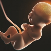
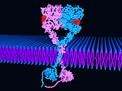
The placenta is an organ that provides the fetus with oxygen and nutrients and removes waste products. It also produces hormones, including high levels of ALLO in late pregnancy that may influence brain development. Penn, now the L. Stanley James Associate Professor of Pediatrics at Columbia University Vagelos College of Physicians and Surgeons and chief of neonatology at Columbia and New York-Presbyterian Morgan Stanley Children’s Hospital, coined the term “neuroplacentology” to describe this new field of research connecting placental function to brain development.

About one in 10 infants is born prematurely (and is thus deprived of normal levels of ALLO and other hormones), and many more pregnancies have poor placental function.

For this study, the researchers created a pre-clinical model in which they were able to selectively decrease the production of ALLO during pregnancy so that some developing pups were exposed to sufficient placental ALLO while others were not. Although male and female fetuses were both subjected to ALLO deficiency, only male subjects showed autism-like behaviors after birth. Working with collaborators in Washington, D.C., France, and Canada, the Penn laboratory analyzed brain development and long-term behavioral outcomes in the offspring.

ALLO reduction led to cerebellum changes, autism-like behaviors

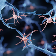
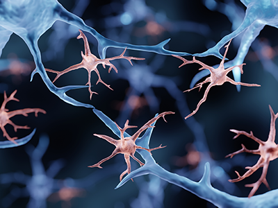
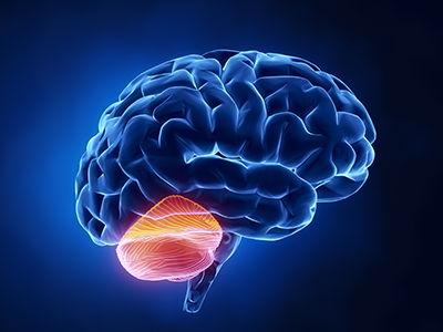
The male subjects that lacked placental ALLO had structural changes in the cerebellum, a brain region that coordinates movement and has been linked to autism, while their littermates did not.

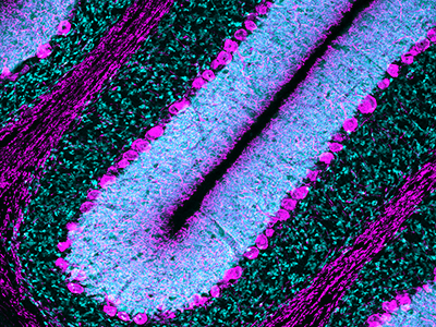
“In particular, we observed thickening of the myelin sheaths, the lipid coating that protects nerve fibers and speeds up neural signaling,” Vacher says. The same type of thickening is also known to occur transiently in the cerebellum of some boys with autism.

The degree of myelin thickening in juvenile male subjects correlated with abnormal behavior, the researchers also found. The more the sheath was thickened (as measured by myelin protein levels), the more the male subjects exhibited autism-like behaviors, such as decreased sociability and repetitive activities.

“Our experimental model demonstrates that losing placental ALLO alters cerebellar development, including white matter development. Cerebellar white matter development occurs primarily after birth, so connecting a change in placental function during pregnancy with lingering impacts on later brain development is a particularly striking result,” says Penn.

“The findings provide a new way to understand poor placental function. Subtle but important changes during pregnancy or after delivery may set in motion neurodevelopmental disorders that children experience later in life.”

Similarities with human tissue

To determine if similar changes occur in infants, the researchers also examined post-mortem cerebellar tissues from preterm and full-term infants who had died soon after birth. Analysis of these human tissues showed similar changes in brain proteins when cerebellum from male babies born preterm were compared to male full-term babies.

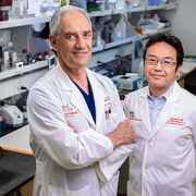
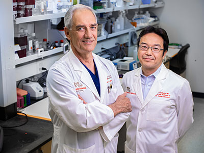
“This study is an important first step in understanding how placental hormones may contribute to specific human neurobehavioral outcomes. We look forward to continuing our collaboration with Dr. Penn and her team to help define how cerebellar neurons and glia respond to environmental factors, including placental function, that can compromise the developing brain,” says study co-author Vittorio Gallo, Ph.D., interim chief academic officer at Children’s National Hospital and interim director of the Children’s National Research Institute.

Hormone injection reduced autism symptoms

ALLO’s therapeutic potential was then tested in the preclinical model.

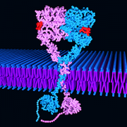
Male offspring of the pre-clinical model given a single injection of ALLO in late pregnancy had fewer autism-like behaviors, the researchers found. Similar results were seen after an injection of muscimol, a drug that enhances the function of GABA receptors—the same receptors that respond to ALLO. Myelin protein levels in the developing cerebellum also normalized with the treatment.

“Identifying when key hormone levels are abnormal, and figuring out how and when to adjust these levels, provides an opportunity to intervene,” Penn says. “Performing additional studies with our pre-clinical model, and measuring hormone levels in moms and babies, may lead to earlier treatment to reduce or prevent long-term cognitive and behavioral impairments in high-risk fetuses and newborns.”

A version of this story appeared on the Columbia University newsroom.

The study is titled “Placental endocrine function shapes cerebellar development and social behavior.” The other contributors: Helene Lacaille (Columbia), Jiaqi J. O’Reilly (Columbia), Jacquelyn Salzbank (Columbia), Dana Bakalar (National Institutes of Health, Bethesda, MD), Sonia Sebaoui (Children’s National Hospital, Washington, DC), Philippe Liere (University Paris Saclay, Le Kremlin‐Bicêtre Cedex, France), Cheryl Clarkson-Paredes (George Washington University, Washington, DC), Toru Sasaki (Children’s National Hospital), Aaron Sathyanesan (Children’s National Hospital), Panagiotis Kratimenos (Children’s National Hospital), Jacob Ellegood (Hospital for Sick Children, Toronto, ON), Jason Lerch (Hospital for Sick Children and University of Oxford, John Radcliffe Hospital, Oxford, UK), Yuka Imamura (Pennsylvania State University College of Medicine, PA), Anastas Popratiloff (George Washington University), Kazue Hashimoto-Torii (Children’s National Hospital and George Washington University), and Michael Schumacher (University Paris Saclay).

Purkinje cell

Premature birth disrupts Purkinje cell function, resulting in locomotor learning deficits

Purkinje cell

Children’s National Hospital researchers explored how preterm birth disrupts Purkinje cell function, resulting in locomotor learning deficits.

As the care of preterm babies continues to improve, neonatologists face new challenges to ensure babies are protected from injury during critical development of the cerebellum during birth and immediately after birth. How does this early injury affect locomotor function, and to what extent are clinicians able to protect the brain of preterm babies?

A new peer-reviewed study by Aaron Sathyanesan, Ph.D., Panagiotis Kratimenos, M.D., Ph.D., and Vittorio Gallo, Ph.D., published in the Proceedings of the National Academy of Sciences of the United States of America (PNAS), explores exactly what neural circuitry of the cerebellum is affected due to complications that occur around the time of birth causing these learning deficits, and finds a specific type of neurons — Purkinje cells — to play a central role.

Up until now, there has been a sparsity of techniques available to measure neuronal activity during locomotor learning tasks that engage the cerebellum. To surmount this challenge, Children’s National used a multidisciplinary approach, bringing together a team of neuroscientists with neonatologists who leveraged their joint expertise to devise a novel and unique way to measure real-time Purkinje cell activity in a pre-clinical model with clinical relevance to humans.

Researchers measured neural circuit function by pairing GCaMP6f fiber photometry, used to measure neuronal activity in the brain of a free moving subject, with an ErasmusLadder, in which it needs to travel from point A to point B on a horizontal ladder with touch-sensitive rungs that register the type and length of steps. By introducing a sudden obstacle to movement, researchers observed how the subject coped and learned accordingly to avoid this obstacle. By playing a high-pitch tone just before the obstacle was introduced, researchers were able to measure how quickly the subjects were able to anticipate the obstacle and adjust their steps accordingly. Subjects with neonatal brain injury and normal models were run through a series of learning trials while simultaneously monitoring brain activity. In this way, the team was able to quantify cerebellum-dependent locomotor learning and adaptive behavior, unlocking a functional and mechanistic understanding of behavioral pathology that was previously unseen in this field.

In addition to showing that normal Purkinje cells are highly active during movement on the ErasmusLadder, the team explored the question of whether Purkinje cells of injured pre-clinical models were generally non-responsive to any kind of stimuli. They found that while Purkinje cells in injured subjects responded to puffs of air, which generally cue the subject to start moving on the ErasmusLadder, dysfunction in these cells was specific to the period of adaptive learning. Lastly, through chemogenetic inhibition, which specifically silences neonatal Purkinje cell activity, the team was able to mimic the effects of perinatal cerebellar injury, further solidifying the role of these cells in learning deficits.

The study results have implications for clinical practice. As the care of premature babies continues to improve, neonatologists face new challenges to ensure that babies not only survive but thrive. They need to find ways to prevent against the lifelong impacts that preterm birth would otherwise have on the cerebellum and developing brain.

Read the full press release here.

Read the full journal article here.

structure of EGFR

Study suggests EGFR inhibition reverses alterations induced by hypoxia

structure of EGFR

The study suggests that specific molecular responses modulated by EGFR (seen here) may be targeted as a therapeutic strategy for HX injury in the neonatal brain.

Hypoxic (HX) encephalopathy is a major cause of death and neurodevelopmental disability in newborns. While it is known that decreased oxygen and energy failure in the brain lead to neuronal cell death, the cellular and molecular mechanisms of HX-induced neuronal and glial cell damage are still largely undefined.

Panagiotis Kratimenos, M.D., and colleagues from the Center for Neuroscience Research at the Children’s National Research Institute, discovered increased expression of activated-epidermal growth factor receptor (EGFR) in affected cortical areas of neonates with HX and decided to further investigate the functional role of EGFR-related signaling pathways in the cellular and molecular changes induced by HX in the cerebral cortex.

The researchers found that HX-induced activation of EGFR and Ca2+/calmodulin kinase IV (CaMKIV) caused cell death and pathological alterations in neurons and glia. EGFR blockade inhibited CaMKIV activation, attenuated neuronal loss, increased oligodendrocyte proliferation and reversed HX-induced astrogliosis.

The researchers also performed, for the first time, high-throughput transcriptomic analysis of the cortex to define molecular responses to HX and to uncover genes specifically involved in EGFR signaling in brain injury. Their results indicate that specific molecular responses modulated by EGFR may be targeted as a therapeutic strategy for HX injury in the neonatal brain.

This study defines many new exciting avenues of scientific exploration to further elucidate the beneficial impact of EGFR blockade on perinatal brain injury at the cellular and molecular levels. This analysis could potentially result in the identification of new therapeutic targets associated with EGFR signaling in the developing mammalian brain that are linked with specific long-term abnormalities caused by perinatal brain injury.

Children’s National researchers who contributed to this study include Panagiotis Kratimenos, M.D., Ioannis Koutroulis, M.D., Ph.D., M.B.A., Susan Knoblach, Ph.D., Payal Banerjee, Surajit Bhattacharya, Ph.D., Maria Almira-Suarez, M.D., and Vittorio Gallo, Ph.D.

Read the full article in iScience.

Drs. Dewesh Agrawal, Andrew Dauber, Robert Freishtat, Vittorio Gallo

Four Children’s National Hospital leaders named to APS

Drs. Dewesh Agrawal, Andrew Dauber, Robert Freishtat, Vittorio Gallo

Drs. Dewesh Agrawal, Andrew Dauber, Robert Freishtat and Vittorio Gallo were named as 2021 American Pediatric Society members.

The American Pediatric Society (APS) has announced 55 new members, four of which are experts from Children’s National Hospital. Founded in 1888, the APS is the first and most prestigious academic pediatric organization in North America.

APS members are recognized child health leaders of extraordinary achievement who work together to shape the future of academic pediatrics. New members are nominated by current members through a process that recognizes individuals who have distinguished themselves as child health leaders, teachers, scholars, policymakers and/or clinicians.

“Our members represent the most distinguished and accomplished academic leaders in pediatrics whose outstanding work has advanced child health,” said APS President Steven Abman, M.D. “I am honored to welcome this exceptional group of individuals to the APS. The APS is especially looking forward to the active engagement of our membership with many exciting programs within the organization that are directed towards improvements in academic pediatric medicine, including more vigorous approaches to express our values of anti-racism, equity, diversity and inclusion.”

APS 2021 active new members from Children’s National are:

  • Dewesh Agrawal, M.D., vice-chair for Medical Education at Children’s National. Agrawal’s career has been marked by academic honors and teaching awards at every stage of his training and faculty employment. He has relentlessly devoted his energy to improving the educational experience for students, residents and fellows at Children’s National.
  • Andrew Dauber, M.D., M.M.Sc., chief of Endocrinology at Children’s National. Dr. Dauber’s leadership is reflected, nationally and internationally, in his ability to create research consortia, bringing together investigators to tackle complex questions. For example, he leads an NIH-funded consortium on the genetics of short statures, with multiple top children’s hospitals as partners. He also leads a large clinical trial testing a novel therapeutic agent for genetic short stature.
  • Robert Freishtat, M.D., M.P.H., senior investigator in the Center for Genetic Medicine of the Children’s National Research Institute (CNRI). Dr. Freishtat has authored or co-authored more than 100 articles and book chapters in the fields of pediatric lung injury, asthma, obesity, exosomes and emergency medicine. His research has been continuously funded by the NIH since 2003.
  • Vittorio Gallo, Ph.D., chief research officer at Children’s National and scientific director of CNRI. Dr. Gallo’s scientific success is attested to by over 130 peer-reviewed publications, many in very high-profile journals, as well as over 30 review articles and book chapters. He has received many national and international awards, including the NINDS Javits award in Neuroscience in 2018. Dr. Gallo has served on the editorial boards of many neuroscience journals, including Glia and the Annual Review in Neuroscience, and has been reviewing editor for the Journal of Neuroscience, all of which is a testament to the tremendous impact that his studies have had on the advancement of neurosciences.

“These new members represent multiple areas of Children’s National and have all leveraged the intersection of science, medicine and clinical education to make advances in their field of study,” said Stephen J. Teach, M.D., M.P.H., chair of the Department of Pediatrics at Children’s National. “Their work has, and will continue to, advance pediatric health care, and I congratulate them on their APS membership.”

illustration of brain showing cerebellum

NIH grant supports research on locomotor dysfunction in Down Syndrome

illustration of brain showing cerebellum

The National Institutes of Health (NIH) has granted the Children’s National Research Institute (CNRI) nearly $500,000 to better understand and identify specific alterations in the circuitry of the cerebellum that results in locomotor dysfunction in down syndrome.

Down syndrome (DS), the most commonly diagnosed chromosomal condition, affects a range of behavioral domains in children including motor and cognitive function. Cerebellar pathology has been consistently observed in DS, and is thought to contribute to dysfunction in locomotor and adaptive motor skills. However, the specific neural pathways underlying locomotor learning that are disrupted in DS remain poorly understood.

The National Institutes of Health (NIH) has granted the Children’s National Research Institute (CNRI) nearly $500,000 through their NIH-wide initiative INCLUDE – INvestigating Co-occurring conditions across the Lifespan to Understand Down syndrome – to better understand and identify specific alterations in the circuitry of the cerebellum that results in locomotor dysfunction in DS. The INCLUDE initiative aims to support the most promising high risk-high reward basic science.

“There is still a lot unknown about Down syndrome, in particular how fundamental cellular and physiological mechanisms of neural circuit function are altered in this syndrome,” says Vittorio Gallo, Ph.D., chief research officer at Children’s National Hospital and scientific director of CNRI. “Grant funding is particularly important to have the resources to develop and apply new cutting-edge methodology to study this neurodevelopmental disorder.”

The main goal of this research is to identify specific alterations in the circuitry of the cerebellum that result in locomotor dysfunction in DS. Defining specific abnormalities in motor behavior, and identifying the brain regions and neurons which are functionally involved will provide the basis for developing potential therapies for treating motor problems in individuals with DS.

“The last decade has brought rapid advances in neurotechnology to address questions at the ‘systems-level’ understanding of brain function,” says Aaron Sathyanesan, Ph.D., a Children’s National postdoctoral research fellow. “This technology has rarely been applied to preclinical models of neurodevelopmental disorders, and even more rarely to models of Down syndrome.”

An example is the use of fiber-optics to probe changes in neural circuitry during behavior. Using this technology, researchers can now directly correlate the changes in circuitry to deficits in behavior.

“Along with the other approaches in our proposal, this represents the synthesis of a new experimental paradigm that we hope will push the field forward,” says Dr. Sathyanesan.

In 1960, the average life expectancy of a baby with Down syndrome was around 10 years. Today, that life expectancy has increased to more than 47 years. That significant increase reflects critical advances in medicine, however, kids with DS still live with long-term challenges in motor and cognitive ability.

Children’s National strongly supports translation and innovation, and recently recruited internationally renowned DS researcher, Tarik Haydar, Ph.D., as its new director of the Center for Neuroscience Research.

“We’re building significant strength in this area of research. This grant helps open new avenues of investigation to define which cells and circuits are impacted by this common neurodevelopmental disorder,” says Dr. Gallo. “Our cutting-edge approach will help us answer questions that we could not answer before.”

Nobuyuki Ishibashi

R01 grant funds white matter protection study for congenital heart disease

Nobuyuki Ishibashi

Nobuyuki Ishibashi, M.D., is the principal investigator on a $3.2 million NIH R01 to study white matter growth and repair in utero for fetal brains affected by congenital heart disease.

Many of the neurological deficits seen in children with congenital heart disease (CHD) are related to abnormal white matter development early in life caused by reduced oxygen supply to the brain while in utero. Children with immature white matter at birth also commonly sustain additional white matter injuries following cardiac surgery.

The NIH recently awarded a prestigious R01 grant totaling more than $3.2 million to a collaborative project led by the Center for Neuroscience Research, the Sheikh Zayed Institute for Pediatric Surgical Innovation and the Children’s National Heart Institute at Children’s National Hospital as well as MedStar Washington Hospital Center.

The research, titled “White matter protection in the fetus with congenital heart disease,” looks specifically at whether providing a supplemental amount of the naturally occurring tetrahydrobiopterin (BH4) for pregnant women could rescue white matter development of fetuses with congenital heart disease whose brains aren’t receiving enough oxygen – or suffering from hypoxic-ischemic events.

Previous preclinical studies have shown that this lack of oxygen depletes the brain’s natural BH4 level, and the researchers hypothesize that BH4 levels play a critical role in the growth and development of white matter in the fetal brain by triggering key cellular/molecular processes. Specifically, the study will focus on three aims:

  1. Establish in a preclinical model the optimal protective regiment for women pregnant with a fetus who has CHD to receive BH4.
  2. Determine the appropriate approach to deliver BH4 to this population
  3. Leverage genetic tools and biochemical techniques in the laboratory to better understand where and how BH4 levels play a role in the growth (or lack thereof) of oligodendrocytes—the primary cells of white matter.

This laboratory-based work is the first step to determining if the neurodevelopment of babies born with CHD can be preserved or recovered by addressing key brain development that occurs before the baby is even born. Findings related to congenital heart disease may also translate to other populations where white matter development is affected by hypoxia-ischemia, including premature infants.

The project is led by principal investigator Nobuyuki Ishibashi, M.D., with co-investigators Vittorio Gallo, Ph.D., Joseph Scafidi, D.O., and Mary Donofrio, M.D. as well as colleagues at MedStar Washington Hospital Center.

Vittorio Gallo and Mark Batshaw

Children’s National Research Institute releases annual report

Vittorio Gallo and Marc Batshaw

Children’s National Research Institute directors Vittorio Gallo, Ph.D., and Mark Batshaw, M.D.

The Children’s National Research Institute recently released its 2019-2020 academic annual report, titled 150 Years Stronger Through Discovery and Care to mark the hospital’s 150th birthday. Not only does the annual report give an overview of the institute’s research and education efforts, but it also gives a peek in to how the institute has mobilized to address the coronavirus pandemic.

“Our inaugural research program in 1947 began with a budget of less than $10,000 for the study of polio — a pressing health problem for Washington’s children at the time and a pandemic that many of us remember from our own childhoods,” says Vittorio Gallo, Ph.D., chief research officer at Children’s National Hospital and scientific director at Children’s National Research Institute. “Today, our research portfolio has grown to more than $75 million, and our 314 research faculty and their staff are dedicated to finding answers to many of the health challenges in childhood.”

Highlights from the Children’s National Research Institute annual report

  • In 2018, Children’s National began construction of its new Research & Innovation Campus (CNRIC) on 12 acres of land transferred by the U.S. Army as part of the decommissioning of the former Walter Reed Army Medical Center campus. In 2020, construction on the CNRIC will be complete, and in 2012, the Children’s National Research Institute will begin to transition to the campus.
  • In late 2019, a team of scientists led by Eric Vilain, M.D., Ph.D., director of the Center for Genetic Medicine Research, traveled to the Democratic Republic of Congo to collect samples from 60 individuals that will form the basis of a new reference genome data set. The researchers hope their project will generate better reference genome data for diverse populations, starting with those of Central African descent.
  • A gift of $5.7 million received by the Center for Translational Research’s director, Lisa Guay-Woodford, M.D., will reinforce close collaboration between research and clinical care to improve the care and treatment of children with polycystic kidney disease and other inherited renal disorders.
  • The Center for Neuroscience Research’s integration into the infrastructure of Children’s National Hospital has created a unique set of opportunities for scientists and clinicians to work together on pressing problems in children’s health.
  • Children’s National and the National Institute of Allergy and Infectious Diseases are tackling pediatric research across three main areas of mutual interest: primary immune deficiencies, food allergies and post-Lyme disease syndrome. Their shared goal is to conduct clinical and translational research that improves what we know about those conditions and how we care for children who have them.
  • An immunotherapy trial has allowed a little boy to be a kid again. In the two years since he received cellular immunotherapy, Matthew has shown no signs of a returning tumor — the longest span of time he’s been tumor-free since age 3.
  • In the past 6 years, the 104 device projects that came through the National Capital Consortium for Pediatric Device Innovation accelerator program raised $148,680,256 in follow-on funding.
  • Even though he’s watched more than 500 aspiring physicians pass through the Children’s National pediatric residency program, program director Dewesh Agrawal, M.D., still gets teary at every graduation.

Understanding and treating the novel coronavirus (COVID-19)

In a short period of time, Children’s National Research Institute has mobilized its scientists to address COVID-19, focusing on understanding the virus and advancing solutions to ameliorate the impact today and for future generations. Children’s National Research Institute Director Mark Batshaw, M.D., highlighted some of these efforts in the annual report:

  • Eric Vilain, M.D., Ph.D., director of the Center for Genetic Medicine Research, is looking at whether or not the microbiome of bacteria in the human nasal tract acts as a defensive shield against COVID-19.
  • Catherine Bollard, M.D., MBChB, director of the Center for Cancer and Immunology Research, and her team are seeing if they can “train” T cells to attack the invading coronavirus.
  • Sarah Mulkey, M.D., Ph.D., an investigator in the Center for Neuroscience Research and the Fetal Medicine Institute, is studying the effects of, and possible interventions for, coronavirus on the developing brain.

You can view the entire Children’s National Research Institute academic annual report online.

Vittorio Gallo

Special issue of “Neurochemical Research” honors Vittorio Gallo, Ph.D.

Vittorio Gallo

Investigators from around the world penned manuscripts that were assembled in a special issue of “Neurochemical Research” that honors Vittorio Gallo, Ph.D., for his leadership in the field of neural development and regeneration.

At a pivotal moment early in his career, Vittorio Gallo, Ph.D., was accepted to work with Professor Giulio Levi at the Institute for Cell Biology in Rome, a position that leveraged courses Gallo had taken in neurobiology and neurochemistry, and allowed him to work in the top research institute in Italy directed by the Nobel laureate, Professor Rita Levi-Montalcini.

For four years as a student and later as Levi’s collaborator, Gallo focused on amino acid neurotransmitters in the brain and mechanisms of glutamate and GABA release from nerve terminals. Those early years cemented a research focus on glutamate neurotransmission that would lead to a number of pivotal publications and research collaborations that have spanned decades.

Now, investigators from around the world who have worked most closely with Gallo penned tributes in the form of manuscripts that were assembled in a special issue of “Neurochemical Research” that honors Gallo “for his contributions to our understanding of glutamatergic and GABAergic transmission during brain development and to his leadership in the field of neural development and regeneration,” writes guest editor Arne Schousboe, of the University of Copenhagen in Denmark.

Dr. Gallo as a grad student

Vittorio Gallo, Ph.D. as a 21-year-old mustachioed graduate student.

“In spite of news headlines about competition in research and many of the negative things we hear about the research world, this shows that research is also able to create a community around us,” says Gallo, chief research officer at Children’s National Hospital and scientific director for the Children’s National Research Institute.

As just one example, he first met Schousboe 44 years ago when Gallo was a 21-year-old mustachioed graduate student.

“Research can really create a sense of community that we carry on from the time we are in training, nurture as we meet our colleagues at periodic conferences, and continue up to the present. Creating community is bi-directional: influencing people and being influenced by people. People were willing to contribute these 17 articles because they value me,” Gallo says. “This is a lot of work for the editor and the people who prepared papers for this special issue.”

In addition to Gallo publishing more than 140 peer-reviewed papers, 30 review articles and book chapters, Schousboe notes a number of Gallo’s accomplishments, including:

  • He helped to develop the cerebellar granule cell cultures as a model system to study how electrical activity and voltage-dependent calcium channels modulate granule neuron development and glutamate release.
  • He developed a biochemical/neuropharmacological assay to monitor the effects of GABA receptor modulators on the activity of GABA chloride channels in living neurons.
  • He and Maria Usowicz used patch-clamp recording and single channel analysis to demonstrate for the first time that astrocytes express glutamate-activated channels that display functional properties similar to neuronal counterparts.
  • He characterized one of the spliced isoforms of the AMPA receptor subunit gene Gria4 and demonstrated that this isoform was highly expressed in the cerebellum.
  • He and his Children’s National colleagues demonstrated that glutamate and GABA regulate oligodendrocyte progenitor cell proliferation and differentiation.
Purkinje cells

Purkinje cells are large neurons located in the cerebellum that are elaborately branched like interlocking tree limbs and represent the only source of output for the entire cerebellar cortex.

Even the image selected to grace the special issue’s cover continues the theme of continuity and leaving behind a legacy. That image of Purkinje cells was created by a young scientist who works in Gallo’s lab, Aaron Sathyanesan, Ph.D. Gallo began his career working on the cerebellum – a region of the brain important for motor control – and now studies with a team of scientists and clinician-scientists Purkinje cells’ role in locomotor adaptive behavior and how that is disrupted after neonatal brain injury.

“These cells are the main players in cerebellar circuitry,” Gallo says. “It’s a meaningful image because goes back to my roots as a graduate student and is also an image that someone produced in my lab early in his career. It’s very meaningful to me that Aaron agreed to provide this image for the cover of the special issue.”

Dr. Jonas and research collaborator Nobuyuki Ishibashi in the laboratory.

Cardiac surgery chief recognized for studies of surgery’s impacts on neurodevelopment

Dr. Jonas and research collaborator Nobuyuki Ishibashi in the laboratory.

Dr. Jonas and research collaborator Nobuyuki Ishibashi in the laboratory.

Richard Jonas, M.D., is this year’s recipient of the Newburger-Bellinger Cardiac Neurodevelopmental Award in recognition of his lifelong research into understanding the impact of cardiac surgery on the growth and development of the brain. The award was established in 2013 by the Cardiac Neurodevelopmental Outcome Collaborative (CNOC) to honor Jane Newburger and David Bellinger, pioneers in research designed to understand and improve neurodevelopmental outcomes for children with heart disease.

At Children’s National, Dr. Jonas’ laboratory studies of neuroprotection have been conducted in conjunction with Dr. Vittorio Gallo, director of neuroscience research at Children’s National, and Dr. Nobuyuki Ishibashi, director of the cardiac surgery research laboratory. Their NIH-supported studies have investigated the impact of congenital heart disease and cardiopulmonary bypass on the development of the brain, with particular focus on impacts to white matter, in people with congenital heart disease.

Dr. Jonas’s focus on neurodevelopment after cardiac surgery has spanned his entire career in medicine, starting with early studies in the Harvard psychology department where he developed models of ischemic brain injury. He subsequently undertook a series of highly productive pre-clinical cardiopulmonary bypass studies at the National Magnet Laboratory at MIT. These studies suggested that some of the bypass techniques used at the time were suboptimal. The findings helped spur a series of retrospective clinical studies and subsequently several prospective randomized clinical trials at Boston Children’s Hospital examining the neurodevelopmental consequences of various bypass techniques. These studies were conducted by Dr. Jonas and others, in collaboration with Dr. Jane Newburger and Dr. David Bellinger, for whom this award is named.

Dr. Jonas has been the chief of cardiac surgery and co-director of the Children’s National Heart Institute since 2004. He previously spent 20 years on staff at Children’s Hospital Boston including 10 years as department chief and as the William E. Ladd Chair of Surgery at Harvard Medical School.

As the recipient of the 2019 award, Dr. Jonas will deliver a keynote address at the 8th Annual Scientific Sessions of the Cardiac Neurodevelopmental Outcome Collaborative in Toronto, Ontario, October 11-13, 2019.

illustration of brain showing cerebellum

Focusing on the “little brain” to rescue cognition

illustration of brain showing cerebellum

Research faculty at Children’s National in Washington, D.C., with colleagues recently published a review article in Nature Reviews Neuroscience that covers the latest research about how abnormal development of the cerebellum leads to a variety of neurodevelopmental disorders.

Cerebellum translates as “little brain” in Latin. This piece of anatomy – that appears almost separate from the rest of the brain, tucked under the two cerebral hemispheres – long has been known to play a pivotal role in voluntary motor functions, such as walking or reaching for objects, as well as involuntary ones, such as maintaining posture.

But more recently, says Aaron Sathyanesan, Ph.D., a postdoctoral research fellow at the Children’s Research Institute, the research arm of Children’s National  in Washington, D.C., researchers have discovered that the cerebellum is also critically important for a variety of non-motor functions, including cognition and emotion.

Sathyanesan, who studies this brain region in the laboratory of Vittorio Gallo, Ph.D., Chief Research Officer at Children’s National and scientific director of the Children’s Research Institute, recently published a review article with colleagues in Nature Reviews Neuroscience covering the latest research about how altered development of the cerebellum contributes to a variety of neurodevelopmental disorders.

These disorders, he explains, are marked by problems in the nervous system that arise while it’s maturing, leading to effects on emotion, learning ability, self-control, or memory, or any combination of these. They include diagnoses as diverse as intellectual disability, autism spectrum disorder (ASD), attention-deficit/hyperactivity disorder and Down syndrome.

“One reason why the cerebellum might be critically involved in each of these disorders,” Sathyanesan says, “is because its developmental trajectory takes so long.”

Unlike other brain structures, which have relatively short windows of development spanning weeks or months, the principal cells of the cerebellum – known as Purkinje cells – start to differentiate from stem cell precursors at the beginning of the seventh gestational week, with new cells continuing to appear until babies are nearly one year old.  In contrast, cells in the neocortex, a part of the brain involved in higher-order brain functions such as cognition, sensory perception and language is mostly finished forming while fetuses are still gestating in the womb.

This long window for maturation allows the cerebellum to make connections with other regions throughout the brain, such as extensive connections with the cerebral cortex, the outer layer of the cerebrum that plays a key role in perception, attention, awareness, thought, memory, language and consciousness. It also allows ample time for things to go wrong.

“Together,” Sathyanesan says, “these two characteristics are at the root of the cerebellum’s involvement in a host of neurodevelopmental disorders.”

For example, the review article notes, researchers have discovered both structural and functional abnormalities in the cerebellums of patients with ASD. Functional magnetic resonance imaging (MRI), an imaging technique that measures activity in different parts of the brain, suggests that significant differences exist between connectivity between the cerebellum and cortex in people with ASD compared with neurotypical individuals. Differences in cerebellar connectivity are also evident in resting-state functional connectivity MRI, an imaging technique that measures brain activity in subjects when they are not performing a specific task. Some of these differences appear to involve patterns of overconnectivity to different brain regions, explains Sathyanesan; other differences suggest that the cerebellums of patients with ASD don’t have enough connections to other brain regions.

These findings could clarify research from Children’s National and elsewhere that has shown that babies born prematurely often sustain cerebellar injuries due to multiple hits, including a lack of oxygen supplied by infants’ immature lungs, he adds. Besides having a sibling with ASD, premature birth is the most prevalent risk factor for an ASD diagnosis.

The review also notes that researchers have discovered structural changes in the cerebellums of patients with Down syndrome, who tend to have smaller cerebellar volumes than neurotypical individuals. Experimental models of this trisomy recapitulate this difference, along with abnormal connectivity to the cerebral cortex and other brain regions.

Although the cerebellum is a pivotal contributor toward these conditions, Sathyanesan says, learning more about this brain region helps make it an important target for treating these neurodevelopmental disorders. For example, he says, researchers are investigating whether problems with the cerebellum and abnormal connectivity could be lessened through a non-invasive form of brain stimulation called transcranial direct current stimulation or an invasive one known as deep brain stimulation. Similarly, a variety of existing pharmaceuticals or new ones in development could modify the cerebellum’s biochemistry and, consequently, its function.

“If we can rescue the cerebellum’s normal activity in these disorders, we may be able to alleviate the problems with cognition that pervade them all,” he says.

In addition to Sathyanesan and Senior Author Gallo, Children’s National study co-authors include Joseph Scafidi, D.O., neonatal neurologist; Joy Zhou and Roy V. Sillitoe, Baylor College of Medicine; and Detlef H. Heck, of University of Tennessee Health Science Center.

Financial support for research described in this post was provided by the National Institute of Neurological Disorders and Stroke under grant numbers 5R01NS099461, R01NS089664, R01NS100874, R01NS105138 and R37NS109478; the Hamill Foundation; the Baylor College of Medicine Intellectual and Developmental Disabilities Research Center under grant number U54HD083092; the University of Tennessee Health Science Center (UTHSC) Neuroscience Institute; the UTHSC Cornet Award; the National Institute of Mental Health under grant number R01MH112143; and the District of Columbia Intellectual and Developmental Disabilities Research Center under grant number U54 HD090257.

Vittorio Gallo Alpha Omega Alpha Award

Vittorio Gallo, Ph.D., inducted into Alpha Omega Alpha

Vittorio Gallo Alpha Omega Alpha Award

Vittorio Gallo, Ph.D., Chief Research Officer at Children’s National, was inducted into Alpha Omega Alpha (AΩA), a national medical honor society that since 1902 has recognized excellence, leadership and research in the medical profession.

“I think it’s great to receive this recognition. I was very excited and surprised,” Gallo says of being nominated to join the honor society.

“Traditionally AΩA membership is based on professionalism, academic and clinical excellence, research, and community service – all in the name of ‘being worthy to serve the suffering,’ which is what the Greek letters AΩA stand for,” says Panagiotis Kratimenos, M.D., Ph.D., an ΑΩΑ member and attending neonatologist at Children’s National who conducts neuroscience research under Gallo’s mentorship. Dr. Kratimenos nominated his mentor for induction.

“Being his mentee, I thought Gallo was an excellent choice for AΩΑ faculty member,” Dr. Kratimenos says. “He is an outstanding scientist, an excellent mentor and his research is focused on improving the quality of life of children with brain injury and developmental disabilities – so he serves the suffering. He also has mentored numerous physicians over the course of his career.”

Gallo’s formal induction occurred in late May 2019, just prior to the medical school graduation at the George Washington University School of Medicine & Health Sciences (GWSMHS) and was strongly supported by Jeffrey S. Akman, Vice President for Health Affairs and Dean of the university’s medical school.

“I’ve been part of Children’s National and in the medical field for almost 18 years. That’s what I’m passionate about: being able to enhance translational research in a clinical environment,” Gallo says. “In a way, this recognition from the medical field is a perfect match for what I do. As Chief Research Officer at Children’s National, I am charged with continuing to expand our research program in one of the top U.S. children’s hospitals. And, as Associate Dean for Child Health Research at GWSMHS, I enhance research collaboration between the two institutions.”

Vittorio Gallo

Neurodevelopmental disorders: Developing medical treatments

Vittorio Gallo

Vittorio Gallo, Ph.D., Chief Research Officer, participates in the world’s largest general scientific gathering, leading panelists in a timely conversation about progress made so far with neurodevelopmental disorders and challenges that lie ahead.

The human brain is the body’s operating system. Imagine if rogue code worked its way into its hardware and software, delaying some processes, disrupting others, wreaking general havoc.

Neurodevelopmental disorders are like that errant code. They can occur early in life and impact brain development for the rest of the person’s life. Not only can fundamental brain development go awry, processes that refine the brain also can become abnormal, creating a double neural hit.  Adding to those complications, children with neurodevelopmental disorders like autism spectrum disorder (ASD) and Fragile X syndrome often contend with multiple, overlapping cognitive impairments and learning disabilities.

The multiple layers of complexities for these disorders can make developing effective medical treatments particularly challenging, says Vittorio Gallo, Ph.D., Chief Research Officer at Children’s National Health System and recipient of a coveted Senator Jacob Javits Award in the Neurosciences.

During the Feb. 16, 2019, “Neurodevelopmental Disorders: Developing Medical Treatments” symposium, Gallo will guide esteemed panelists in a timely conversation about progress made so far and challenges that lie ahead during the AAAS Annual Meeting in Washington, the world’s largest general scientific gathering.

“This is a very important symposium; we’re going to put all of the open questions on the table,” says Gallo. “We’re going to present a snapshot of where the field is right now: We’ve made incredible advances in developmental neuroscience, neonatology, neurology, diagnostic imaging and other related fields. The essential building blocks are in place. Where are we now in developing therapeutics for these complex disorders?”

For select disorders, many genes have been identified, and each new gene has the potential to become a target for improved therapies. However, for other neurodevelopmental disorders, like ASD, an array of new genes continue to be discovered, leaving an unfinished picture of which genetic networks are of most importance.

Gallo says the assembled experts also plan to explore major research questions that remain unanswered as well as how to learn from past experiences to make future studies more powerful and insightful.

“One topic up for discussion will be new preclinical models that have the potential to help in identifying specific mechanisms that cause these disorders. A combination of genetic, biological, psychosocial and environmental risk factors are being combined in these preclinical models,” Gallo says.

“Our studies of the future need to move beyond describing and observing in order to transform into studies that establish causality between the aberrant developmental processes and these constellations of neurodevelopmental disorders.”

Study authors Aaron Sathyanesan, Ph.D., Joseph Abbah, B.Pharm., Ph.D., Srikanya Kundu, Ph.D. and Vittorio Gallo, Ph.D.

Children’s perinatal hypoxia research lauded

Study authors Aaron Sathyanesan, Ph.D., Joseph Abbah, B.Pharm., Ph.D., Srikanya Kundu, Ph.D. and Vittorio Gallo, Ph.D.

Study authors Aaron Sathyanesan, Ph.D., Joseph Abbah, B.Pharm., Ph.D., Srikanya Kundu, Ph.D. and Vittorio Gallo, Ph.D.

Chronic sublethal hypoxia is associated with locomotor miscoordination and long-term cerebellar learning deficits in a clinically relevant model of neonatal brain injury, according to a study led by Children’s National Health System researchers published by Nature Communications. Using high-tech optical and physiological methods that allow researchers to turn neurons on and off and an advanced behavioral tool, the research team found that Purkinje cells fire significantly less often after injury due to perinatal hypoxia.

The research team leveraged a fully automated, computerized apparatus – an Erasmus Ladder – to test experimental models’ adaptive cerebellar locomotor learning skills, tracking their missteps as well as how long it took the models to learn the course.

The research project, led by Aaron Sathyanesan, Ph.D., a Children’s postdoctoral research fellow, was honored with a F1000 prime “very good rating.” The Children’s research team used both quantitative behavior tests and electrophysiological assays, “a valuable and objective platform for functional assessment of targeted therapeutics in neurological disorders,” according to the recommendation on a digital forum in which the world’s leading scientists and clinicians highlight the best articles published in the field.

Calling the Erasmus Ladder an “elegant” behavioral system, Richard Lu, Ph.D., and Kalen Berry write that the Children’s National Health System research team “revealed locomotor behavior and cerebellar learning deficits, and further utilized multielectrode recording/optogenetics approaches to define critical pathophysiological features, such as defects in Purkinje cell firing after neonatal brain injury.”

Lu, Beatrice C. Lampkin Endowed Chair in Cancer Epigenetics, and Berry, an associate faculty member in the Cancer and Blood Diseases Institute, both at Cincinnati Children’s, note that the Children’s results “suggest that GABA signaling may represent a potential therapeutic target for hypoxia-related neonatal brain injury that, if provided at the correct time during development post-injury, could offer lifelong improvements.”

In addition to Sathyanesan, Children’s co-authors include Co-Lead Author, Srikanya Kundu, Ph.D., and Joseph Abbah, both of Children’s Center for Neuroscience Research, and Vittorio Gallo, Ph.D., Children’s Chief Research Officer and the study’s senior author.

Research covered in this story was supported by the Intellectual and Developmental Disability Research Center under award number U54HD090257.