Researchers found that both insufficient beta-lactam pharmacokinetics (PK) and broad-spectrum antibiotics were associated with a greater decrease in species richness at the end of antibiotic therapy compared to pulmonary exacerbations onset.
There are more than 70,000 children and adults living with cystic fibrosis (CF) worldwide. Those with this progressive disease frequently suffer from recurrent episodes of lung infection and inflammation called pulmonary exacerbations.
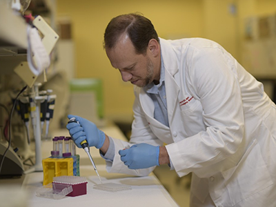
In a new observational study led by Andrea Hahn, M.D., infectious diseases specialist at Children’s National Hospital, researchers found that both insufficient beta-lactam pharmacokinetics (PK) and broad-spectrum antibiotics were associated with a greater decrease in species richness at the end of antibiotic therapy compared to pulmonary exacerbations onset.
In prior studies evaluating the association between beta-lactam PK, insufficient beta-lactam PK was associated with reduced short-term decreases in microbial diversity compared to sufficient beta-lactam dosing. In this study researchers found that insufficient beta-lactam PK was associated with a greater short-term decrease in microbial diversity.
Dr. Hahn’s team also found that an increased presence of beta-lactam antibiotic resistance genes was associated with lower microbial diversity and lower lung function.
These studies suggest that community-level antibiotic resistance, rather than the resistance patterns of the most prevalent bacteria identified in cultures, may serve as a useful predictor of lung function recovery in individuals with cystic fibrosis (CF). This finding may aid clinicians in selecting the most effective antibiotics to treat pulmonary exacerbations in CF patients, thus enhancing their clinical outcomes.
Read the full study in Nature’s Scientific Reports.
https://innovationdistrict.childrensnational.org/wp-content/uploads/2023/02/pills-in-hand-feature.png300400Innovation Districthttps://innovationdistrict.childrensnational.org/wp-content/uploads/2023/12/innovationdistrict_logo-1-1030x165.pngInnovation District2023-02-28 17:31:012023-07-03 10:55:48Therapeutic antibiotics associated with reductions in microbial diversity in CF
The study suggests that the use of antibiotics to treat PEx in children with CF may not be as harmful to the airway microbiome as previously believed.
Cystic fibrosis (CF) is a disease that affects many people, especially children. Pulmonary exacerbations (PEx) are common in people with CF and can cause a decline in lung function. These PEx are often treated with antibiotics, but little is known about how antibiotics affect the airway microbiome (the collection of microorganisms in the lungs) of people with CF over time.
Experts from Children’s National Hospital took part in a recent study which looked at how the airway microbiome and lung function of children with CF changed over the course of a year following an initial PEx. The study found that the diversity of the airway microbiome increased over the year despite a decrease in lung function associated with repeated PEx events requiring antibiotic therapy. This suggests that repeated treatment with antibiotics may not have a negative impact on the overall diversity of microorganisms in the lungs.
It is important for pediatricians to understand how antibiotics affect the airway microbiome in children with CF because it can help them make more informed decisions about treatment options. The findings of this study suggest that the use of antibiotics to treat PEx in children with CF may not be as detrimental to the airway microbiome as previously thought. This information can help pediatricians provide better care for children with CF and ultimately improve their overall health outcomes.
https://innovationdistrict.childrensnational.org/wp-content/uploads/2023/02/girl-looking-at-medicine-bottle-feature.png300400Innovation Districthttps://innovationdistrict.childrensnational.org/wp-content/uploads/2023/12/innovationdistrict_logo-1-1030x165.pngInnovation District2023-02-28 16:43:442023-07-03 10:56:36Effect of antibiotics on microorganisms and lung function in children with CF
“The Connor Family Professorship will allow my team to act rapidly upon potential transformative discoveries for children’s health” said Dr. Freishtat. “There is no greater honor than to carry the Connor family name as we follow in Dr. Edward Connor’s footsteps to drive breakthroughs that will benefit all children. I am eternally grateful for their support.”
Children’s National Hospital named Robert Freishtat, M.D., M.P.H., as the first Connor Family Professor in Research and Innovation at Children’s National Hospital.
Dr. Freishtat serves as Chief Biotechnology Officer and Senior Investigator, Center for Genetic Medicine Research in the Children’s National Research Institute. He is also a Professor with Tenure in Pediatrics, Emergency Medicine, Genomics and Precision Medicine at The George Washington University School of Medicine and Health Sciences.
About the award
Dr. Freishtat joins a distinguished group of 42 Children’s National physicians and scientists who hold an endowed chair. Professorships at Children’s National support groundbreaking work on behalf of children and their families and foster new discoveries and innovations in pediatric medicine. These appointments carry prestige and honor that reflect the recipient’s achievements and donor’s forethought to advance and sustain knowledge.
Dr. Freishtat is an internationally recognized translational researcher. He is the principal investigator for multiple international collaboratives studying intercellular communication in organ injury/repair. He has authored or co-authored more than 140 articles and book chapters in the fields of lung injury, asthma, obesity, exosomes and emergency medicine.
In 2020, Dr. Freishtat founded the Office of Biotechnology at Children’s National to fast-track novel ideas and forge industry partnerships so solutions can reach patients sooner.
“The Connor Family Professorship will allow my team to act rapidly upon potential transformative discoveries for children’s health” said Dr. Freishtat. “There is no greater honor than to carry the Connor family name as we follow in Dr. Edward Connor’s footsteps to drive breakthroughs that will benefit all children. I am eternally grateful for their support.”
The Connor family, through their vision and generosity, are ensuring that Dr. Freishtat and future holders of this professorship will launch bold, new initiatives to rapidly advance the field of pediatric research and innovation, elevate our leadership and improve the lifetimes of children.
About the donors
Dr. and Mrs. Connor are longtime donors and members of the Children’s National community. Dr. Connor previously served as Director of the Office of Innovation Development and a member of the executive team at the Clinical and Translational Science Institute. His institutional involvement continues through service, formerly as a board member for the Children’s National Research Institute and more recently as a member of the Research, Education, and Innovation Advisory Board. Mrs. Connor, a clinical microbiologist and educator, has worked throughout her career creating a legacy of young people in science.
“We strongly believe in the power of academic entrepreneurship to improve the health and wellbeing of children. This endowment is our way of supporting Children’s National’s work in research and innovation and recognizing Dr. Freishtat’s leadership as an outstanding physician-scientist and role model in clinical and translational pediatrics.”
https://innovationdistrict.childrensnational.org/wp-content/uploads/2022/07/Robert-Freishtat.png300400Innovation Districthttps://innovationdistrict.childrensnational.org/wp-content/uploads/2023/12/innovationdistrict_logo-1-1030x165.pngInnovation District2022-07-20 14:58:202023-07-03 10:59:53Robert Freishtat, M.D., M.P.H., named as Connor Family Professor in Research and Innovation
Researchers from Children’s National Hospital found that broad spectrum antianaerobic therapy had greater and longer lasting effects on the lung microbiome of persons with cystic fibrosis.
Cystic fibrosis (CF) is an autosomal recessive disease caused by mutations in the chloride ion channel encoding CF transmembrane conductance regulator gene, leading to multiple morbidities and early mortality. In a new clinical study, researchers from Children’s National Hospital found that broad spectrum antianaerobic therapy had greater and longer lasting effects on the lung microbiome of persons with CF.
They found this difference when comparing the microbiology and clinical outcomes in children with CF who were treated with “broad” or “narrow” antianaerobic antibiotics for exacerbations of their disease. While there are many factors that determine whether “narrow” or “broad” spectrum antibiotics are used, the data showed that the recovery of pulmonary function was similar between those groups.
“The findings prove that most providers are following best practices when treating patients with cystic fibrosis using the narrowest spectrum of antibiotics possible, and reserving broad spectrum agents for more advanced disease when culture data shows more resistant bacteria,” says Michael Bozzella, the study’s lead author.
The study, published in the Pediatric Infectious Disease Journal, analyzed how the spectrum of antibiotics prescribed to patients with cystic fibrosis impacts the population of bacteria in their lungs how it ties back to lung function.
“Research like this improves antibiotic and antimicrobial stewardship,” said Bozzella. “When speaking with families and patients with cystic fibrosis, providers can be more aware of the relationship between lung microbiome, disease state, and antibiotics and create more holistic treatment plans.”
The study, published in the Journal of Investigative Medicine, examined the hypotheses that beta-lactam antibiotic PK and PD is associated with changes in richness and alpha diversity following treatment of a pulmonary exacerbations and determined associations between antibiotic PK, PD, antibiotic resistance and lung function.
Cystic fibrosis (CF) is a chronic lung disease that affects more than 30,000 people in the United States and 70,000 people worldwide. While this chronic disease is characterized by acute pulmonary exacerbations that are frequently treated with antibiotics, the impact of antibiotics on airway microbial diversity remains a critical knowledge gap.
A new study led by researchers at Children’s National Hospital found that beta-lactam antibiotic pharmacokinetic (PK) and pharmacodynamic (PD) target attainment during treatment is associated with suppressed recovery of microbial diversity, following a pulmonary exacerbation in children and adolescents with CF.
“By laying the groundwork for understanding how antibiotic PK may influence microbial diversity following pulmonary exacerbation, we hope to identify improved ways to guide antibiotic therapy in persons with CF,” says Andrea Hahn, M.D., M.S., an infectious diseases specialist at Children’s National and lead author of the study.
The study, published in the Journal of Investigative Medicine, examined the hypotheses that beta-lactam antibiotic PK and PD is associated with changes in richness and alpha diversity following treatment of a pulmonary exacerbations and determined associations between antibiotic PK, PD, antibiotic resistance and lung function.
“Beta-lactam antibiotics are frequently used to treat pulmonary exacerbations in persons with CF, yet are not routinely optimized,” says Dr. Hahn. “This study demonstrates the importance of beta-lactam PK’s on changes within the airway microbiome and provides context for care providers regarding the potential long-term impacts of antibiotic use in persons with CF, to ensure that we are optimizing therapy with each pulmonary exacerbation.”
“I am honored to be recognized by Pediatric Research and the Society of Pediatric Research (SPR) at large,” said Dr. Hahn. “SPR is an amazing organization filled with excellent scientists, and to be highlighted by them for my work is truly affirming.”
For her work on the impact of bacterial functional and metabolic activity on acute episodes of cystic fibrosis, the journal Pediatric Research recognized Andrea Hahn, M.D., M.S., as Pediatric Research’s Early Career Investigator.
Cystic fibrosis is an autosomal recessive genetic disease, affecting more than 70,000 people worldwide. The condition’s morbidity and mortality are recurrent and result in a progressive decline of lung function.
“I am honored to be recognized by Pediatric Research and the Society of Pediatric Research (SPR) at large,” said Dr. Hahn. “SPR is an amazing organization filled with excellent scientists, and to be highlighted by them for my work is truly affirming.”
The exact mechanisms of the bacteria that chronically infect the airway triggering acute cystic fibrosis episodes, also known as pulmonary exacerbations, remain unclear. Dr. Hahn’s research is one of the few to explore this gap and found an association with long-chain fatty acid production in cystic fibrosis inflammation.
“As a physician-scientist, there are many competing priorities between developing and executing good science — including writing manuscripts and grants — and providing excellent patient care both directly and through hospital-wide quality improvement initiatives,” said Dr. Hahn. “It is often easier to have successes and feel both effective and appreciated on the clinical side. This recognition of my scientific contributions to the medical community is motivating me to continue pushing forward despite the setbacks that often come up on the research side.”
The exposure to many programs and institutions gave Dr. Hahn the foundation to create a research program at Children’s National that helps decipher the complexities of antibiotic treatment and how it changes the airway microbiome of people with cystic fibrosis. The program also explores the impacts of antibiotic resistance and beta-lactam pharmacokinetics/pharmacodynamics (PK/PD) — the oldest class of antibiotics used to treat infections.
Dr. Hahn believes that the people and environment at Children’s National Hospital allowed her to grow and thrive as a physician-scientist.
“I was initially funded through an internal K12 mechanism, which was followed up by Foundation support, which was only possible because of the strong mentorship teams I have been able to build here at Children’s National,” said Dr. Hahn. “My division chief has also been very supportive, providing me with both protected time as well as additional resources to build my research lab.”
She is particularly appreciative of Robert Freishtat, M.D., M.P.H, senior investigator at the Center for Genetic Medicine Research, and Mary Callaghan Rose (1943-2016).
“Robert Freishtat has been a great advocate for me, and I am indebted to him for my success thus far in my career,” said Dr. Hahn. “Likewise, I want to specifically recognize Mary Rose. She was a great scientist at Children’s National until her death in 2016. She gave me the initial opportunity and support to begin a career studying cystic fibrosis, and she is missed dearly.”
https://innovationdistrict.childrensnational.org/wp-content/uploads/2021/04/Andrea_Hahn.png300400Innovation Districthttps://innovationdistrict.childrensnational.org/wp-content/uploads/2023/12/innovationdistrict_logo-1-1030x165.pngInnovation District2021-04-19 10:49:282023-07-03 10:56:51Pediatric Research names Andrea Hahn, M.D., M.S., early career investigator
A recent study sheds light on the microbiologic triggers for lung inflammation and pulmonary exacerbations in cystic fibrosis.
Cystic fibrosis is an autosomal recessive disease that affects more than 70,000 people worldwide and results in a progressive decline of lung function. Patients with cystic fibrosis experience intermittent episodes of acute worsening of symptoms, commonly referred to as pulmonary exacerbations. While Staphylococcus aureus and Pseudomonas aeruginosa are thought to contribute to both lung inflammation and pulmonary exacerbations, the microbiologic trigger for these events remains unknown. Andrea Hahn, M.D., M.S., and her colleagues at Children’s National Hospital recently shed light on this matter by studying the changes in bacterial metabolic pathways associated with clinical status and intravenous (IV) antibiotic exposure in cystic fibrosis patients.
The researchers found increased levels of long-chain fatty acids (LCFAs) after IV antibiotic treatment in patients with cystic fibrosis. LCFAs have previously been associated with increased lung inflammation in asthma, but this is the first report of LCFAs in the airway of people with cystic fibrosis. This research indicates that bacterial production of LCFAs may be a contributor to inflammation in people with cystic fibrosis and suggests that future studies should evaluate LCFAs as predictors of pulmonary exacerbations.
https://innovationdistrict.childrensnational.org/wp-content/uploads/2021/03/girl-with-cystic-fibrosis-getting-breathing-treatment.jpg300400Innovation Districthttps://innovationdistrict.childrensnational.org/wp-content/uploads/2023/12/innovationdistrict_logo-1-1030x165.pngInnovation District2021-03-02 11:34:582023-07-03 10:56:59The role of long-chain fatty acids in cystic fibrosis inflammation
Drs. Dewesh Agrawal, Andrew Dauber, Robert Freishtat and Vittorio Gallo were named as 2021 American Pediatric Society members.
The American Pediatric Society (APS) has announced 55 new members, four of which are experts from Children’s National Hospital. Founded in 1888, the APS is the first and most prestigious academic pediatric organization in North America.
APS members are recognized child health leaders of extraordinary achievement who work together to shape the future of academic pediatrics. New members are nominated by current members through a process that recognizes individuals who have distinguished themselves as child health leaders, teachers, scholars, policymakers and/or clinicians.
“Our members represent the most distinguished and accomplished academic leaders in pediatrics whose outstanding work has advanced child health,” said APS President Steven Abman, M.D. “I am honored to welcome this exceptional group of individuals to the APS. The APS is especially looking forward to the active engagement of our membership with many exciting programs within the organization that are directed towards improvements in academic pediatric medicine, including more vigorous approaches to express our values of anti-racism, equity, diversity and inclusion.”
APS 2021 active new members from Children’s National are:
Dewesh Agrawal, M.D., vice-chair for Medical Education at Children’s National. Agrawal’s career has been marked by academic honors and teaching awards at every stage of his training and faculty employment. He has relentlessly devoted his energy to improving the educational experience for students, residents and fellows at Children’s National.
Andrew Dauber, M.D., M.M.Sc., chief of Endocrinology at Children’s National. Dr. Dauber’s leadership is reflected, nationally and internationally, in his ability to create research consortia, bringing together investigators to tackle complex questions. For example, he leads an NIH-funded consortium on the genetics of short statures, with multiple top children’s hospitals as partners. He also leads a large clinical trial testing a novel therapeutic agent for genetic short stature.
Robert Freishtat, M.D., M.P.H., senior investigator in the Center for Genetic Medicine of the Children’s National Research Institute (CNRI). Dr. Freishtat has authored or co-authored more than 100 articles and book chapters in the fields of pediatric lung injury, asthma, obesity, exosomes and emergency medicine. His research has been continuously funded by the NIH since 2003.
Vittorio Gallo, Ph.D., chief research officer at Children’s National and scientific director of CNRI. Dr. Gallo’s scientific success is attested to by over 130 peer-reviewed publications, many in very high-profile journals, as well as over 30 review articles and book chapters. He has received many national and international awards, including the NINDS Javits award in Neuroscience in 2018. Dr. Gallo has served on the editorial boards of many neuroscience journals, including Glia and the Annual Review in Neuroscience, and has been reviewing editor for the Journal of Neuroscience, all of which is a testament to the tremendous impact that his studies have had on the advancement of neurosciences.
“These new members represent multiple areas of Children’s National and have all leveraged the intersection of science, medicine and clinical education to make advances in their field of study,” said Stephen J. Teach, M.D., M.P.H., chair of the Department of Pediatrics at Children’s National. “Their work has, and will continue to, advance pediatric health care, and I congratulate them on their APS membership.”
https://innovationdistrict.childrensnational.org/wp-content/uploads/2020/12/APS-Members-2021.png300400Innovation Districthttps://innovationdistrict.childrensnational.org/wp-content/uploads/2023/12/innovationdistrict_logo-1-1030x165.pngInnovation District2020-12-01 15:02:332024-11-27 09:44:31Four Children’s National Hospital leaders named to APS
Despite having less overall microbial richness, children with Cystic Fibrosis displayed a greater presence of Staphylococcus species.
Cystic Fibrosis (CF) is a disease that mainly affects the lungs and arises from mutations in the cystic fibrosis transmembrane conductance regulator (CFTR) gene that encodes for the CFTR membrane protein located on certain secretory cells. CFTR dysfunction leads to complications such as the production of abnormally viscous mucus which causes chronic suppurative lung infections that require antibiotics to treat. New drugs called CFTR modulators can help improve CFTR protein function and some are even FDA-approved for use in children. In addition to CFTR protein function, the lung’s resident microbiota and its richness of diversity, plays an important role in both health and disease, including CF.
In a new study published in Heliyon, scientists from Children’s National Hospital examined the difference in the upper airway microbiome between children with CF and healthy controls. Age-related differences among children with CF and the impact of CFTR modulators on microbial diversity were also assessed. Seventy-five children between 0-6 years of age participated in the study, including 25 children with CF and 50 healthy controls. For CF participants, oropharyngeal swabs and clinical data were obtained from the biorepository, while data for controls were obtained during a single clinical visit.
Analysis revealed that CF patients had less microbial diversity and different composition of the upper airway microbiome compared to age similar controls, a finding that is consistent with research on the lower airways. Despite having less overall microbial richness, children with CF displayed a greater presence of Staphylococcus species, (a main driver of the pulmonary exacerbations characteristic of CF), three Rothia operational taxonomic units (OTUs) and two Streptococcus OTUs. CF patients received a significantly higher number of antibiotics courses within the previous year compared to healthy controls, and further investigation will be necessary to understand the impact of antibiotics on the upper airway microbiome of infants and children with CF.
Longitudinal comparisons to study effects of age and CFTR modulation on the microbiome of children with CF were also undertaken. Younger CF patients (those 0 to <3 years of age at study enrollment), were more likely to have culturally-normal respiratory flora and more stable microbial composition over time than older CF patients (those ≥ 3–6 years of age at study enrollment), with no significant differences in alpha or beta diversity. Older CF patients were significantly more likely to be receiving a CFTR modulator than younger patients. CF patients receiving CFTR modulators had higher microbial diversity measures than those not receiving CFTR modulators and were closer (but still significantly lower) in microbial richness to healthy controls. No significant differences in beta diversity were found between the three groups.
This study adds to the growing body of evidentiary support for the use of CFTR modulators in improving airway microbial diversity in CF patients. Future studies with a larger cohort and greater focus on the impact on early initiation of CFTR modulators on microbial diversity and clinical outcomes is necessary.
https://innovationdistrict.childrensnational.org/wp-content/uploads/2018/05/Staphylococcus.jpg300400Innovation Districthttps://innovationdistrict.childrensnational.org/wp-content/uploads/2023/12/innovationdistrict_logo-1-1030x165.pngInnovation District2020-06-18 16:44:072023-07-03 10:57:20Airway microbial diversity in children with Cystic Fibrosis
Physician-scientists from Children’s National Hospital are unlocking new insights into Cystic Fibrosis by studying the type and number of bacteria in the lungs.
Cystic Fibrosis (CF) is a genetic disorder that chiefly affects the lungs and results in the production of abnormally dehydrated, viscous mucus. The inability to adequately clear this mucus leads to bacterial retention and both intermittent and chronic lung infections which require antibiotic therapy to treat. Researchers have used 16S rDNA amplicon sequencing for years in the attempts to characterize the airway microbiomes of CF patients, and more recently have used shotgun whole genome sequencing (WGS) techniques to obtain further details regarding bacterial species and strains. Previous studies on the airway microbiomes of CF patients have revealed that inter-person variability is high and can sometimes exceed intra-person variability. This can preclude generalizations regarding the CF population as a whole, which includes more than 30,000 Americans.
A recently published case study examined a young child with advanced and severely aggressive CF over a 12-month period, during which five pulmonary exacerbations occurred. A total of 14 sputum samples were collected across three clinical periods- baseline, exacerbation, and treatment. Samples were subsequently genetically sequenced (via 16s rDNA sequencing and, in three instances, WGS) and volatile metabolites were analyzed. The researchers hypothesized that if signature microbiome and metabolome characteristics correlated with one other and could be identified for each disease state, this data could serve as conglomerate biomarkers for the continuum of CF clinical states within an individual. In turn, this could inform future study design in a larger cohort.
Across all sputum samples, 109 individual operational taxonomic units (OTUs) and 466 distinct volatile metabolites were identified. 16s rDNA sequencing and WGS revealed that Escherichia coli and Staphylococcus aureus were the predominant bacteria during most baseline and exacerbation samples, despite some significant fluctuations in relative abundances. After the patient’s fifth antibacterial course, however, Achromobacter xylosoxidans became the new dominant bacterium.
Analysis revealed that the phylum Bacteroidetes and the genus Stenotrophomonas were significantly more abundant in treatment periods compared to baseline and exacerbation periods. WGS revealed the presence of bacteriophages as well as antibiotic resistance genes (mostly due to multi-drug resistance mechanisms), which can have important clinical ramifications and adds some dimensionality to the genetic analysis.
Volatile metabolite analysis found that observable fluctuations in metabolome composition coincided with fluctuations in the sputum microbiome. In this case, the microbiome and volatile metabolites produced by these bacteria provided an accurate assessment of the child’s clinical state. More specifically, the authors saw a distinct shift in both the microbiome and volatile metabolites with antibiotic treatment across the five independent pulmonary exacerbations. These additional assessments of the bacteria within the CF airway could provide an additional technique beyond standard bacterial cultures to better understand how the patient is responding to antibiotic treatment. Future studies in a larger group of children with CF may provide further insights into bacteria and volatile metabolite combinations that predict pulmonary exacerbation.
Children’s National Hospital researchers for the first time have isolated bacterial extracellular vesicles from the blood of healthy donors. The team theorizes that the solar eclipse lookalikes contain important signaling proteins and chromatin, DNA from the human host.
Children’s National Hospital researchers for the first time have isolated bacterial extracellular vesicles from the blood of healthy donors, a critical step to better understanding the way gut bacteria communicate with the rest of the body via the bloodstream.
For decades, researchers considered circulating bacterial extracellular vesicles as bothersome flotsam to be jettisoned as they sought to tease out how bacteria that reside in the gut whisper messages to the brain.
There is a growing appreciation that extracellular vesicles – particles that cells naturally release – actually facilitate intracellular communication.
“In the past, we thought they were garbage or noise,” says Robert J. Freishtat, M.D., MPH, associate director, Center for Genetic Medicine Research at Children’s National Research Institute. “It turns out what we throw away is not trash.”
Kylie Krohmaly, a graduate student in Dr. Freishtat’s laboratory, has isolated from blood, extracellular vesicles from Escherichia coli and Haemophilus influenzae, common bacteria that colonize the gut, and validated the results via electron microscopy.
“The images are interesting because they look like they have a bit of a halo around them or penumbra,” Krohmaly says.
The team theorizes that the solar eclipse lookalikes contain important signaling proteins and chromatin, DNA from the human host.
“It’s the first time anyone has pulled them out of blood. Detecting them is one thing. Pulling them out is a critical step to understanding the language the microbiome uses as it speaks with its human host,” Dr. Freishtat adds.
Krohmaly’s technique is so promising that the Children’s National team filed a provisional patent.
The Children’s research team has devised a way to gum up the cellular works so that bacteria no longer become antibiotic resistant. Targeted bacteria retain the ability to make antibiotic-resistance RNA, but like a relay runner dropping rather than passing a baton, the bacteria are thwarted from advancing beyond that step. And, because that gene is turned off, the bacteria are newly sensitive to antibiotics – instead of resistant bacteria multiplying like clockwork these bacteria get killed.
“Our plan is to hijack this process in order to turn off antibiotic-resistance genes in bacteria,” Dr. Freishtat says. “Ultimately, if a child who has an ear infection can no longer take amoxicillin, the antibiotic would be given in tandem with the bacteria-derived booster to turn off bacteria’s ability to become antibiotic resistant. This one-two punch could become a novel way of addressing the antibiotic resistance process.”
ISEV2020 Annual Meeting presentation
(Timing may be subject to change due to COVID-19 safety precautions)
Oral with poster session 3: Neurological & ID
Saturday May 23, 2020, 5 p.m. to 5:05 p.m. (ET)
“Detection of bacterial extracellular vesicles in blood from healthy volunteers”
Kylie Krohmaly, lead author; Claire Hoptay, co-author; Andrea Hahn, M.D., MS, infectious disease specialist and co-author; Robert J. Freishtat, M.D., MPH, associate director, Center for Genetic Medicine Research at Children’s National Research Institute and senior author.
AlgometRx, which joins JPOD @ Philadelphia, was founded by Julia Finkel, M.D., pediatric anesthesiologist and director of Pain Medicine and Research at Children’s Sheikh Zayed Institute.
AlgometRx and Adipomics, two companies that spun out of innovations discovered at Children’s National Health System, have been selected by Johnson & Johnson Innovation – JLABS to join JPOD @ Philadelphia and JPOD @ Boston, respectively.
JLABS is a global network of no-strings-attached incubators for innovative companies from across the pharmaceutical, medical device, consumer and health technology sectors. Start-up companies are free to pursue their own research priorities independently, with access to state-of-the-art facilities to develop new drugs, medical devices, precision diagnostics and health technologies for people around the world.
AlgometRx, which joins JPOD @ Philadelphia, was founded by Julia Finkel, M.D., pediatric anesthesiologist and director of Pain Medicine and Research at Children’s Sheikh Zayed Institute. The AlgometRx device is a first-of-its-kind platform technology that aims to objectively measure pain intensity, type and drug effects in real time by capturing a digital image of a patient’s pupillary light response and applying a series of proprietary algorithms to various characteristics.
AlgometRx is designed to provide an objective pain measurement that aims to help physicians select the correct analgesic class of drug and dosage. By optimizing pain assessment, drug selection and drug management, AlgometRx aims to impact the opioid epidemic and the monitoring and management of Opioid Use Disorder.
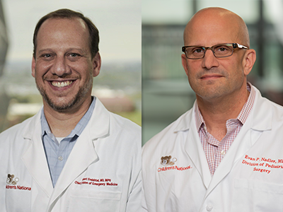
Adipomics, which joins JPOD @ Boston, was co-founded by Robert Freishtat, M.D., M.P.H., senior investigator in the Center for Genetic Medicine of the Children’s Research Institute and chief of the Division of Emergency Medicine at Children’s National, and pediatric surgeon Evan P. Nadler, M.D., co-director of the Obesity Program and director of the Bariatric Surgery Program at Children’s National.
Adipomics, which joins JPOD @ Boston, was co-founded by pediatric surgeon Evan P. Nadler, M.D., co-director of the Obesity Program and director of the Bariatric Surgery Program at Children’s National, and Robert Freishtat, M.D., M.P.H., senior investigator in the Center for Genetic Medicine of the Children’s Research Institute and chief of the Division of Emergency Medicine at Children’s National. Adipomics was founded with the aim to address the global epidemic of obesity-related diseases including Type 2 diabetes and cardiovascular diseases. World health experts predict that one billion people worldwide will be obese by 2030.
Drs. Nadler and Freishtat discovered that exosomes released from fat cells (adipocytes) carry genetic material that can mediate various diseases related to obesity. Through their research, they developed a proprietary method that aims to detect how obesity affects an individual patient’s metabolism before the onset of overt disease. Adipomics aims to create the first non-invasive, “anticipatory medicine” diagnostic that detects risk for obesity-related diseases prior to the onset of clinical signs or even biochemical abnormalities. If successful, this predictive methodology would enable treatment much earlier in the disease process, which is likely to improve effectiveness.
A recent news release from Children’s National provides more details on these innovations.
As organizations that share a commitment to improving the pace of healthcare innovation, Children’s National and Johnson & Johnson Innovation – JLABS also recently announced their collaboration to launch JLABS @ Washington, DC, a 32,000-square foot facility to be located at the new Children’s National Research & Innovation Campus in Washington, D.C. The JLABS @ Washington, DC will have the capacity to house up to 50 pharmaceutical, medical device, consumer and health technology companies that are aiming to advance the development of new drugs, medical devices, precision diagnostics and health technologies, including applications in pediatrics. The campus is located on a 12-acre portion of the former Walter Reed Army Medical Center campus in the nation’s capital and is slated to open in 2020, coinciding with the 150th Anniversary of Children’s National Health System.
https://innovationdistrict.childrensnational.org/wp-content/uploads/2019/09/Julia-Finkel.png300400Innovation Districthttps://innovationdistrict.childrensnational.org/wp-content/uploads/2023/12/innovationdistrict_logo-1-1030x165.pngInnovation District2019-09-11 10:51:282024-05-16 14:45:56Two Children’s National spin-outs join Johnson & Johnson–JLABS
Obesity is a major risk factor for insulin resistance and type 2 diabetes. Now researchers understand the pathogenesis better among teens with mid-level obesity, thanks to clues released from circulating adipocyte-derived exosomes.
Researchers know that exosomes, tiny nanoparticles released from fat cells, travel through the bloodstream and body, regulating a variety of processes, from growth and development to metabolism. The exosomes are important in lean, healthy individuals in maintaining homeostasis, but when fat gets ‘sick’ – the most common reason for this is too much weight gain – it can change its phenotype, becoming inflammatory, and disrupts how our organs function, from how our skeletal muscle and liver metabolize sugar to how our blood vessels process cholesterol.
Robert J. Freishtat, M.D., M.P.H., the chief of emergency medicine at Children’s National Health System and a professor of precision medicine and genomics at the George Washington University School of Medicine and Health Sciences, and Sheela N. Magge M.D., M.S.C.E., who is now the director of pediatric endocrinology and an associate professor of medicine at the Johns Hopkins School of Medicine, were curious about what this process looked like in teens who fell in the mid-range of obesity.
Obesity is a major risk factor for insulin resistance and type 2 diabetes, but Dr. Freishtat and Dr. Magge wanted to know: Why do some teens with obesity develop type 2 diabetes over others? Why are some teens in this mid-range of obesity metabolically healthy while others have metabolic syndrome? Can fat in obese people become sick and drive disease?
To test this, Dr. Freishtat and Dr. Magge worked with 55 obese adolescents, ages 12 to 17, as part of a study at Children’s National. The participants – 32 obese normoglycemic youth and 23 obese hyperglycemic youth – were similar in age, sex, race, pubertal stage, body mass index and overall fat mass. The distinguishing factor: The hyperglycemic study participants, the teens with elevated blood sugar, differed in where they stored fat. They had extra visceral fat (or adipose tissue) storage, the type of fat that surrounds the liver, pancreas and intestines, a known risk factor for type 2 diabetes.
Dr. Magge and Dr. Freishtat predicted that circulating exosomes from the teens with elevated blood sugar are enriched for microRNAs targeting carbohydrate metabolism.
They used three tests to examine study participants’ metabolism, body composition and circulating exosomes. The first test, an oral glucose tolerance test, measures how efficiently the body metabolizes sugar; the second test is the whole body DXA, or dual-energy x-ray absorptiometry, which analyzes body composition, including lean tissue, fat mass and bone mineral density; and the third test, the serum adipocyte-derived exosomal microRNA assays, is an analysis of circulating fat signals in the bloodstream.
They found that teens with elevated blood sugar and increased visceral fat had different circulating adipocyte-derived exosomes. These study participants’ exosomes were enriched for 14 microRNAs, targeting 1,304 mRNAs and corresponding to 179 canonical pathways – many of which are directly associated with carbohydrate metabolism and visceral fat.
Dr. Magge will present this research, entitled “Changes in Adipocyte-Derived Exosomal MicroRNAs May Play a Role in the Progression from Obese Normoglycemia to Hyperglycemia/Diabetes,” as an oral abstract at the American Diabetes Association’s 79th Scientific Sessions on Saturday, June 8.
Dr. Freishtat envisions having this information will be especially helpful for a patient in a mid-range of obesity. Exosomes primarily consist of small non-coding RNAs. In the current study, the altered RNAs affect P13K/AKT and STAT3 signaling, vital pathways for metabolic and immune function.
“Instead of waiting until someone has the biochemical changes associated with type 2 diabetes, such as hyperglycemia, hyperlipidemia and insulin resistance, we’re hoping physicians will use this information to work with patients earlier,” says Dr. Freishtat. “Through earlier detection, clinicians can intervene when fat shows sign of illness, as opposed to when the overt disease has occurred. This could be intervening with diet and lifestyle for an obese individual or intervening with medication earlier. The goal is to work with children and teens when their system is more plastic and responds better to intervention.”
As this research evolves, Dr. Freishtat continues to look at the intergenerational effects of circulating adipocyte-derived exosomes. Through ongoing NIH-funded research in India, he finds these exosomes, similar in size to lipoproteins, can travel across the placenta, affecting development of the fetus in utero.
“What we’re finding in our initial work is that these exosomes, or ‘sick’ fat, cross the placenta and affect fetal development,” Dr. Freishtat says. “Some of the things that we’re seeing are a change in body composition of the fetus to a more adipose phenotype. Some of our work in cell cultures shows changes in stem cell function and differentiation, but what’s even more interesting to us is that if the fetus is a female sex that means her ovaries are developing while she’s in utero, which means a mother’s adipocyte-derived exosomes could theoretically be affecting her grandchild’s phenotype – influencing the health of three generations.”
While this research is underway, Dr. Freishtat is working with JPOD @ Boston, co-located with the Cambridge Innovation Center in Cambridge, Massachusetts, to develop a test to provide analyses of adipocyte-derived exosomal microRNAs.
“It’s important for families to know that these studies are designed to help researchers and doctors better understand the development of disease in its earliest stages, but there’s no need for patients to wait for the completion of our studies,” says Dr. Freishtat. “Reaching and maintaining a healthy body weight and exercising are important things teens and families can do today to reduce their risk for obesity and diabetes.”
https://innovationdistrict.childrensnational.org/wp-content/uploads/2019/05/Robert-J.-Freishtat-working-in-the-lab.png300400Innovation Districthttps://innovationdistrict.childrensnational.org/wp-content/uploads/2023/12/innovationdistrict_logo-1-1030x165.pngInnovation District2019-06-09 07:00:522023-07-03 10:34:06Detecting early signs of type 2 diabetes through microRNA
A new study shows that steroids might work for rhinovirus but not for respiratory syncytial virus.
Every winter, doctors’ offices and hospital emergency rooms fill with children who have bronchiolitis, an inflammation of the small airways in the lung. It’s responsible for about 130,000 admissions each year. Sometimes these young patients have symptoms reminiscent of a bad cold with a fever, cough and runny nose. Other times, bronchiolitis causes breathing troubles so severe that these children end up in the intensive care unit.
“The reality is that we don’t have anything to treat these patients aside from supportive care, such as intravenous fluids or respiratory support,” says Robert J. Freishtat, M.D., M.P.H., chief of emergency medicine at Children’s National Health System. “That’s really unacceptable because some kids get very, very sick.”
Several years ago, Dr. Freishtat says a clinical trial tested using steroids as a potential treatment for bronchiolitis. The thinking was that these drugs might reduce the inflammation that’s a hallmark of this condition. However, he says, the results weren’t a slam-dunk for steroids: The drugs didn’t seem to improve outcomes any better than a placebo.
But the trial had a critical flaw, he explains. Rather than having one homogenous cause, bronchiolitis is an umbrella term for a set of symptoms that can be caused by a number of different viruses. The most common ones are respiratory syncytial virus (RSV) and rhinovirus, the latter itself being an assortment of more than 100 different but related viruses. By treating bronchiolitis as a single disease, Dr. Freishtat says researchers might be ignoring the subtleties of each virus that influence whether a particular medication is useful.
“By treating all bronchiolitis patients with a single agent, we could be comparing apples with oranges,” he says. “The treatment may be completely different depending on the underlying cause.”
To test this idea, Dr. Freishtat and colleagues examined nasal secretions from 32 infants who had been hospitalized with bronchiolitis from 2011 to 2014 at 17 medical centers across the country that participate in a consortium called the 35th Multicenter Airway Research Collaboration. In half of these patients, lab tests confirmed that their bronchiolitis was caused by RSV; in the other half, the cause was rhinovirus.
From these nasal secretions, the researchers extracted nucleic acids called microRNAs. These molecules regulate the effects of different genes through a variety of different mechanisms, usually resulting in the effects of target genes being silenced. A single microRNA typically targets multiple genes by affecting messenger RNA, a molecule that’s key for producing proteins.
Comparing results between patients with RSV or rhinovirus, the researchers found 386 microRNAs that differed in concentration. Using bioinformatic software, they traced these microRNAs to thousands of messenger RNAs, looking for any interesting clues to important mechanisms of illness that might vary between the two viruses.
Their findings eventually turned up important differences between the two viruses in the NF-kB (nuclear factor kappa-light-chain-enhancer of activated B cells) pathway, a protein cascade that’s intimately involved in the inflammatory response and is a target for many types of steroids. Rhinovirus appears to upregulate the expression of many members of this protein family, driving cells to make more of them, and downregulate inhibitors of this cascade. On the other hand, RSV didn’t seem to have much of an effect on this critical pathway.
To see if these effects translated into cells making more inflammatory molecules in this pathway, the researchers searched for various members of this protein cascade in the nasal secretions. They found an increase in two, known as RelA and NFkB2.
Based on these findings, published online Jan. 17, 2018, in Pediatric Research, steroids might work for rhinovirus but not for RSV, notes Dr. Freishtat the study’s senior author.
“We’re pretty close to saying that you’d need to conduct a clinical trial with respect to the virus, rather than the symptoms, to measure any effect from a given drug,” he says.
Future clinical trials might test the arsenal of currently available medicines to see if any has an effect on bronchiolitis caused by either of these two viruses. Further research into the mechanisms of each type of illness also might turn up new targets that researchers could develop new medicines to hit.
“Instead of determining the disease based on symptoms,” he says, “we can eventually treat the root cause.”
Study co-authors include Kohei Hasegawa, study lead author, and Carlos A. Camargo Jr., Massachusetts General Hospital; Marcos Pérez-Losada, The George Washington University School of Medicine and Health Sciences; Claire E. Hoptay, Samuel Epstein and Stephen J. Teach, M.D., M.P.H., Children’s National; Jonathan M. Mansbach, Boston Children’s Hospital; and Pedro A. Piedra, Baylor College of Medicine.
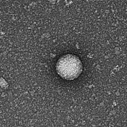
https://innovationdistrict.childrensnational.org/wp-content/uploads/2017/12/Human-Rhinovirus.jpg300400Innovation Districthttps://innovationdistrict.childrensnational.org/wp-content/uploads/2023/12/innovationdistrict_logo-1-1030x165.pngInnovation District2018-01-24 14:31:232024-10-29 15:25:38Finding the root cause of bronchiolitis symptoms
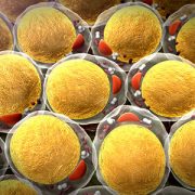
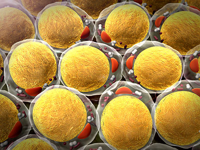
Fat cells from obese patients have the ability to send signals that can accelerate biological processes leading to atherosclerosis.
Obesity has been linked to a variety of adverse health conditions, including Type 2 diabetes, cancer, heart attack and stroke – conditions that may begin as early as childhood in patients whose obesity also begins early. While this much is known, it has been unclear how extra fat mass might lead to these chronic health conditions.
New research from Children’s National Health System scientists might help answer this question. In findings presented at the 2017 annual meeting of the Pediatric Academic Societies, the research team shows that exosomes – nanosized chemical messages that cells send to each other to regulate protein production – isolated from very obese teenage patients behave very differently from those derived from lean patients and could be key players in heightening the risk of developing atherosclerosis. This hardening of the arteries can, in turn, increase the risk of heart disease and stroke in adulthood.
A research team led by Robert J. Freishtat, M.D., M.P.H., chief of emergency medicine at Children’s National, is exploring possible links between extra belly fat and obesity-related diseases, such as atherosclerosis, a buildup of plaque in arteries that can harden and restrict blood flow. More precise knowledge of the mechanisms by which obesity ratchets up heart risks holds the promise of helping the next generation of kids avoid experiencing chronic disease.
The working theory is that exosomes derived from belly fat from obese patients have the distinct ability to accelerate biological processes leading to atherosclerosis.
The research team isolated exosomes from five obese teenagers and compared them to five sex-matched lean adolescents. It turns out that exosomes derived from fat pick up their marching orders from microRNA content likely to target cholesterol efflux genes, which help reduce cholesterol buildup in cells.
The research team looked at differences in cholesterol efflux gene expression in THP-1 macrophages. Uptake of low-density lipoprotein cholesterol, “bad” cholesterol, was 92 percent higher than in those exposed to exosomes from obese patients compared with their lean counterparts. Exposure to obese exosomes also reduced cholesterol efflux.
“Atherogenic properties of fat-cell derived exosomes from obese patients differ markedly from the non-atherogenic profile of exosomes from lean patients. It is especially concerning that we see biological clues of heightened risk in teenagers, and the finding underscores how the seeds for atherosclerosis can be planted very early in life,” Dr. Freishtat says.
The presentation is the latest finding from a research team that, over years of work, is unraveling the mechanisms of cellular signaling by fat cells. By closely examining very obese children – who have the most severe cardiometabolic disease – the team identified strong molecular signals of disease risk that they can search for in leaner patients who may be at risk for disease years from now.
“We know that morbidly obese patients have cardiovascular issues,” explains Dr. Freishtat. “An unanswered question is for patients with no clinical symptoms who are a little overweight. Can we look at them and say whether they are at risk for developing atherosclerosis, insulin resistance or Type 2 diabetes five or 10 years down the line? That’s the whole rationale for doing this work.”
The critical issue is what exosomes are up to. Dr. Freishtat says in lean people, they’re active and are very important in maintaining stable metabolism and homeostatic processes.
“When a person becomes obese, however, exosomes evolve,” he says. “They no longer support insulin signaling, which is helpful, and drive processes in the reverse direction, repressing insulin signaling – which can be harmful,” he adds.
Ultimately, the research team aims to revolutionize how chronic diseases like Type 2 diabetes are diagnosed. For far too long, clinicians have relied on symptoms like high glucose levels and excess urination to diagnose diabetes.
“By the time you have symptoms, it’s too late,” says Dr. Freishtat. “In many cases, damage has been done by relentless exposure to high sugar levels. The biological processes that underlie the Type 2 diabetes process began five, 10, 15 years earlier. If we can detect it earlier, before symptoms arise, intervention is going to have a more significant impact on improving and extending patients’ lives.”
https://innovationdistrict.childrensnational.org/wp-content/uploads/2017/05/Fat-Cells.jpg300400Innovation Districthttps://innovationdistrict.childrensnational.org/wp-content/uploads/2023/12/innovationdistrict_logo-1-1030x165.pngInnovation District2017-05-22 10:02:172023-07-03 10:34:13Cellular signals may increase atherosclerosis risk
Study findings offer hope to the nearly 2 billion adults who are overweight or obese worldwide that detrimental effects of carrying too much weight can recede. (Image source: Centers for Disease Control and Prevention)
Losing weight appears to reset the chemical messages that fat cells send to other parts of the body that otherwise would encourage the development of Type 2 diabetes, substantially reducing the risk of that disease, a team led by Children’s National Health System researchers report in a new study. The findings offer hope to the nearly 2 billion adults who are overweight or obese worldwide that many of the detrimental effects of carrying too much weight can recede, even on the molecular level, once they lose weight.
In 2015, Robert J. Freishtat, M.D., M.P.H., Chief of Emergency Medicine at Children’s National and Associate Professor of Pediatrics, Emergency Medicine and Integrative Systems Biology at The George Washington University School of Medicine & Health Sciences, and colleagues showed that fat cells (also known as adipocytes) from people who are obese send messages to other cells that worsen metabolic function. These messages are in the form of exosomes, nanosized blobs whose contents regulate which proteins are produced by genes. Exosomes are like “biological tweets,” Dr. Freishtat explains — short signals designed to travel long distances throughout the body.
Dr. Freishtat’s earlier research showed that the messages contained in exosomes from patients who are obese alter how the body processes insulin, setting the stage for Type 2 diabetes. However, says Dr. Freishtat, it has remained unclear since that publication whether these aberrant messages from adipocytes improve after weight loss.
“We’ve known for a long time that too much adipose tissue is bad for you, but it’s all moot if you lose the weight and it’s still bad for you,” he explains. “We wanted to know whether these negative changes are reversible. If you reduce fat, does the disease risk that goes along with excess fat also go away?”
Details of the study
To investigate this question, Dr. Freishtat and colleagues worked with six African American adults scheduled to receive gastric bypass surgery — a nearly surefire way to quickly lose a large amount of weight. The volunteers, whose average age was 38 years, started out with an average body mass index (BMI) of 51.2 kg/m2. (The Centers for Disease Control and Prevention considers a healthy BMI to range between 18.5 to 24.9.)
Two weeks before these volunteers underwent surgery, researchers collected blood samples and took a variety of measurements. The researchers then performed a repeat blood draw and measurements one year after the surgery took place, when the volunteers’ average BMI had dropped to 32.6.
Dr. Freishtat and colleagues drew out the adipocyte-derived exosomes from both sets of blood samples and analyzed their contents. The team reports in the January 2017 issue of Obesity that at least 168 microRNAs — the molecules responsible for sending specific messages — had changed before and after surgery. Further analyses showed that many of these microRNAs were involved in insulin signaling, the pathways that the body uses to regulate blood sugar. By changing these outgoing microRNAs for the better, Dr. Freishtat says, adipocytes actively were encouraging higher insulin sensitivity in other cells, warding off Type 2 diabetes.
Sure enough, each volunteer had better insulin sensitivity and other improved markers of metabolic health post-surgery, including lower branched chain amino acids and a two-fold reduction in their glutamate to glutamine ratio.
“These volunteers were essentially cured of their diabetes after surgery. The changes we saw in their surgery-responsive microRNAS correlated with the changes we saw in their metabolic health,” Dr. Freishtat says.
A glimpse into the future
Dr. Freishtat and colleagues plan to study this phenomenon in other types of weight loss, including the slower and steadier paths that most individuals take, such as improving diet and doing more exercise. The team expects to see similar changes in exosomes of patients who lose weight in non-surgical ways.
By further examining the aberrant messages in microRNAs being sent out from adipocytes, he says, researchers eventually might be able to develop treatments to reverse metabolic problems in overweight and obese patients before they lose the weight, improving their health even before the often challenging process of weight loss begins.
“Then, if you can disrupt this harmful signaling in combination with weight-loss strategies,” Dr. Freishtat says, “you’re really getting the best of both worlds.”
Eventually, he adds, tests might be available so that doctors can warn patients that their fat cells are sending out harmful messages before disease symptoms start. By giving patients an early heads up, Dr. Freishtat says, patients might be more likely to heed advice from physicians and make changes before it’s too late.
“If doctors could warn patients that their fat is telling their blood vessels to fill up with plaque and trigger a heart attack in 10 to 20 years,” he says, “patients might be more compliant with treatment regimens.”
The work that Children’s National Health System physician-scientist Robert J. Freishtat, M.D., M.P.H., and colleagues are doing could soon be a game changer when it comes to early intervention and prevention of obesity-related illnesses.
They already knew there’s a direct relationship between the amount of visceral adipose, or belly fat, a person has and development of some of the most common and life-threatening complications of obesity, including cardiovascular disease and the insulin resistance that leads to diabetes. What remained unclear, until recently, were the precise mechanisms for how the increase in belly fat triggers the onset of additional disease.
Dr. Freishtat, senior author of “Adipocyte-Derived Exosomal miRNAs: A Novel Mechanism for Obesity-Related Disease,” published by Pediatric Research, studies the adipocytes, or fat cells, of visceral adipose in both lean and obese patients to understand exactly how these fat cells can and do wreak havoc — not just locally but throughout the body. Cells leverage exosomes to communicate among themselves, but in overweight patients those cellular communications can go awry.
“As the body’s visceral fat grows, somewhere on the path to obesity the fat cells change and begin to release different exosomes than lean adipose cells do. These new messages disrupt some important processes that eventually prevent the body from effectively dealing with sugar and cholesterol,” says Dr. Freishtat, chief of Emergency Medicine at Children’s National, and associate professor of Pediatrics, Emergency Medicine, and Integrative Systems Biology at the George Washington University.
Dr. Freishtat describes exosomes as “biological tweets”— short messages shed by all cells that allow for intercellular communication and alter gene expression. In the case of the adipocytes that exist in large quantities of visceral fat, these “tweets” actually cause the downregulation of proteins impacting two key signaling pathways — TGF-β and Wnt/β-catenin — associated with controlling chronic inflammation and fibrotic disease throughout the body. These signaling changes make morbidly obese patients more vulnerable to systemwide issues, such as cholesterol accumulation and changes to how the liver processes fat.
Details of the study
The study authors surgically collected fat tissue from lean and obese female patients aged 11 to 19 and used modified bead-based flow cytometry to separate, identify, and compare the exosomal RNA shed by the fat cells in both lean and obese samples. To confirm the unique impact of the obese adipose exosomes on gene expression, the research team then exposed lung cells in vivo to the exosomes shed by both lean and obese adipose. They measured the impact of exposure and uptake on a single receptor type — activin receptor type-2B — known to have a major influence on the TGF-β pathway. The exosomes from obese adipose caused the receptor to slow down, leading to significant changes in the function of the TGF-β pathway.
The team continues to explore how the exosomes shed from excess amounts of visceral adipose spread throughout the body and how the function of organs such as the liver, the heart, and the brain are impacted by the migrating fat cells.
A Look into the future
Successfully identifying and isolating these exosomes also has opened the door to developing a test to detect them, an idea that may permit even earlier intervention to delay or prevent the onset of obesity-related illnesses.
“It is entirely plausible, and is on its way to happening very soon, that someone could walk into their physician’s office for a routine physical and, via a urine test, find out that they are on the road to some dangerous additional side effects of significant weight gain,” says Dr. Freishtat. “That type of early detection could really be a game changer for the millions of Americans who are on track to developing heart, liver, and other diseases resulting from morbid obesity.”
https://innovationdistrict.childrensnational.org/wp-content/uploads/2016/10/xxOct16_GameChangerR@GImage.jpg1195797Innovation Districthttps://innovationdistrict.childrensnational.org/wp-content/uploads/2023/12/innovationdistrict_logo-1-1030x165.pngInnovation District2016-10-13 11:53:072023-07-03 11:00:18A game changer for detecting complications from obesity