Tag Archive for: Featured

Rare Diseases Institute sign

Children’s National Rare Disease Institute named a Center of Excellence

Rare Diseases Institute sign

RDI, which includes the largest clinical group of pediatric geneticists in the nation, focuses on developing the clinical care field of more than 8,000 rare diseases currently recognized and advancing the best possible treatments for children with these diseases.

The Rare Disease Institute (RDI) at Children’s National Hospital announced its designation as a NORD Rare Disease Center of Excellence, joining a highly select group of 31 medical centers nationwide. This new, innovative network seeks to expand access and advance care and research for rare disease patients in the United States. The program is being led by the National Organization for Rare Disorders (NORD), with a goal to foster knowledge sharing between experts across the country, connect patients to appropriate specialists regardless of disease or geography, and to improve the pace of progress in rare disease diagnosis, treatment and research.

“Children’s National has worked closely with NORD to move this program forward and is very proud to be amongst the first group of recognized centers,” said Marshall Summar, M.D., chief of the Division of Genetics and Metabolism and the director of RDI at Children’s National. “This is a recognition of the institutional efforts, as we take care of patients with the rare disease and help set the standard for the field.”

RDI, which includes the largest clinical group of pediatric geneticists in the nation, focuses on developing the clinical care field of more than 8,000 rare diseases currently recognized and advancing the best possible treatments for children with these diseases.

In February 2021, RDI became the first occupant of the new Children’s National Research & Innovation Campus, a first-of-its-kind pediatric research and innovation hub. The campus now also houses the Center for Genetic Medicine Research, and together researchers are constantly pursuing high-impact opportunities in pediatric genomic and precision medicine. Both centers combine its strengths with public and private partners, including industry, universities, federal agencies, start-up companies and academic medical centers. They also serve as an international referral site for rare disorders.

People living with rare diseases frequently face many challenges in finding a diagnosis and quality clinical care. In establishing the Centers of Excellence program, NORD has designated clinical centers across the U.S. that provide exceptional rare disease care and have demonstrated a deep commitment to serving rare disease patients and their families using a holistic, state of the art approach.

“Right now, far too many rare diseases are without an established standard of care. The Centers of Excellence program will help set that standard – for patients, clinicians, and medical centers alike,” said Ed Neilan, chief scientific and medical officer of NORD. “We are proud to announce Children’s National as a NORD Rare Disease Center of Excellence and look forward to their many further contributions as we collectively seek to improve health equity, care and research to support all individuals with rare diseases.”

Each center was selected by NORD in a competitive application process requiring evidence of staffing with experts across multiple specialties to meet the needs of rare disease patients and significant contributions to rare disease patient education, physician training and research.

Dr. Javad Nazarian

Q&A with Dr. Javad Nazarian on his upcoming work on low-grade gliomas

Dr. Javad Nazarian

Supported by the Gilbert Family Foundation, Dr. Nazarian’s return is part of a special research program within the Gilbert Family Neurofibromatosis Institute that focuses on NF1 research.

Javad Nazarian, Ph.D., M.Sc., associate professor of Pediatrics at George Washington University and professor at the University of Zurich, has expanded his research group at Children’s National to focus on Neurofibromatosis type 1 (NF1) transformed low-grade gliomas (LGGs). Dr. Nazarian will apply his expertise from establishing a successful DIPG (diffuse intrinsic pontine glioma) and DMG (diffuse midline glioma) program in Zurich Switzerland and previously at Children’s National.

In addition to his continued research in Zurich, as a principal investigator at the Department of Genomics and Precision Medicine at Children’s National Dr. Nazarian plans on aggregating his knowledge to the new research and work spearheaded at Children’s National. As one of the first research teams to move to the Children’s National Research & Innovation Campus, Dr. Nazarian’s group is excited to use the opportunity to establish cutting-edge and clinically translational platforms.

Supported by the Gilbert Family Foundation, Dr. Nazarian’s return is part of a special research program within the Gilbert Family Neurofibromatosis Institute that focuses on NF1 research. This research includes associated gliomas with a special emphasis on NF1-associated transformed anaplastic LGGs. His team will develop new avenues of research into childhood and young adult NF-associated LGGs with a special emphasis on transformed high-grade gliomas.

Dr. Nazarian is excited for what’s to come and his goals are clear and set. Here, Dr. Nazarian tells us more about his main objectives and what it means for the future of pediatric neuro-oncology care at Children’s National.

  1. What excites you most about being back at Children’s National?

I have received most of my training at Children’s National, so this is home for me. Being one of the nation’s top children’s hospitals gives a unique advantage and ability to advocate for childhood diseases and cancers. It is always exciting to play a part in the vision of Children’s National.

  1. What are some of the lessons learned during your time working in Zurich? And how do you think these will compliment your work at Children’s National?

We developed a focused group with basic research activities intertwined with clinical needs.  The result was the launch of two clinical trials. I also helped in developing the Diffuse Midline Glioma-Adaptive Combinatory Trial (DMG-ACT) working group that spans across the world with over 18-member institutions that will help to design the next generation clinical trials. I will continue leading the research component of these efforts, which will have a positive impact on our research activities at Children’s National.

  1. How does your work focusing on low-grade gliomas formulating into high-grade gliomas expand and place Children’s National as a leader in the field?

Scientifically speaking, transformed LLGs are very intriguing. I became interested in the field because these tumors share molecular signatures similar to high-grade gliomas (HGGs). Our team has done a great job at Children’s National to develop tools – including biorepositories, avatar models, drug screening platforms, focused working groups, etc. – for HGGs. We will apply the same model to transformed LGGs with the goal of developing biology-derived clinical therapeutics for this patient population.

  1. How will this work support families and patients seeking specific neuro-oncology care?

We will develop new and high thruput tools so that we can better study cancer formation or transformation. These tools and platforms will allow us to screen candidate drugs that will be clinically effective. The main focus is to accelerate discovery, push drugs to the clinic, feed information back to the lab from clinical and subsequently design better therapies.

  1. You are one of the first scientists to move to the Children’s National Research & Innovation Campus. What are some of the valuable changes or advancements you hope to see as a result of the move?

The campus will provide high-end facilities, including cutting-edge preclinical space, and allow for team expansion. The close proximity to Virginia Tech will also provide an environment for cross-discipline interactions.

  1. Anything else you think peers in your field should know about you, the field or our program?

The team at Children’s National includes Drs. Roger Packer and Miriam Bornhorst. Both have provided constant clinical support, innovation and clinical translation of our findings. I look forward to working with them.

brain network illustration

Cardiopulmonary bypass may cause significant changes to developing brain and nerve cells

brain network illustration

Cardiopulmonary bypass, more commonly known as heart-and-lung bypass, has some unique impacts on the creation and growth of brain cells in the area of a child’s brain called the subventricular zone (SVZ), according to a study in the Annals of Neurology. The SVZ is a critical area for the growth and migration of neurons and nerve cells called neuroblasts, both of which ultimately contribute to the proper development of key brain structures and functions during the early years of life.

The findings, from a study conducted in the Cardiac Surgery Research Laboratory at Children’s National Hospital, provide new insight into the cellular impacts of the cardiopulmonary bypass machine on brain growth and development for newborn infants with congenital heart disease. They will have an important role in the refinement of strategies to help protect the fragile brains of children who require lifesaving cardiac surgery with cardiopulmonary bypass immediately after birth.

Specifically, the research team found that during cardiopulmonary bypass:

  • Creation of neurons (neurogenesis) in the neonatal and infant subventricular zone is altered.
  • Migration of nerve cells, called neuroblasts, to the frontal lobe is potentially disrupted.
  • Changes to the growth and movement of neurons in the SVZ are prolonged.
  • Cortical development and expansion is impaired.
  • Specific types of neurons found only in the brain and spinal cord, called interneurons, are also affected.

The study uses an innovative pre-clinical model of the developing brain that is more anatomically and physiologically similar to human neonates and infants than those used in prior studies and in most neurological laboratory-based research.

Cardiopulmonary bypass is one of several key factors thought to cause children with congenital heart disease to sometimes demonstrate delays in the development of cognitive and motor skills. These disabilities often persist into adolescence and adulthood and can ultimately represent long-term neurocognitive disabilities. It is also believed that genetic factors, abnormal blood flow to the brain while in utero or low cardiac output after surgical procedures on the heart may contribute to these challenges.

“Unraveling cellular and molecular events during surgery using this preclinical model will allow us to design therapeutic approaches that can be restorative or reparative to the neurogenic potential of the neuronal stem precursor cells found in the subventricular zone of the neonatal or infant brain,” says Nobuyuki Ishibashi. M.D., Foglia-Hills Professor of Pediatric Cardiac Research, director of the Cardiac Surgery Research Laboratory at Children’s National and senior author on the study. “In particular, previous studies in our laboratory have shown improvement in the neurogenic activities of these precursor cells when they are treated with mesenchymal stromal cells (MSCs).”

The findings from this study further support the work already underway in the NIH-funded MeDCaP clinical trial for neonates and infants undergoing cardiac surgery using the cardiopulmonary bypass machine. That trial uses the heart and lung machine itself to deliver MSCs directly into the main arteries that carry blood to the brain.

masked kids giving thumbs up in front of school bus

Pediatricians and public health officials should unite against controversial school masking bans

masked kids giving thumbs up in front of school bus

To keep in-person learning and protect students in schools, pediatricians and public health officials must advocate for evidence-based mitigation strategies that can reduce COVID-19 transmission — especially the Delta variant, which overwhelmed pediatric emergency rooms and hospitals, argued Yang et al. in a Perspective published in the journal Pediatrics.

To keep in-person learning and protect students in schools, pediatricians and public health officials must advocate for evidence-based mitigation strategies that can reduce COVID-19 transmission — especially the Delta variant, which overwhelmed pediatric emergency rooms and hospitals, argued Yang et al. in a Perspective published in the journal Pediatrics.

The authors propose that pediatricians and their associated institutions actively advocate for masking in schools and debunk myths and misinformation during well and sick visits. In addition, they encourage doctors to develop and disseminate behavioral strategies to support children’s compliance with masking based on individual abilities and needs. Finally, providers can partner with educators at the local, district, state and national levels to advocate for evidence-based masking policies.

“As pediatricians, it is our responsibility to advocate for universal masking to facilitate safe in-person schooling for all children,” said Sarah Schaffer DeRoo, M.D., pediatrician at Children’s National Hospital and co-author of the Perspective. “Children have readily adapted to masking during the pandemic and continuing this practice in schools is not a significant change from their recent experience.”

To date, nine states have enacted policies to prohibit school masking mandates, disregarding evidence that masking is a crucial COVID-19 preventive measure, Yang et al. wrote. The court overturned these mandates in four states out of the nine because they either exceeded the governor’s executive authority or did not comply with the law granting the executive order’s authority. In other instances, judges have only placed a temporary block.

“Despite politically charged rhetoric and headline-grabbing lawsuits, evidence shows that schools without mask mandates are more likely to have COVID-19 outbreaks,” said Y. Tony Yang, Sc.D., endowed professor of health policy and executive director of the Center for Health Policy and Media Engagement at the George Washington University, and lead author of the Perspective. “Pediatricians have generally commanded a heightened level of public trust, which suggests that pediatricians who make the case for policies that advance sound medical and public health science may have a greater chance than other advocates of generating the public and political will needed to make evidence-based policy ideas, such as school mask mandates, a reality.”

Some localities have found creative ways to circumvent state mask mandate bans by altering the school dress code to include face coverings and finding loopholes that do not apply to individual cities. Parents have also tried to challenge the policies in court, asserting that mask mandate bans violate federal anti-discrimination laws.

“Continued efforts are needed to ensure schools are able to promote reasonable, evidence-based strategies to promote the health of their students, teachers and communities, and we, as advocates for children, are obligated to emphatically support these efforts,” said Yang et al.

illustration of Research & Innovation Campus

NIH awards $6.7M to build additional lab space at Children’s National Research & Innovation Campus

Children’s National Hospital today announced a $6.7 million award from the National Institute of Health (NIH) for the new Children’s National Research & Innovation Campus (RIC). The funds will help transform a historic building on the former site of Walter Reed Army Medical Center into new research labs. The NIH construction grant marks the first secured grant funding for Phase II of the campus project, signaling continued momentum for the first-of-its-kind pediatric research and innovation hub.

The funding was announced as D.C. Mayor Muriel Bowser, D.C. Deputy Mayor for Planning and Economic Development John Falcicchio and D.C. Council Chair Phil Mendelson took their first tour of the already-renovated Phase I of the RIC. The campus began opening in early 2021 and brings together Children’s National with top-tier research and innovation partners: Johnson & Johnson Innovation – JLABS @ Washington, DC and Virginia Tech. They come together with a focus on driving discoveries and innovation that will save and improve the lives of children.

“This NIH award is the latest confirmation that we are creating something very special at the Children’s National Research & Innovation Campus,” said Kurt Newman, M.D., president and CEO of Children’s National. “Only the D.C. region can offer this proximity to federal science agencies and policy makers. When you pair our location with these incredible campus partners, I know the RIC will be a truly transformational space where we develop new and better ways to care for kids everywhere.”

The campus is an enormous addition to the BioHealth Capital Region, the fourth largest research and biotech cluster in the U.S., with the goal of becoming a top-three hub by 2023. The RIC exemplifies the city’s commitment to building the partnerships necessary to drive discoveries, create jobs, promote economic growth, treat underserved populations, improve health outcomes, and keep D.C. at the forefront of innovation and change.

“We are proud to officially welcome the Children’s National Research & Innovation Campus to the District and to the Ward 4 community,” said Mayor Bowser, after touring the campus. “This partnership pairs a world-class hospital with a top university and a premier business incubator – right here in the capital of inclusive innovation. Not only will our community benefit from the jobs and opportunities on this campus, but the ideas and innovation that are born here will benefit children and families right here in D.C. and all around the world.”

The NIH grant funding announced today will go toward the expansion and relocation of the DC Intellectual and Developmental Disabilities Research Center (DC-IDDRC). This research center will increase the efforts to improve the understanding and treatment of children with developmental disabilities, including autism, cerebral palsy, epilepsy, inherited metabolic disorders and intellectual disability.

The space where the new lab will be built used to be the Armed Forces Institute of Pathology Building, a portion of the Walter Reed Army Medical Center. The site closed and Children’s National secured 12 acres in 2016, breaking ground on Phase I construction in 2018.

The new space will offer highly cost-effective services and unique state-of-the-art research cores that are not available at other institutions, boosting the interdisciplinary and inter-institutional collaboration between Children’s National, George Washington University, Georgetown University and Howard University. Investigators from the four institutions will access the center, which includes hoteling laboratory space for investigators whose laboratories are not on-site but are utilizing the core facilities — Cell and Tissue Microscopy, Genomics and Bioinformatics, and Inducible Pluripotent Stem Cells.

“While we have explored outsourcing some of these cores, especially genomics, we found that expertise, management, training and technical support needed for pediatric research requires on-site cores,” said Vittorio Gallo, Ph.D., interim chief academic officer, interim director of the Children’s National Research Institute, and principal investigator for the DC-IDDRC. “The facility is designed to support pediatric studies that are intimately connected with our community. We operate in a highly diverse environment, addressing issues of health equity through research.”

The RIC provides graduate students, postdocs and trainees with unique training opportunities, expanding the workforce and talent of new investigators in the D.C. area. Young investigators will have job opportunities as research assistants and facility managers as well. The new labs will support these researchers so they can tackle pressing questions in pediatric research by integrating pre-clinical and clinical models.

Phase II will place genetic and neuroscience research initiatives of the DC-IDDRC at the forefront to treat a variety of pediatric developmental disorders. Other Children’s National research centers will also benefit from this additional space. The clinical and research campuses will be physically and electronically integrated with new informatics and video-communication systems.

The total projected cost of Phase II is $180 million, with design and construction to take up to three years to complete once started.

illustration of Research & Innovation Campus

Phase II will place genetic and neuroscience research initiatives of the DC-IDDRC at the forefront to treat a variety of pediatric developmental disorders. Other Children’s National research centers will also benefit from this additional space. The clinical and research campuses will be physically and electronically integrated with new informatics and video-communication systems.

Dr. Matthew Bramble, Vincent Kambale, and Neerja Vashist

Gut microbiome may impact susceptibility to konzo

Dr. Matthew Bramble, Vincent Kambale, and Neerja Vashist

From left to right: Dr. Matthew Bramble, Vincent Kambale, and Neerja Vashist. Here, the team is processing samples in the field collected from the study cohort prior to storage in liquid nitrogen. Bramble et al. Nature Communications (2021).

Differences between gut flora and genes from konzo-prone regions of the Democratic Republic of Congo (DRC) may affect the release of cyanide after poorly processed cassava is consumed, according to a study with 180 children. Cassava is a food security crop for over half a billion people in the developing world. Children living in high-risk konzo areas have high glucosidase (linamarase) microbes and low rhodanese microbes in their gut, which could mean more susceptibility and less protection against the disease, suggest Children’s National Hospital researchers who led the study published in Nature Communications.

Konzo is a severe, irreversible neurologic disease that results in paralysis. It occurs after consuming poorly processed cassava — a manioc root and essential crop for DRC and other low-income nations. Poorly processed cassava contains linamarin, a cyanogenic compound. While enzymes with glucosidase activity convert starch to simple sugars, they also break down linamarin, which then releases cyanide into the body.

Neerja Vashist learning how to make fufu

Neerja Vashist is learning how to make fufu. Fufu is a traditional food made from cassava flour, and the cassava flour used in the making of the fufu here has gone through the wetting method to further remove toxins from the cassava flour prior to consumption. Bramble et al. Nature Communications (2021).

“Knowing who is more at risk could result in targeted interventions to process cassava better or try to diversify the diet,” said Eric Vilain, M.D., Ph.D., director of the Center for Genetic Medicine Research at Children’s National. “An alternative intervention is to modify the microbiome to increase the level of protection. This is, however, a difficult task which may have unintended consequences and other side effects.”

The exact biological mechanisms underlying konzo disease susceptibility and severity remained poorly understood until now. This is the first study to shed light on the gut microbiome of populations that rely on toxic cassava as their primary food source.

“While the gut microbiome is not the sole cause of disease given that environment and malnourishment play a role, it is a required modulator,” said Matthew S. Bramble, Ph.D., staff scientist at Children’s National. “Simply stated, without gut microbes, linamarin and other cyanogenic glucosides would pose little to no risk to humans.”

To understand the influence of a detrimental subsistence on the gut flora and its relationship to this debilitating multifactorial neurological disease, the researchers compared the gut microbiome profiles in 180 children from the DRC using shotgun metagenomic sequencing. This approach evaluates bacterial diversity and detects the abundance of microbes and microbial genes in various environments.

The samples were collected in Kinshasa, an urban area with diversified diet and without konzo; Masi-Manimba, a rural area with predominant cassava diet and low prevalence of konzo; and Kahemba, a region with predominant cassava diet and high prevalence of konzo.

Dr. Nicole Mashukano and Dr. Matthew Bramble wetting cassava flour

From left to right: Dr. Nicole Mashukano and Dr. Matthew Bramble. Dr. Mashukano leads the efforts in Kahemba to teach the wetting method to individuals in different health zones. The wetting method is used as an additional step to further detoxify toxins from cassava flour prior to consumption. Here, Dr. Mashukano and Dr. Bramble are spreading out the wet mixture of cassava flour and water into a thin layer on a tarp for drying in the sun, which allows cyanogen breakdown and release in the form of hydrogen cyanide gas. Bramble et al. Nature Communications (2021).

“This study overcame many challenges of doing research in low-resource settings,” said Desire Tshala-Katumbay, M.D., M.P.H., Ph.D., FANA, co-senior author and expert scientist at Institut National de Recherche Biomédicale in Kinshasa, DRC, and professor of neurology at Oregon Health & Science University. “It will open novel avenues to prevent konzo, a devastating disease for many children in Sub-Saharan Africa.”

For next steps, the researchers will study sibling pairs from konzo-prone regions of Kahemba where only one sibling is affected with the disease.

“Studying siblings will help us control for factors that cannot be controlled otherwise, such as the cassava preparation in the household,” said Neerja Vashist, Ph.D. candidate and research trainee at Children’s National. “In this work, each sample had approximately 5 million DNA reads each, so for our follow-up, we plan to increase that to greater than 40 million reads per sample and the overall study cohort size. This study design will allow us to confirm that the trends we observed hold on a larger scale, while enhancing our ability to comprehensively characterize the gut microbiome.”

Hodgkin lymphoma cells

T-cell therapy alone or combined with nivolumab is safe and persistent in attacking Hodgkin’s lymphoma cells

Hodgkin lymphoma cells

Hodgkin’s lymphoma is a type of cancer that attacks part of the immune system and expresses tumor-associated antigens (TAA) that are potential targets for cellular therapies.

It is safe for patients with relapsed or refractory Hodgkin’s lymphoma (HL) to receive a novel tumor-associated antigen specific T-cell therapy (TAA-T) either alone or combined with a checkpoint inhibitor, nivolumab — a medication used to treat several types of cancer. The study, published in Blood Advances, further suggests that nivolumab aids in T-cell persistence and expansion to ultimately enhance anti-tumor activity. This offers a potential option for patients who do not have a durable remission with checkpoint inhibitors alone or are at a high risk of relapse after a transplant.

“The fact that this combination therapy is so safe was very encouraging for the treatment of patients with lymphomas,” said Catherine Bollard, M.D., M.B.Ch.B., director of the Center for Cancer and Immunology Research at Children’s National Hospital. “In addition, this data allows us to consider this combination immunotherapy for other patients, including those with solid tumors.”

HL is a type of cancer that attacks part of the immune system and expresses tumor-associated antigens (TAA) that are potential targets for cellular therapies. While it may affect children and adults, it is most common in those who are between 20 and 40 years old. The survival rate for this condition has improved due to scientific advances.

A new approach in cancer therapy is the use of “checkpoint inhibitors,” which are a class of drugs that block some of the inhibitory pathways of the immune system to unleash a powerful tumor killing immune response. Similarly, T-cell therapies have also shown to enhance anti-tumor immune response. Therefore, combining these novel immune therapies is an attractive and targeted alternative to conventional untargeted therapies – such as chemotherapy and radiation – which not only kill the tumor cells but also can kill healthy cells and tissues.

“In five to 10 years we can get rid of chemotherapy and radiation therapy and have an immunotherapy focused treatment for this disease,” said Dr. Bollard.

To determine the safety of infusing TAA-T with and without checkpoint inhibitors, eight patients were infused with TAA-specific T-cell products manufactured from their own blood. Two other patients received TAA-T generated from matched healthy donors as adjuvant therapy after hematopoietic stem cell transplant. According to Dave et al., the TAA-T infusions were safe and patients who received TAA-T as adjuvant therapy after transplant remained in continued remission for over two years.

Of the eight patients with active disease, one patient had a complete response, and seven had stable disease at three months, three of whom remained with stable disease during the first year.

“Treating Hodgkin’s lymphoma with cellular therapy has not yet achieved the same success that we have seen for other lymphoma subtypes,” said Keri Toner, M.D., attending physician at Children’s National. “This study brings us closer to overcoming some of the current barriers by developing methods to improve the persistence and function of the tumor-specific T-cells.”

This study builds upon the researchers’ latest findings in another study, which demonstrated that TAA-T manufactured from patients were safe and associated with prolonged time to progression in solid tumors.

“The addition of a checkpoint inhibitor like Nivolumab to the TAA-T treatment is a powerful next step, but previously, the safety of this combination was unknown,” said Patrick Hanley, Ph.D., chief and director of the Cellular Therapy Program at Children’s National, leader of the GMP laboratory and co-author of the study. “Now that we have demonstrated a safety profile, the next step will be to evaluate the efficacy of this combination in a larger subset of patients.”

control population and population with Williams-Beuren syndrome.

Machine learning tool detects the risk of genetic syndromes

control population and population with Williams-Beuren syndrome.

(A) Control population. (B) Population with Williams-Beuren syndrome. Average faces were generated for each demographic group after automatic face pose correction.

With an average accuracy of 88%, a deep learning technology offers rapid genetic screening that could accelerate the diagnosis of genetic syndromes, recommending further investigation or referral to a specialist in seconds, according to a study published in The Lancet Digital Health. Trained with data from 2,800 pediatric patients from 28 countries, the technology also considers the face variability related to sex, age, racial and ethnic background, according to the study led by Children’s National Hospital researchers.

“We built a software device to increase access to care and a machine learning technology to identify the disease patterns not immediately obvious to the human eye or intuition, and to help physicians non-specialized in genetics,” said Marius George Linguraru, D.Phil., M.A., M.Sc., principal investigator in the Sheikh Zayed Institute for Pediatric Surgical Innovation at Children’s National Hospital and senior author of the study. “This technological innovation can help children without access to specialized clinics, which are unavailable in most of the world. Ultimately, it can help reduce health inequality in under-resourced societies.”

This machine learning technology indicates the presence of a genetic syndrome from a facial photograph captured at the point-of-care, such as in pediatrician offices, maternity wards and general practitioner clinics.

“Unlike other technologies, the strength of this program is distinguishing ‘normal’ from ‘not-normal,’ which makes it an effective screening tool in the hands of community caregivers,” said Marshall L. Summar, M.D., director of the Rare Disease Institute at Children’s National. “This can substantially accelerate the time to diagnosis by providing a robust indicator for patients that need further workup. This first step is often the greatest barrier to moving towards a diagnosis. Once a patient is in the workup system, then the likelihood of diagnosis (by many means) is significantly increased.”

Every year, millions of children are born with genetic disorders — including Down syndrome, a condition in which a child is born with an extra copy of their 21st chromosome causing developmental delays and disabilities, Williams-Beuren syndrome, a rare multisystem condition caused by a submicroscopic deletion from a region of chromosome 7, and Noonan syndrome, a genetic disorder caused by a faulty gene that prevents normal development in various parts of the body.

Most children with genetic syndromes live in regions with limited resources and access to genetic services. The genetic screening may come with a hefty price tag. There are also insufficient specialists to help identify genetic syndromes early in life when preventive care can save lives, especially in areas of low income, limited resources and isolated communities.

“The presented technology can assist pediatricians, neonatologists and family physicians in the routine or remote evaluation of pediatric patients, especially in areas with limited access to specialized care,” said Porras et al. “Our technology may be a step forward for the democratization of health resources for genetic screening.”

The researchers trained the technology using 2,800 retrospective facial photographs of children, with or without a genetic syndrome, from 28 countries, such as Argentina, Australia, Brazil, China, France, Morocco, Nigeria, Paraguay, Thailand and the U.S. The deep learning architecture was designed to account for the normal variations in the face appearance among populations from diverse demographic groups.

“Facial appearance is influenced by the race and ethnicity of the patients. The large variety of conditions and the diversity of populations are impacting the early identification of these conditions due to the lack of data that can serve as a point of reference,” said Linguraru. “Racial and ethnic disparities still exist in genetic syndrome survival even in some of the most common and best-studied conditions.”

Like all machine learning tools, they are trained with the available dataset. The researchers expect that as more data from underrepresented groups becomes available, they will adapt the model to localize phenotypical variations within more specific demographic groups.

In addition to being an accessible tool that could be used in telehealth services to assess genetic risk, there are other potentials for this technology.

“I am also excited about the potential of the technology in newborn screening,” said Linguraru. “There are approximately 140 million newborns every year worldwide of which eight million are born with a serious birth defect of genetic or partially genetic origin, many of which are discovered late.”

Children’s National as well recently announced that it has entered into a licensing agreement with MGeneRx Inc. for its patented pediatric medical device technology. MGeneRx is a spinoff from BreakThrough BioAssets LLC, a life sciences technology operating company focused on accelerating and commercializing new innovations, such as this technology, with an emphasis on positive social impact.

“The social impact of this technology cannot be underestimated,” said Nasser Hassan, acting chief executive officer of MGeneRx Inc. “We are excited about this licensing agreement with Children’s National Hospital and the opportunity to enhance this technology and expand its application to populations where precision medicine and the earliest possible interventions are sorely needed in order to save and improve children’s lives.”

Sickle-Cell-Blood-Cells

Children’s National joins ASH RC Sickle Cell Disease Clinical Trials Network

Sickle-Cell-Blood-Cells

The American Society of Hematology Research Collaborative (ASH RC) has announced the first 10 clinical research consortia to join the ASH RC Sickle Cell Disease Clinical Trials Network. Children’s National Hospital will be one of the clinical trials units to serve in the DMV Sickle Cell Disease Consortium (DMVSCDC).

The sites will be able to enroll children and adults living with sickle cell disease (SCD) within their patient populations in clinical trials as part of an unprecedented national effort to streamline operations and facilitate data sharing to expedite the development of new treatments for this disease.

“As part of the ASH RC SCD clinical trials network, we will learn regionally and nationally how sickle cell patients respond differently to therapies, hopefully giving us clues to provide more successful targeted and individualized treatments that will improve the morbidity and mortality in sickle cell disease patients,” said Andrew Campbell, M.D., director of Comprehensive Sickle Cell Disease Program at Children’s National.

SCD is a chronic, progressive, life-threatening, inherited blood disorder that affects more than 100,000 Americans and an estimated 100 million persons worldwide. Clinical trials hold incredible promise for the development of much-needed new treatments, and possibly even a cure. While there are currently only four U.S. Food and Drug Administration (FDA)-approved drugs to treat the disease, there is a robust SCD drug development pipeline that will drive demand for clinical trials to a new level, providing a prime opportunity to advance treatment and care of those affected by SCD.

“We are proud that the DMV Sickle Cell Disease Consortium will contribute regionally, allowing our patients and families to benefit from new clinical trials investigating new therapies that may improve the clinical course and quality of life of patients living with sickle cell disease in the DMV region,” Dr. Campbell added. “We will also have an integrated Community Advisory Board who will continue to provide guidance and expertise for our consortium including patients, families and caregivers.”

Read the full list of other hospitals joining the network.

A transient low-dose MEKi treatment in a pre-clinical model prevents NF1-OPG formation

Using targeted signaling pathway therapy to prevent pediatric glioma formation

Researchers at Children’s National Hospital identified a vulnerability in a developmental signaling pathway that can be hijacked to drive pediatric low-grade glioma (pLGG) formation, according to a pre-clinical study published in Developmental Cell. The study demonstrated that targeted treatment prevents tumor formation, long before irreversible damage to the optic nerve can cause permanent loss of vision. This finding will inform chemo-prevention therapeutic trials in the future.

Brain tumors are the most common solid tumors in children, the most prevalent of which are pLGGs. Approximately 10% to 15% of pLGGs arise in patients with the familial cancer predisposition syndrome known as neurofibromatosis type 1 (NF1). This is a genetic condition that increases risks of developing tumors along the nerves and in the brain.

Nearly 20% of children with NF1 develop pLGGs along the optic pathway, also known as NF1-associated optic pathway glioma (NF1-OPG). Despite many advances in cancer therapy, there are no definitive therapies available that prevent or alleviate the neurological deficits (i.e. vision loss) and that could improve the quality of life.

“The evidence presented can inform chemoprevention therapeutic trials for children with NF1-OPG,” said Yuan Zhu, Ph.D., scientific director and Gilbert Family Endowed professor at the Gilbert Family Neurofibromatosis Institute and associate director of the Center for Cancer and Immunology Research, both part of Children’s National. “This therapeutic strategy may also be applicable to children with the developmental disorders that are at high risk of developing pediatric tumors, such as other RASopathies.”

The mechanism of vulnerability to pLGGs during development is not fully understood. It has been implied that the cell population of origin for this debilitating tumor is transiently proliferative during development. The NF1 gene produces a protein that helps regulate normal cell proliferation, survival and differentiation by inhibiting MEK/ERK signaling. When there is loss of function in NF1, it abnormally activates the MEK/ERK signaling pathway and leads to tumor formation.

Certain cells that exist transiently during the normal development of the brain and optic nerve are vulnerable to tumor formation because they depend on the MEK/ERK signaling. In this study, researchers in Zhu’s lab identified cells that were MEK/ERK pathway dependent and grew during a transient developmental window as the lineage-of-origin for NF1-OPG in the optic nerve. The researchers used a genetically engineered pre-clinical model to design a transient, low-dose chemo-preventative strategy, which prevented these tumors entirely.

“When we provided a dose-dependent inhibition of MEK/ERK signaling, it rescued the emergence and increase of brain lipid binding protein-expressing (BLBP+) migrating GPs glial progenitors, preventing NF1-OPG formation,” wrote Jecrois et al. “Equally importantly, the degree of ERK inhibition required for preventing NF1-OPG formation also greatly improved the health and survival of the NF1-deficient model.”

Ongoing clinical trials using MEK inhibitors (MEKi) are being performed for children as young as 1 month old. Thus, it becomes increasingly feasible to design a chemo-preventative trial using a MEKi to treat children with NF1. These treatment paradigms may have the potential to not only prevent OPG formation, but also other NF1-associated and RASopathies-associated developmental defects and tumors.

A transient low-dose MEKi treatment in a pre-clinical model prevents NF1-OPG formation

A transient low-dose MEKi treatment in a pre-clinical model prevents NF1-OPG formation. The middle panels highlighted by a red dashed box show an OPG in the optic nerve (arrows, top), exhibiting abnormal triply-labeled tumor cells, inflammation and nerve damage (the bottom three panels), which are absent in the normal (left panels) or MEKi-treated Nf1-deficient optic nerves (right panels). [Credit: Jecrois et al., Developmental Cell, (2021)]

Could whole-exome sequencing become a standard part of state newborn screening?

smiling baby boy

There are concerns about implementing whole-exome sequencing since it takes away the child’s right to decide if they want to know — or not — about their specific inherited disease.

It is still premature to standardize an innovative methodology known as whole-exome sequencing (WES) as part of state newborn screening programs, argues Beth A. Tarini, M.D., M.S., associate director for the Center of Translational Research at Children’s National Hospital, in a new editorial published in JAMA Pediatrics.

About 4 million infants are born annually in the United States. Newborn screening is a mandatory state-run public health program that screens infants for inherited diseases in the first days of life so they can receive treatment before irreversible damage occurs. Several of these screening tests are done on blood drawn from an infant’s heel.

WES holds the potential to screen infants for thousands of disorders and traits, including those that appear in adulthood. But there are concerns about implementing WES since it takes away the child’s right to decide if they want to know — or not — about their specific inherited disease. There is also the unknown effect that it could have on their ability to obtain health insurance.

“As caretakers for their children, parents have the challenge of deciding what kind of information, including genetic, will be valuable for their child,” says Dr. Tarini. “As a society, we have the responsibility of deciding where the healthcare dollars get the best return – especially when it comes to children. We need to start that conversation for universal genomic sequencing of newborns sooner rather than later.”

The Pereira et al. study, appearing in the new edition of JAMA Pediatrics and referenced in Dr. Tarini’s editorial, is the first to demonstrate no significant harm in the initial 10 months of life after performing WES under the best conditions of access to resources and a controlled environment.

While the Pereira et al. study has limited data on the effects of WES on families from underrepresented backgrounds, Dr. Tarini notes that it does provide a critical first step in this area of pediatric genomic research and for policy decision-making about the widespread implementation of WES in newborns.

“Moving forward, the U.S. will have to make a collective decision about the value of WES for newborns,” says Dr. Tarini. That value calculus cannot be made without consideration of the general state of healthcare for infants. As she points out, “This is not an easy question to answer in a country whose infant mortality ranks 34th according to the Organization for Economic Co-operation and Development (OECD).”

Dr. Tarini’s research identifies ways to optimize the delivery of genetic services to families and children, particularly newborn screening. She has also chaired state newborn screening committees and served on several federal newborn screening committees.

Dr. Eric Vilain and researcher in a lab

Children’s National Hospital joins the Mendelian Genomics Research Consortium, receiving $12.8 million

Dr. Eric Vilain and researcher in a lab

Dr. Eric Vilain accompanied by a fellow researcher at the new Research & Innovation Campus.

Children’s National Hospital announces a $12.8 million award from the National Institutes of Health’s National Human Genome Research Institute (NHGRI) to establish the only Pediatric Mendelian Genomics Research Center (PMGRC) as part of a new Mendelian Genomics Research Consortium. Researchers at Children’s National and Invitae — a leading medical genetics company — will identify novel causes of rare inherited diseases, investigate the mechanisms of undiagnosed conditions, enhance data sharing, and generally interrogate Mendelian phenotypes, which are conditions that run in families.

“Our overall approach provides an efficient and direct path for pediatric patients affected with undiagnosed inherited conditions through a combination of innovative approaches, allowing individuals, families and health care providers to improve the management of the disease,” says Eric Vilain, M.D., Ph.D., director of the Center for Genetic Medicine Research at Children’s National.

To accelerate gene discovery for Mendelian phenotypes and the clinical implementation of diagnosis, the consortium will leverage the broad pediatric clinical and research expertise of the Children’s National Research Institute and laboratories in partnership with Invitae. The Molecular Diagnostics Laboratory at Children’s National will provide genetic testing for patients in the Washington, D.C., metropolitan area. Invitae will provide genetic testing for patients from elsewhere in the U.S., giving the project a national reach and allowing researchers to leverage more robust data. Integrative analyses will be performed jointly with scientists at Children’s National and Invitae.

“Some patients have genetic test results that are ‘negative,’ meaning the results do not explain their condition. When a patient receives a negative result, it is challenging for parents and doctors to know what to do next,” says Meghan Delaney, D.O., M.P.H., chief of the Division of Pathology and Laboratory Medicine and Molecular Diagnostics Laboratory at Children’s National. “The project will provide an avenue to possibly find an explanation of their child’s condition. Besides filling an important clinical gap, the results will add new knowledge for future patients and the scientific community.”

“Too often parents of children suffering from a rare condition find themselves in a protracted diagnostic odyssey when early intervention could mean better overall outcomes,” says Robert Nussbaum, M.D., chief medical officer of Invitae. “We are proud to partner with Children’s National Research Institute on this important effort to identify the genetic cause of these rare conditions earlier and improve the chances that children with such conditions can receive the appropriate treatments and live healthier lives.”

Deciphering Mendelian conditions will help diagnose more of the estimated 7,000 rare inherited diseases and predict the tremendous variability of clinical presentations in both rare and common conditions caused by the same gene.

There is also a need to establish a new standard of care to bridge the gap in the use of genomic information from diagnosis to improved outcomes. The consortium will establish best practices for obtaining a genetic diagnosis, offering an explanation for the condition to affected patients, and is likely to provide additional explanations for basic biological mechanisms, increasing the knowledge of physiopathology and possibly leading to better condition management.

The PMGRC will enroll an average of 2,600 participants per year with suspected Mendelian phenotypes and previously non-diagnostic tests and their family members. The integration of multiple genomic technologies, including short and long read genome sequencing, optical genome mapping and RNA-sequencing, will enable these discoveries. To disambiguate uncertain variants and candidate genes, the PMGRC will use whole transcriptome analysis, RNA-sequencing, CRE-sequencing and functional modeling.

Since many Mendelian conditions first appear prenatally or during infancy, Children’s National will have a unique bed-to-bench-to-bed symbiosis. Patients eligible for the study will come from across the multiple specialty divisions of Children’s National, including the Children’s National Rare Disease Institute, and nationally through the partnership with Invitae. From there, experts from the Children’s National Center for Genetic Medicine Research will enroll patients and integrate the initial clinical test results with broad-based genomic interrogation, leading to new diagnoses and novel discoveries. Finally, the results will be verified and returned to clinicians, which will help inform targeted therapies.

Typically, the patients eligible for this study jump from specialist to specialist without an answer, have a condition that appears in other family members or they have symptoms involving more than one affected organ, which suggests a complex developmental condition. The PMGRC at Children’s National will help find answers to the causes of many puzzling pediatric conditions, providing faster clinical diagnoses and opening up pathways to potentially better treatments.

Dr. Vilain’s work will be based at the Children’s National Research & Innovation Campus on the grounds of the former Walter Reed Army Medical Center in Washington, D.C. The campus is also home to the Children’s National Rare Disease institute — one of the largest clinical genetics program in the United State that provides care to more than 8,500 rare disease patients.

Jia-Ray Yu

Virginia Tech announces cancer biologist to launch lab at Children’s National Research & Innovation Campus

Jia-Ray Yu

Jia-Ray Yu, Ph.D., will be an assistant professor at Virginia Tech’s Fralin Biomedical Research Institute at Virginia Tech Carilion and in the Department of Biomedical Sciences and Pathobiology in the Virginia-Maryland College of Veterinary Medicine, as well as an adjunct assistant professor at Children’s National Hospital starting Sept. 1.

Every year, 790 Americans are diagnosed with a rare and deadly form of brain cancer called a diffuse midline glioma, according to the National Cancer Institute. Tragically, only 2% of children with this disease will survive five years.

Jia-Ray Yu, Ph.D., a new assistant professor joining the Fralin Biomedical Research Institute at Virginia Tech Carilion and the Department of Biomedical Sciences and Pathobiology on Sept. 1, studies these fast-growing, treatment-resistant brain tumors, which commonly affect children, with hopes of identifying new therapeutic approaches. Yu will be the first of several cancer researchers to work in Virginia Tech’s brand-new research facility on the Children’s National Research & Innovation Campus in Washington, D.C.

“This disease is fatal and there is no cure. Any hint at a potential therapeutic pathway could be helpful,” said Yu, who will also hold an adjunct faculty position in the Children’s National Hospital Center for Cancer and Immunology Research.

Michael Friedlander, Virginia Tech’s vice president for health sciences and technology, and executive director of the Fralin Biomedical Research Institute, led Yu’s recruitment.

“Jia-Ray Yu is one of the rising leaders in understanding the molecular substrates of aggressive forms of pediatric brain cancer that can contribute to the identification of innovative therapeutic approaches. Moreover, his fundamental research into chromatin remodeling is at the very forefront of this area of emerging area importance in molecular biology,” Friedlander said. “We are very fortunate to have been able to attract Dr. Yu to Virginia Tech as we grow our greater cancer research community and our partnership with one of the nation’s pre-eminent children’s health care delivery and research systems, Children’s National Hospital.”

Yu studies how genes change when an ordinary brain cell develops malignant traits.

In particular, he examines changes in proteins called histones that spool strands of DNA molecules into a substance called chromatin, which forms chromosomes. In addition to packing genetic material into cells, these structures also play a key role in telling genes when to turn on or off.

Faulty histone proteins alter the chromatin’s structure, which in turn garbles the genetic instructions that regulate a cell’s behavior, growth rate, and identity. Furthermore, when this defective cell divides, its two daughter cells inherit the original cell’s chromatin, the malignant traits are passed on, and the cancer grows.

“These epigenetic features of chromatin are distinct from the DNA itself, yet they are inherited during cellular division,” said Yu.

Yu said 80% of tumors from diffuse midline gliomas begin with one cell that has a histone gene defect. He found when this tiny piece of a specific histone, called H3K27, stops working properly, it creates a series of domino-like reactions that cause normal cells to become cancerous.

Yu recently examined this molecular cascade as a postdoctoral fellow in the lab of Danny Reinberg, Terry and Mel Karmazin Professor in the NYU Grossman School of Medicine Department of Biochemistry and Molecular Pharmacology, and senior Howard Hughes Medical Institute Investigator.

The research team identified two genes, NSD1 and NSD2, appear to be the molecular fingers that tap the histone domino. When these genes are disabled, diffuse midline gliomas stop growing in a cultured lab dish, and in animal models. They also identified signaling pathways that could be targets for new drug therapies. Their findings are available in pre-print and will be published this summer in Science Advances.

Yu’s laboratory at the new Children’s National Research & Innovation Campus in Washington, D.C., will build on this fundamental question: How can chromatin-associated molecules be targeted to stop aggressive cancers?

Yu says that as he studies the molecular genesis of diffuse midline glioma, he may also identify therapeutic approaches for other diseases, such as leukemia and Sotos syndrome, that involve mutations in these chromatin-associated molecules.

His research team will combine biochemistry, single-molecule imaging, next-generation sequencing, biophysics, and preclinical research to develop and test new pharmaceutical alternatives to chemotherapy and radiation.

Yu was awarded a three-year American Cancer Society Postdoctoral Fellowship while working in Reinberg’s laboratory.

He completed a bachelor’s degree in biological science and technology at National Chiao Tung University in Taiwan, and his doctoral degree in genetics at Stony Brook University and Cold Spring Harbor Laboratory, where he studied signaling pathways in lung adenocarcinoma metastasis.

Recruitment for research positions in the Yu lab begins this summer.

cancer cell

Muller Fabbri, M.D., Ph.D.: The microRNA journey and the future of cancer therapy

cancer cell

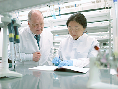
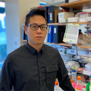
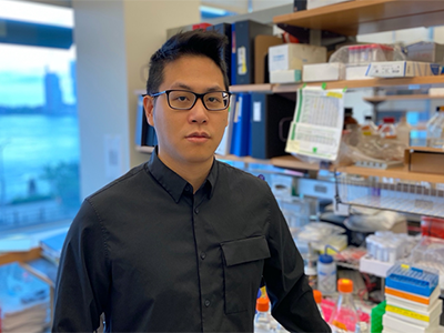
Children’s National Hospital welcomes Muller Fabbri, M.D. Ph.D., as associate director for the Center for Cancer and Immunology Research at the Children’s National Research Institute. In this role, he will build and lead the Cancer Biology Program while developing and conducting basic and translational research. Dr. Fabbri will also develop multidisciplinary research projects with various clinical divisions, including oncology, blood and marrow transplantation, pathology and hematology.

Dr. Fabbri shares his journey working with microRNAs, how his work is advancing the field and his vision for the Center for Cancer and Immunology Research at Children’s National.

Q: You have been working with microRNAs for quite some time. How are you exploring the role of microRNAs in cancer?

A: It was well established within the scientific community that a gene, which is a piece of DNA, becomes a piece of RNA and then becomes a protein. This thought process was pretty much a one-way flow of information that we had, going from DNA to protein as part of a cell function. But, almost 30 years ago, it was discovered that this is not entirely true because what happens is that some of these genes that are transcribed into RNA do not become a protein. Instead, they stay as RNA. Some of these RNAs are tiny and have short sequences, which is why they are called microRNAs.

I work primarily on microRNAs and non-coding RNAs and my research studies focus on the role that microRNAs play in cancer. I can take a cancer cell and a healthy cell and observe how these microRNAs are expressed in the two different cell populations. In this way, the microRNAs expressed in cancer cells are profoundly different from the microRNAs expressed in healthy cells.

We conducted a series of studies to observe what happens to a cancer cell if we restore normal levels of certain microRNAs like the ones you would see in a normal cell. We discovered that by restoring some of these microRNAs levels it led to the death of the cancer cells, suggesting that this approach may be used as a cancer treatment. This is one of the research areas that I will further develop at Children’s National as I seek to understand the mechanisms that control microRNA expression and subsequently affect cancer cell proliferation. With this information, we can target these mechanisms and create drugs that interfere with this function and, hopefully, stop cancer cell growth.

Q: Can you tell us about that eureka moment with your best friend during a lunch break?

A: This was a bit of a crazy idea. I will never forget. I shared a theory during a lunch break with a friend. I dared to ask, what if microRNAs worked like hormones? MicroRNAs can be detected in the blood of patients with cancer, and they can be transferred from one cell to another inside of little vesicles called exosomes. If you think about it, I further asked, what other molecules in our body behave like that — i.e. are secreted, circulate in the blood and then transferred to a target cell? My friend replied, “well, those would be hormones.” To which, I added, yes, exactly! Then, why do we not think of RNAs as hormones? And I quote him now, “you are crazy, but if it works it is huge.”

I felt that I had some validation from my best friend, so I decided to invest in this crazy idea, carving extra time on the side while working on my “safe” projects. It was one of those rare cases in science, where in a little over a year, we showed for the first time that microRNAs do not only work the traditional way, but they can also work as hormones. They do have a receptor protein to attach to, and by binding to this protein, they trigger a response in a cell that can be pro-tumoral or anti-tumoral.

Even today, if you open a textbook of endocrinology, under the chapter of hormones, it mentions that there are only two categories, proteins and lipids. Well, it turns out there is a third category, which is nucleic acids because of RNAs.

Q: You mentioned other research areas of interest as it relates to cancer cell biology. What are they?

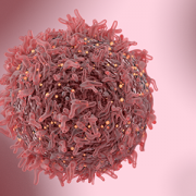
A: The other line of research that I am developing stems from the original observation that I made in 2012. Cancer cells release tiny vesicles that I like to compare to envelopes containing a written message — the RNA and microRNA. These vesicles released in the surrounding environment contain a message captured by immune cells, known as macrophages. Macrophages act as scavengers in our bodies. In cancer, macrophages are supposed to digest and destroy the cancer cell. However, it turns out that they also have the proper receptor to receive and read the message enclosed in the vesicles. Then, something shocking happens. The macrophage stops fighting the cancer cell and starts producing proteins called cytokines that promote cancer growth. This finding means that we are 180 degrees apart from what we thought at the beginning. A lot of macrophages in the cancer are good news for the patient because they are supposed to kill cancer cells, but because of this mechanism, a lot of macrophages can be bad news since they can also help the cancer cell grow.

My contribution to this discovery was to investigate how the macrophage response is mediated. I discovered that macrophages operate, at least in part, by expressing receptors that bind to microRNAs released by the cancer cell, thereby favoring cancer growth. In the pediatric cancer field we discovered that because of this microRNA–receptor interaction, the pediatric tumor neuroblastoma becomes resistant to chemotherapy. Therefore, one of the strategies we are working on now is to interfere or impair these negative communications between the cancer cell and immune cell. We want to disrupt these communications so the macrophage cannot read the message from the cancer cell anymore and instead keeps doing its job to fight the cancer. We hope that we can leverage this approach to develop novel cancer treatments or create strategies that improves immune cell function in the presence of the patient’s current therapy to enhance an anti-cancer treatment response.

Q: What is your vision for the Center of Cancer and Immunology Research?

A: I am very excited about what I saw at Children’s NationalI was delighted to talk to many faculty members, and I recognized the immense talent within the Center. I would like to help elevate and enhance the cancer biology program focused on solid tumors, and augment the work being done in this space by the cell therapy program. The clinicians are clearly eager to collaborate with the basic scientists including the sharing of samples and ideas, which is not typical of many scientific environments. My other goal is to ensure that the Cancer Biology Program plays a central role in acquiring an NCI-Designated Cancer Center recognition often given to institutions that stand out in scientific leadership and clinical research. Finally, I want to create the first national center that develops extracellular vesicles as an innovative treatment strategy for cancer. Importantly, I think that we have all the resources and connections at Children’s National that are necessary to realize this vision!


little boy at doctor

Demographic, clinical and biomarker features of MIS-C

little boy at doctor

In a new observational study, researchers provide insight into key features distinguishing MIS-C patients to provide a more realistic picture of the burden of disease in the pediatric population and aid with the early detection of disease and treatment for optimal outcomes.

Multisystem Inflammatory Syndrome in Children (MIS-C) significantly affected more Black and Latino children than white children, with Black children at the highest risk, according to a new observational study of 124 pediatric patients treated at Children’s National Hospital in Washington, D.C. Researchers also found cardiac complications, including systolic myocardial dysfunction and valvular regurgitation, were more common in MIS-C patients who were critically ill. Of the 124 patients, 63 were ultimately diagnosed with MIS-C and were compared with 61 patients deemed controls who presented with similar symptoms but ultimately had an alternative diagnosis.

In the study, published in The Journal of Pediatrics, researchers provide insight into key features distinguishing MIS-C patients to provide a more realistic picture of the burden of disease in the pediatric population and aid with the early detection of disease and treatment for optimal outcomes. The COVID-linked syndrome has affected nearly 4,000 children in the United States in the past year. Early reports showed severe illness, substantial variation in treatment and mortality associated with MIS-C. However, this study demonstrated that with early recognition and standardized treatment, short-term mortality can be nearly eliminated.

“Data like this will be critical for the development of clinical trials around the long-term implications of MIS-C,” says Dr. Roberta DeBiasi, M.D., lead author and chief of the Division of Pediatric Infectious Diseases at Children’s National. “Our study sheds light on the demographic, clinical and biomarker features of this disease, as well as viral load and viral sequencing.”

Of the 63 children with MIS-C, 52% were critically ill, and additional subtypes of MIS-C were identified including those with and without still detectable virus, those with and without features meeting criteria for Kawasaki Disease, and those with and without detectable cardiac abnormalities. While median age (7.25 years) and sex were similar between the MIS-C cohort and control group, Black (46%) and Latino (35%) children were overrepresented in the MIS-C group, especially those who required critical care. Heart complications were also more frequent in children who became critically ill with MIS-C (55% vs. 28%). Findings also showed MIS-C patients demonstrated a distinct cytokine signature, with significantly higher levels of certain cytokines than those of controls. This may help in the understanding of what drives the disease and which potential treatments may be most effective.

In reviewing viral load and antibody biomarkers, researchers found MIS-C cases with detectable virus had a lower viral load than in primary SARS-CoV-2 infection cases, but similar to MIS-C controls who had alternative diagnoses, but who also had detectable virus. A larger proportion of patients with MIS-C had detectable SARS-CoV-2 antibodies than controls. This is consistent with current thinking that MIS-C occurs a few weeks after a primary COVID-19 infection as part of an overzealous immune response.

Viral sequencing was also performed in the MIS-C cohort and compared to cases of primary COVID-19 infection in the Children’s National geographic population. 88% of the samples analyzed fell into the GH clade consistent with the high frequency of the GH clade circulating earlier in the pandemic in the U.S. and Canada, and first observed in France.

“The fact that there were no notable sequencing differences between our MIS-C and primary COVID cohorts suggests that variations in host genetics and/or immune response are more likely primary determinants of how MIS-C presents itself, rather than virus-specific factors,” says Dr. DeBiasi. “As we’ve seen new variants continue to emerge, it will be important to study their effect on the frequency and severity of MIS-C.”

Researchers are still looking for consensus on the most efficacious treatments for MIS-C. In a recent editorial in the New England Journal of Medicine, Dr. DeBiasi calls for well-characterized large prospective cohort studies at single centers, and systematic and long-term follow-up for cardiac and non-cardiac outcomes in children with MIS-C. Data from these studies will be a crucial determinant of the best set of treatment guidelines for immunotherapies to treat MIS-C.

girl with smart brain imagination doodle

Children’s National provides clinical validation, IP for health challenge designed to advance pediatric innovation

girl with smart brain imagination doodle

Reinforcing its commitment to expanding innovation in pediatric care, Children’s National Hospital has joined a strategic partnership with the Center for Advancing Innovation (CAI) , along with collaborators Resonance Philanthropies and Digital Infuzion, to launch the 2021-2022 Innovate Children’s Health Challenge. This year’s event, Innovate Children’s Health II, focuses on technologies that address pandemic resiliency and prevention in the pediatric population and seeks to advance diagnostics, therapeutics and digital health tools that address pediatric mental health.

The initiative matches entrepreneurial talent with breakthrough inventions to launch startups and connect them with capital. For this challenge, more than 15 startups will compete for the opportunity to commercialize promising mental health solutions from a variety of research partners, including Children’s National. Nationally recognized for its expertise and commitment to innovation in pediatric care, Children’s National will contribute to the clinical validation of selected technologies.

“In addition to our role in providing clinical validation, this initiative provides the opportunity for intellectual property (IP) developed by leading clinicians at Children’s National Hospital, as well as other great pediatric institutions, to be considered for partnership with entrepreneurs who can help bring these technologies to market,” says Kolaleh Eskandanian, PhD, MBA, PMP, vice president and chief innovation officer at Children’s National Hospital. “Our mission is to improve children’s healthcare and Innovate Children’s Health II is a great way to harness this trifecta model — innovation, talent and capital — in order to develop breakthrough solutions that address the unique needs of pediatric patients.”

Kolaleh-Eskandanian

“In addition to our role in providing clinical validation, this initiative provides the opportunity for intellectual property (IP) developed by leading clinicians at Children’s National Hospital, as well as other great pediatric institutions, to be considered for partnership with entrepreneurs who can help bring these technologies to market,” says Kolaleh Eskandanian, PhD, MBA, PMP, vice president and chief innovation officer at Children’s National Hospital.

There are three ways to participate in Innovate Children’s Health II:

  • Entrepreneurial-minded people, alone or as members of multidisciplinary teams, may compete to commercialize vetted inventions;
  • Existing startups may enter the challenge with other public health-related inventions, including their own and/or others to which they have access;
  • Participants may submit ideas that they believe will improve emergency preparedness and pandemic response.

Inventors and technology licensing officers may submit inventions to be evaluated and made available for licensing to challenge winners. Innovate Children’s Health II will accept invention submissions until September 1, 2021. Anyone with an entrepreneurial spirit and interest in stopping current and future pandemics is invited to sign up to learn more about the challenge. Teams may also enroll in the challenge to choose a featured invention, bring in a third-party invention or get matched with an invention based on area of interest.

“The COVID-19 pandemic has made our children anxious, depressed and pessimistic about their futures. Through Innovate Children’s Health II, CAI and our strategic partner Children’s National will strive to give our children hope,” says Rosemarie Truman, founder and CEO of CAI. “We are grateful to Digital Infuzion and Resonance Philanthropies for their support, which makes this challenge possible.”

Eskandanian adds that supporting and expanding pediatric innovation is a key focus of the new Children’s National Research & Innovation Campus, the first-of-its-kind focused on pediatric health care innovation, with the first phase currently open on the former Walter Reed Army Medical Center campus in Washington, D.C. With its proximity to federal research institutions and agencies, universities, academic research centers, as well as on-site incubator Johnson and Johnson Innovation – JLABS, the campus provides a rich ecosystem of public and private partners which will help bolster pediatric innovation and commercialization.