A new AI tool developed by Children’s National and Howard University analyzes brain immune cells 10,000x faster than manual methods.
A new Machine Learning and Artificial Intelligence tool from researchers at Children’s National Hospital (CNH) and Howard University (HU) accelerates discoveries in brain inflammation. Called StainAI, it rapidly and accurately analyzes microglia, the brain’s immune cells. Scientists currently analyze microglia slowly by hand. StainAI automates this process and speeds it up 10,000-fold. Its use will aid discovery of new treatments for inflammatory brain conditions such as infection, autoimmunity, and aging.
Solving a problem
Traditionally, scientists study microglia one cell at a time. They reconstruct each cell’s shape by hand under a microscope. The shape helps classify microglia as “resting” (normal) or “activated” (inflamed). The manual process is tedious and slow. It limits analyses to a few microglia in small brain areas.
StainAI changes that. It uses deep machine learning and artificial intelligence to overcome and exceed the manual method’s limitations. It correctly classifies millions of microglia from standard microscopic images. StainAI also localizes each microglia to its brain region in 3D. These features enable single-cell analyses of immune activity at a scale not feasible before – the entire brain.
A tool with broad impact
The team applied StainAI to two models of brain injury and inflammation to show its utility. In a rodent model of pediatric cardiac arrest, StainAI identified new brain regions susceptible to injury. In a simian model of viral infection, StainAI localized rod-shaped microglia normally found in white matter to an unexpected brain region – the hippocampus. These findings point towards new treatments and highlight StainAI’s value across diseases and species.
StainAI is fast, accurate and adaptable. It uses common laboratory equipment. Its creators, Michael Shoykhet, MD, PhD, at CNH and Dr. Tsang-Wei Tu at HU, are making StainAI available to other researchers. They hope StainAI will help labs worldwide discover new ways to protect children’s brains from inflammation and injury.
Using Fitbit data from the the largest long-term study of brain development and child health in the United States, researchers employed machine learning to test whether physiological markers could accurately predict ADHD diagnoses.
A new study published in Frontiers in Child and Adolescent Psychiatry reveals that common wearable devices like Fitbits may hold the key to improving how we identify Attention-Deficit/Hyperactivity Disorder (ADHD) in adolescents. By analyzing patterns in heart rate, activity levels and energy expenditure, researchers were able to predict ADHD diagnoses with striking accuracy, offering a glimpse into a future where objective, real-time data supports earlier and more personalized mental healthcare.
A fresh approach to a common challenge
ADHD affects approximately 1 in 10 children and adolescents in the United States. It is typically diagnosed based on parent and teacher reports, clinical interviews and behavioral observations. While effective, these methods rely heavily on subjective interpretation and can sometimes miss important nuances in how symptoms appear over time. This study, led by Muhammad Mahbubur Rahman, PhD, and colleagues at Children’s National, sought to determine whether wearable health data could help fill that gap.
Turning Fitbit metrics into meaningful insights
The study used data from 450 adolescents who were part of the larger Adolescent Brain Cognitive Development (ABCD) study, the largest long-term study of brain development and child health in the United States. Each participant wore a Fitbit, which captured three key activity and physiological measures:
Resting Heart Rate (RHR) – the number of heart beats per minute while the body is at rest
Sedentary Time – time spent with little or no physical activity
Energy Expenditure – estimated calories burned through physical activity
When the researchers compared these measures between teens with and without ADHD, they found statistically significant differences. Teens with ADHD had consistently higher resting heart rates and showed distinctive patterns in both their movement and stillness.
To go further, the team applied a machine learning model to test whether these physiological markers could accurately predict ADHD diagnoses. The model performed extremely well with 89% accuracy, 88% precision, 90% recall and a 0.95 area under the curve (AUC). These results suggest that the combination of passive, continuous data and predictive modeling could serve as a valuable screening tool, particularly in settings where full clinical evaluations are difficult to access.
A path toward more accessible mental healthcare
The implications are big. If validated in larger and more diverse populations, wearable-derived data could offer a low-cost, low-burden way to flag teens who might benefit from further ADHD evaluation. This could lead to earlier support, fewer misdiagnoses and more tailored treatment strategies.
Importantly, this approach isn’t about replacing clinicians, it’s about giving them better tools. Real-world, real-time data from wearables could act as an additional layer of insight that supports more precise, individualized care. As wearable technology becomes more embedded in daily life, its role in healthcare, especially adolescent mental health, is poised to grow.
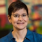
Julia Finkel, MD, is a pediatric anesthesiologist and the director of Pain Medicine Research at the Sheikh Zayed Institute for Pediatric Surgical Innovation.
What if doctors could measure pain as precisely as they measure blood pressure? That’s the vision driving Julia Finkel, MD, a pediatric anesthesiologist and director of Pain Medicine Research at the Sheikh Zayed Institute for Pediatric Surgical Innovation. Recently featured in The Washington Post, Dr. Finkel is pioneering the Nociometer, a new device that could transform how we understand and treat pain.
This new innovation uses a painless electrical current on a finger or toe to gently stimulate the body’s three main sensory nerve fibers. It then measures pupil response — linked to the brain’s pain centers — to identify the type and intensity of pain.
Now heading into clinical trials, the Nociometer shows how federal funding fuels innovation for patients. Here, Dr. Finkel shares the inspiration behind her work and the vital role of federal support in pediatric research.
Q: What started you on the path to developing this device?
A: My father is a theoretical physicist. He often talked in lay terms about everything he was thinking. When I was a child, we’d go for walks at night and look at the stars, and he’d explain the universe. My mother was an artist, but she struggled with constant pain from rheumatoid arthritis. There weren’t very good therapeutics at the time. Pain impacted her quality of life.
As an undergraduate, I was a chemistry major and interested in the mechanisms of how things worked. I planned to be a rheumatologist, but switching to anesthesia was transformative.
Q: What did you love about anesthesiology?
A: I love how it profoundly impacts patients. You induce unconsciousness, you induce wakefulness and you eliminate pain. When pain stops for a child, it also lessens the suffering of the parents. Our research is all about gaining a deeper understanding of the mechanisms of pain.
The Nociometer helps identify the mechanism behind the pain symptom so we can treat it appropriately. Research shows us that pain has a recognizable signature in the body. Because we can recognize it, we can treat it better. It’s like having an X-ray to look at before treating a broken leg.
Q: What is it like to work with children experiencing chronic pain? Why is measuring pain this way more effective than the typical scale?
A: Families become desperate. Often, they have seen many doctors before coming to a specialist. But pain is a single word for many different disease states. Not everyone has the right expertise. We often see children with chronic abdominal pain. Perhaps they struggle in school or can’t go at all. They don’t eat well, they have trouble engaging in regular activities. The Nociometer helps us determine the cause of the pain to better treat the problem. The device also gives us a way to validate pain while the pain scale tells us perception. The Nociometer provides objective data, very similar to a blood pressure reading.
Q: How has federal funding, and support from Children’s National, shaped your work?
A: Federal funding has been absolutely critical. Initially, we had grants from multiple agencies within the National Institutes of Health (NIH). This shows how ubiquitous and problematic pain is across the board. It’s the underpinning of many disease states and an upstream driver of the opioid epidemic.
We have grants from the Small Business Innovation Research program within the National Cancer Institute. We have funding from the National Institute of Arthritis and Musculoskeletal and Skin Diseases and an award from the Advanced Research Projects Agency for Health. This federal funding comes with scientific rigor and the ecosystem at Children’s National has allowed me to accomplish so much.
Q: What makes Children’s National the right place for this kind of innovation?
A: Children’s National has given me the space to think creatively and pursue unconventional ideas. In 2011, I became a founding member of the Sheikh Zayed Institute for Pediatric Surgical Innovation. With early institutional support, I was able to explore questions that led to the discovery of a physiological biomarker for pain — the basis of the Nociometer.
“Creative” isn’t a word you often hear in medicine, but here, it’s part of the process. I was able to spin out a company, secure funding and build a prototype — steps that wouldn’t have been possible elsewhere. The culture at Children’s National embraces discovery and fuels real impact.
Julia Finkel, MD, is a pediatric anesthesiologist and Director of Pain Medicine Research and Development, The Sheikh Zayed Institute for Pediatric Surgical Innovation and Professor of Anesthesiology, Pediatrics and Critical Care Medicine at The George Washington University.
https://innovationdistrict.childrensnational.org/wp-content/uploads/2025/06/Julia-Finkel-CNRI.jpg385685Innovation Districthttps://innovationdistrict.childrensnational.org/wp-content/uploads/2023/12/innovationdistrict_logo-1-1030x165.pngInnovation District2025-06-04 16:47:312025-06-05 12:42:51Behind the Nociometer: Q&A with Julia Finkel, MD
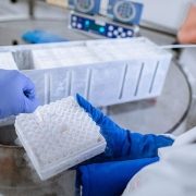
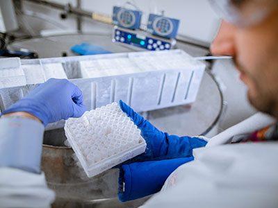
A new HIV-specific T cell therapy, tested in six adults living with HIV, used specially trained immune cells made from each person’s own blood — a personalized therapy designed to target the virus with precision.
An exciting small clinical trial led by the Center for Cancer and Immunology Research at Children’s National Hospital has shown that a new HIV-specific T cell therapy is safe and may help reduce hidden reservoirs of the virus in the body. This approach, tested in six adults living with HIV, uses specially trained immune cells made from each person’s own blood — a personalized therapy designed to target the virus with precision.
The results, published in Nature Communications, represent a step forward in the search for a long-term, drug-free way to control or even cure HIV.
A smarter way to fight HIV
Today, people living with HIV rely on anti-retroviral therapy (ART) to keep the virus under control. These medications are highly effective but must be taken daily and do not eliminate the virus entirely. That is because HIV can hide in a “reservoir” of cells where it remains dormant and invisible to both drugs and the immune system. If ART is stopped, the virus can quickly return.
To change that, scientists at Children’s National and partnering institutions developed a new type of cellular therapy called HST-NEETs — short for “HIV-specific T cells targeting conserved epitopes”. These T cells are trained in the lab to recognize parts of the virus that do not change much, even as HIV mutates. This makes it harder for the virus to escape. The goal is to help the immune system find and destroy the infected cells that are normally hidden.
Safe and promising results
In this phase 1 clinical trial, researchers created personalized HST-NEET therapy from each participant’s own immune cells. After training the cells to recognize HIV, they were infused back into the patients twice over a period of weeks.
The results showed that:
No serious side effects were reported from the infusions.
The treatment was well-tolerated by all six participants.
In two people, the therapy led to stronger HIV-specific immune responses, including more virus-fighting T cells and antibodies.
In two others, researchers saw a drop in the level of HIV hidden in their cells, a sign that the virus reservoir might be shrinking.
In four participants, the infused T cells persisted in the bloodstream for up to 40 weeks, continuing to patrol for signs of HIV.
While not a cure, these findings show early evidence that the therapy may help the body better recognize and fight HIV, even the hidden forms that are hardest to treat.
Building toward a cure
“The fact that we saw HIV-specific T cell responses increase in some participants, even without additional immune-boosting drugs, is very encouraging,” said Catherine Bollard, MBChB, MD, senior author of the study and director of the Center for Cancer and Immunology Research at Children’s National. “It suggests that the immune system can be trained to go after parts of the virus that were previously out of reach.”
Unlike bone marrow transplants, which have led to a cure in a few people with both HIV and cancer but carry high risk, HST-NEET therapy is much safer and more scalable. That is important for the millions of people living with HIV worldwide.
This study also sets the stage for future clinical trials that could combine T cell therapy with other strategies, like latency-reversing drugs that “wake up” hidden HIV, to further shrink the reservoir. It also shows that personalized T cells can be safely made, infused and tracked over time and that they can continue working in the body for many months. Those lessons are valuable not just for HIV but also for developing safer, more targeted cancer immunotherapies in children and adults.
What’s next
The next phase of research will evaluate this therapy in larger groups and under different conditions, including in people undergoing stem cell transplants or with added immune system boosters. Clinical trials are already underway exploring these combinations.
By focusing on preserved parts of the virus, the regions that HIV cannot easily mutate, HST-NEETs could one day become part of a combination approach to eliminate HIV from the body altogether.
“Every step brings us closer to a functional cure,” said Dr. Bollard. “And the lessons we’re learning from HIV may also inform how we treat other chronic viral infections, and even cancer, in the future.”
Authors authors from Children’s National include Danielle K. Sohai, Michael D. Keller, Patrick J. Hanley, Fahmida Hoq, Divyesh Kukadiya, Anushree Datar, Emily Reynolds, Christopher Lazarski, Chase D. McCann, Jay Tanna, Abeer Shibli, Haili Lang, Anqing Zhang, Pamela A. Chansky, Cecilia Motta and Conrad Russell Y. Cruz.
https://innovationdistrict.childrensnational.org/wp-content/uploads/2025/05/tube-rack-in-lab-CNRI.jpg385685Innovation Districthttps://innovationdistrict.childrensnational.org/wp-content/uploads/2023/12/innovationdistrict_logo-1-1030x165.pngInnovation District2025-05-23 12:34:312025-05-23 12:35:29Personalized T cell therapy for HIV shows safety and early signs of impact
“Healthcare is moving very fast. And what often happens in adults, also happens in children. Unfortunately, most of the research is directed initially at adults, and then whittles down to children. At Children’s National, we’re trying to turn that around. We’re trying to do research for children that will expand its way up to adults, turning it on its head.”
Anthony Sandler, MD, senior vice president and surgeon-in-chief, Joseph E. Robert Jr. Center for Surgical Care, and director of the Sheikh Zayed Institute for Pediatric Surgical Innovation highlighted the exciting research and innovation happening at Children’s National – including demonstrating a technology, led by Raj Shekhar, PhD, that uses real-time imaging with augmented reality to project live ultrasound visualization of a patient within the surgeon’s field of view. This enhances surgical precision and ultimately supports positive patient outcomes.
This conversation was a part of Axios’ inaugural Future of Health Summit – an event bringing together the top voices in healthcare, policy and technology to explore the biggest challenges and innovations shaping the future of medicine.
https://innovationdistrict.childrensnational.org/wp-content/uploads/2025/05/Sandler-Axios-CNRI.jpg385685Innovation Districthttps://innovationdistrict.childrensnational.org/wp-content/uploads/2023/12/innovationdistrict_logo-1-1030x165.pngInnovation District2025-05-15 15:55:332025-05-15 16:13:36In the news: Axios’ Future of Health Summit
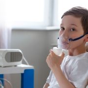
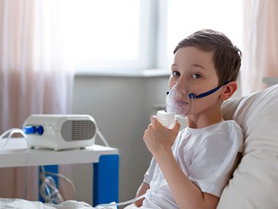
A new international study led by Children’s National Hospital and Lurie Children’s Hospital of Chicago introduces a validated tool to predict pneumonia severity in children, helping emergency clinicians make faster, evidence-based decisions about hospitalization and intensive care.
Pneumonia is one of the most common infections in children. In the U.S., it’s a leading reason why kids are admitted to the hospital. But for emergency doctors, it’s not always easy to know which cases are serious — and which children will get better at home.
Now, a new study led by doctors at Children’s National Hospital and Lurie Children’s Hospital of Chicago has created a tool to help. It’s based on research from over 2,200 children treated at emergency departments in 14 countries.
The tool is simple: it uses symptoms that doctors already look for — like how fast a child is breathing, whether they’re getting enough oxygen, and if they’re drinking fluids — to score how serious their pneumonia might be. The score helps doctors decide whether a child needs to stay in the hospital, go to intensive care or can safely recover at home.
What the study found
The research team looked at children ages 3 months to 13 years who came to emergency departments with community-acquired pneumonia — a kind of pneumonia picked up outside of a hospital. Most had mild cases. But about 1 in 20 developed severe symptoms, like needing breathing support or admission to intensive care.
The team found that certain symptoms (like fast breathing or heart rate, chest retractions (a sign of struggling to breathe), low oxygen levels, refusing to drink and already being on antibiotics before coming to the hospital) were linked to a higher risk of serious illness. On the other hand, children who had a runny nose or congestion were more likely to have mild illness.
Using this data, the team created a point-based score. For example, if a child had low oxygen levels, they’d get 3 to 6 points depending on how low it was. Chest retractions added 3 points. Having a runny nose subtracted a point. The higher the total score, the greater the risk of moderate or severe pneumonia.
How it helps
The model was tested and found to be highly accurate. It performed better than doctors’ judgment alone in earlier studies — especially in spotting the children most at risk. The scorecard gives emergency doctors a fast, evidence-based way to support the decisions they make under pressure.
“Emergency departments around the world see thousands of children with pneumonia every day, but until now, we haven’t had a reliable way to predict who’s truly at risk of getting sicker,” said co-PI and senior author Nathan Kuppermann, MD, MPH, executive vice president, chief academic officer and director of the Children’s National Research Institute. “This model gives clinicians a practical tool, rooted in data, to guide that decision and ultimately improve care and outcomes.”
What’s next
While the tool is ready to be used in hospitals now, the team plans to test it in more locations and study how it affects real-world decisions. They also hope to add biomarkers — lab tests that could improve the score’s accuracy even more.
For now, the study offers something simple and powerful: a better way to know when a child’s pneumonia might become serious — and when it won’t.
The study was published in The Lancet Child & Adolescent Health and is part of a larger effort by the Pediatric Emergency Research Network (PERN), which connects emergency departments in dozens of countries.
https://innovationdistrict.childrensnational.org/wp-content/uploads/2025/05/boy-with-breathing-apparatus-CNRI.jpg385685Innovation Districthttps://innovationdistrict.childrensnational.org/wp-content/uploads/2023/12/innovationdistrict_logo-1-1030x165.pngInnovation District2025-05-15 11:22:392025-05-15 15:49:28New tool helps doctors know when kids with pneumonia need hospital care
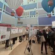
Children’s National Hospital hosted its fifteenth annual Research, Education and Innovation Week from March 31–April 4, 2025, bringing together clinicians, scientists, educators and innovators from across the institution to celebrate discovery and collaboration. This year’s theme, “Empowering the Future in Pediatric Research and Innovation with Equity, Technology and a Global Reach,” served as a call to action for advancing science that improves child health both locally and around the world.
Each day of the week-long event featured thought-provoking lectures — now available to watch — dynamic panel discussions, interactive workshops and vibrant poster sessions, all highlighting the diverse and interdisciplinary work taking place across Children’s National.
Centering the patient and the planet
REI Week began on Monday with a powerful keynote lecture from Lynn R. Goldman, MD, MS, MPH, Michael and Lori Milken dean of the Milken Institute School of Public Health at the George Washington University. In her talk, “Children: Uniquely vulnerable to climate-related threats,” Dr. Goldman underscored the urgent need to protect children from the environmental hazards of a changing climate and to integrate climate science into pediatric care and advocacy.
At mid-morning, Mary-Anne “Annie” Hartley, MD, PhD, MPH, director of the LiGHT Laboratory at École Polytechnique Fédérale de Lausanne, introduced the “MOOVE” platform — Massive Open Online Validation and Evaluation of clinical LLMs. Her talk demonstrated how artificial intelligence, when rigorously validated, has the potential to transform clinical decision-making and global health equity.
Monday’s final keynote, “Zinc and childhood diarrhea,” was presented by Christopher Duggan, MD, MPH, director of the Division of Nutrition at Harvard Medical School. Dr. Duggan highlighted the global health impact of zinc supplementation in reducing childhood mortality — a reminder that simple, evidence-based interventions can save millions of lives.
In that first day, the first poster session of the week showcased projects in adolescent medicine, global health, infectious diseases, oncology and more. The session reflected the full breadth of research taking place across Children’s National.
Ambroise Wonkam, MD, PhD, professor of genetic medicine at Johns Hopkins University, then delivered Tuesday’s Global Health Keynote Lecture, “Harnessing our common African genomes to improve health and equity globally.” His work affirmed that inclusive genomics is key to building a healthier world.
Later, the Global Health Initiative event and GCAF Faculty Seminar encouraged attendees to pursue collaborative opportunities at home and abroad, reflecting the growing global footprint of Children’s National research programs.
Transforming education and care delivery
On Wednesday, Larrie Greenberg, MD, professor emeritus of pediatrics, kicked off the day with a Grand Rounds keynote on educational transformation: “Shouldn’t teachers be more collaborative with their learners?” He followed with a CAPE workshop exploring the effectiveness of case-based learning.
In the Jill Joseph Grand Rounds Lecture, Deena J. Chisolm, PhD, director of the Center for Child Health Equity at Nationwide Children’s Hospital, challenged attendees to move beyond dialogue into action in her talk, “Health equity: A scream to a whisper?,” reminding researchers and clinicians that advocacy and equity must be foundational to care.
The day continued with a poster session spotlighting medical education, neonatology, urology and neuroscience, among other fields.
Posters and pathways to progress
Throughout the week, poster sessions highlighted cutting-edge work across dozens of pediatric disciplines. These sessions gave attendees the opportunity to engage directly with investigators and reflect on the shared mission of discovery across multiple disciplines, including:
The REI Week 2025 Awards Ceremony celebrated outstanding contributions in research, mentorship, education and innovation. The winners in each category were:
POSTER SESSION AWARDS
Basic & Translational Research
Faculty: Benjamin Liu, PhD
“Genetic Conservation and Diversity of SARS-CoV-2 Envelope Gene Across Variants of Concern”
Faculty: Steve Hui, PhD
“Brain Metabolites in Neonates of Mothers with COVID-19 Infection During Pregnancy”
Faculty: Raj Shekhar, PhD
“StrepApp: Deep Learning-Based Identification of Group A Streptococcal (GAS) Pharyngitis”
Post docs/Fellows/Residents: Dae-young Kim, PhD
“mhGPT: A Lightweight Domain-Specific Language Model for Mental Health Analysis”
Post docs/Fellows/Residents: Leandros Boukas, MD, PhD
“De Novo Variant Identification From Duo Long-Read Sequencing: Improving Equitable Variant Interpretation for Diverse Family Structures”
Staff: Naseem Maghzian
“Adoptive T Lymphocyte Administration for Chronic Norovirus Treatment in Immunocompromised Hosts (ATLANTIC)”
Graduate Students: Abigail Haffey
“Synergistic Integration of TCR and CAR T Cell Platforms for Enhanced Adoptive Immunotherapy in Brain Tumors”
High School/Undergraduate Students: Medha Pappula
“An ADHD Diagnostic Interface Based on EEG Spectrograms and Deep Learning Techniques”
Clinical Research
Faculty: Folasade Ogunlesi, MD
“Poor Air Quality in Sub-Saharan Africa is Associated with Increase Health Care Utilization for Pain in Sickle Cell Disease Patients”
Faculty: Ayman Saleh, MD
“Growth Parameters and Treatment Approaches in Pediatric ADHD: Examining Differences Across Race”
Post docs/Fellows/Residents: Nicholas Dimenstein, MD, MPH
“Pre-Exposure Prophylaxis (PrEP) Eligibility in the Pediatric Emergency Department”
Staff: Tayla Smith, MPH
“The Public Health Impact of State-Level Abortion and Firearm Laws on Health Outcomes”
Graduate Students: Natalie Ewing
“Patterns of Bacteriuria and Antimicrobial Resistance in Patients Presenting for Primary Cloacal Repair: Is Assisted Bladder Emptying Associated with Bacteriuria?”
Graduate Students: Manuela Iglesias, MS
“Exploring the Relationship Between Child Opportunity Index and Bayley-III Scores in Young Children”
High School/Undergraduate Students: Nicholas Lohman
“Preliminary Findings: The Efficacy, Feasibility and Acceptability of Group Videoconference Cognitive Behavioral Therapy with Exposure and Response Prevention for Treating Obsessive-Compulsive Disorder Among Children and Young People”
Community-Based Research
Faculty: Sharon Shih, PhD “Assessing Pediatric Behavioral Health Access in DC using Secret Shopper Methodology”
Post docs/Fellows/Residents: Georgios Sanidas, MD “Arrested Neuronal Maturation and Development in the Cerebellum of Preterm Infants”
Staff: Sanam Parwani
“Intersectionality of Gender and Sexuality Diversity in Autistic and Non-Autistic Individuals”
Graduate Student: Margaret Dearey “Assessing the Burden of Period Poverty for Youth and Adolescents in Washington, DC: A Pilot Study”
Quality and Performance Improvement
Faculty: Nichole L. McCollum, MD
“A Quality Improvement Study to Increase Nurse Initiated Care from Triage and Improve Timeliness to Care”
Post docs/Fellows/Residents: Hannah Rodriguez, MD
“Reducing Unnecessary Antibiotic Use in a Level IV NICU”
Staff: Amber K. Shojaie, OTD, OTR/L
“Implementing Dynamic Axilla Splints in a Large Burn Patient”
Meleah Boyle, PhD, MPH
“Understanding and Addressing Environmental Sustainability to Protect the Health of the Children’s National and Global Communities”
Eiman Abdulrahman, MD
“Research Capacity Building to Improve Pediatric Emergency and Critical Care in Ethiopia”
Pilot Awards
Alexander Andrews, MD
“EEG as a Diagnostic and Prognostic Marker in Severe Pediatric Malaria, Blantyre Malawi”
Daniel Donoho, MD & Timothy Singer, MD
“Feasibility Study of a Novel Artificial Intelligence-Based Educational Platform to Improve Neurosurgical Operative Skills in Tanzania”
Hasan Syed, MD
“Bridging the Gap an Educational Needs Assessment for Pediatric Neurosurgery Training in Pakistan”
Sofia Perazzo, MD & Lamia Soghier, MD, MEd, MBA
“QI Mentorship to Improve Pediatric Screening and Follow-up in Rural Argentina”
Benjamin Liu, PhD
“AI-Empowered Real-Time Sequencing Assay for Rapid Detection of Schistosomiasis in Senegal”
Rae Mittal, MD
“Assessment and Enhancement of Proficiency in Emergency Child Neurology Topics for Post-Graduate Emergency Medicine Trainees in India”
Innovation Day ignites bold thinking
Thursday, REI Week shifted to the Children’s National Research & Innovation Campus for Innovation Day, a celebration of how bold ideas and collaborative culture can accelerate progress in pediatric medicine.
REI Week 2025 reaffirmed the values that define Children’s National: a commitment to excellence, collaboration and equity in pediatric research and care. As discoveries continue to emerge from our hospital and our research campuses, the connections built and ideas sparked during this week will help shape the future of pediatric health — locally and globally.
By elevating voices from the bedside to the bench, with the support of the executive sponsors Nathan Kuppermann, MD, MBChB, Catherine Bollard, MBChB, MD, Kerstin Hildebrandt, MSHS, Linda Talley, MS, RN, NE-BC and David Wessel, MD, REI Week demonstrated that we must embrace the community in all aspects of our work. Because we know that there are answers we can only get from the patients that we serve—and we need to be their voice.
Research, Education & Innovation Week will be back next year on April 13-17, 2026.
Posters at the REI Week 2025 Monday, March 31 poster session.
Panelists discuss innovation during REI Week 2025.
Global Health Initiative community engagement event during REI Week 2025.
Chris Rees presents his REI Week 2025 lecture.
Nathan Kuppermann listens to a presenter during the REI Week 2025 Tuesday, April 1, poster session.
Michelle Riley-Brown, Nathan Kuppermann, Catherine Bollard and Naomi Luban on stage during the REI Week 2025 awards ceremony.
Brandy Salmon presents on innovation programs at Virginia Tech during the REI Week 2025 Innovation Day.
Catherine Bollard listens to a presenter during the REI Week 2025 Monday, March 21 poster session.
Ambroise Wonkman poses for a picture with Children’s National staff.
Tanzeem Choudhury presenting during REI Week 2025.
https://innovationdistrict.childrensnational.org/wp-content/uploads/2025/04/REI-Week-2025-Monday-Poster-Session-CNRI.jpg385685Innovation Districthttps://innovationdistrict.childrensnational.org/wp-content/uploads/2023/12/innovationdistrict_logo-1-1030x165.pngInnovation District2025-04-22 10:31:052025-06-10 12:20:52REI Week 2025 empowers the future in pediatric research and innovation
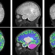
A new study out of the Center for Prenatal, Neonatal & Maternal Health Research, directed by Catherine Limperopoulos, PhD, led by Yao Wu, PhD and Stephanie Merhar, MD, MS, out of Cincinnati Children’s Hospital Medical Center, sought answers to the question: “Do brain volumes differ in opioid-exposed vs. unexposed newborns?” In one of the largest studies of this kind, researchers found that prenatal exposure to opioids is associated with smaller brain volumes in newborns. These findings from the landmark Outcomes of Babies with Opioid Exposure (OBOE) study build on, reinforce smaller studies, and achieve a better understanding of the impacts of prenatal opioid exposure (POE).
Dive Deeper
The Advancing Clinical Trials in Neonatal Opioid Withdrawal (ACT-NOW)’s OBOE study is a multi-site observational study of newborns with prenatal opioid exposure and a control group of unexposed newborns from four different sites in the United States – Case Western Reserve University, Cincinnati Children’s Hospital Medical Center, University of Alabama at Birmingham, and Children’s Hospital of Philadelphia.
In a study involving 173 newborns who were exposed to opioids during pregnancy and 96 newborns not exposed to opioids prenatally showed smaller brain sizes in several key areas. Specifically, these exposed babies had smaller total brain volumes, as shown through MRI, as well as reduced volumes in important parts of the brain, including the cortex (outer layer of the brain), deep gray matter (areas that control movement and emotions), white matter (which helps transmit signals in the brain), cerebellum (responsible for coordination and movement), brainstem (controls basic functions like breathing), and the amygdala (involved in emotions and memory).
Further details showed that newborns exposed to medication for opioid use disorder (MOUD) during pregnancy with methadone, had smaller white matter volumes, while those exposed to MOUD with buprenorphine had smaller volumes specifically in the right amygdala. Additionally, newborns who were exposed to opioids plus additional substances such as THC and gabapentin had smaller volumes in even more brain areas compared to those who were only exposed to opioids.
What’s Next
The OBOE study sets the groundwork for further research into the long-term impact of opioid exposure during pregnancy. Additional work is necessary to expand on these findings and how they relate to functions in childhood – including exploring the way these reduced brain volumes may impact cognitive, behavioral, and motor impairments. The study raises important questions about how current guidelines for MOUD during pregnancy – specifically with methadone and buprenorphine – might evolve considering these findings. This study highlights the need for further research to assess the long-term effects of MOUD regimens on both maternal and infant outcomes.
This significant study underscores the importance of multi-disciplinary collaboration in opioid exposure research, effective regulation, and policy interventions – involving healthcare providers, researchers, policymakers, and affected families – to best mitigate the consequences and improve the health outcomes of children affected by prenatal opioid exposure.
Additional authors from Children’s National include Kushal Kapse, BS, MS, and Josepheen De Asis-Cruz, MD, PhD. Other authors include Carla M. Bann, PhD, Jamie E. Newman, PhD4, Nicole Mack, MS, Sara B. De Mauro, MD, MSCE, Namasivayam Ambalavanan, MD, Jonathan M. Davis, MD, Scott A. Lorch, MD, MSCE5, Deanne Wilson-Costello, MD, Brenda B. Poindexter, MD and Myriam Peralta-Carcelen, MD.
https://innovationdistrict.childrensnational.org/wp-content/uploads/2025/04/prenatal-opiod-exposure-CNRI-feature.jpg385685Innovation Districthttps://innovationdistrict.childrensnational.org/wp-content/uploads/2023/12/innovationdistrict_logo-1-1030x165.pngInnovation District2025-04-15 15:08:582025-04-15 15:08:58New study finds prenatal opioid exposure linked to smaller newborn brain volumes
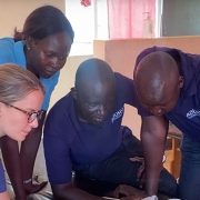
In December of 2024, a team that included experts from Children’s National Hospital traveled to Uganda to continue work on a pilot program applying artificial intelligence (AI) to the diagnosis of rheumatic heart disease (RHD). Ugandan health care providers have been trained and equipped to acquire echocardiograms for their patients but lack expertise in consistently being able to diagnose RHD by detecting leaky heart valves. The team created a tool that uses AI to predict RHD by identifying leaky heart valves on handheld ultrasound devices, then prompts a referral for a full echocardiogram.
The goal is to find ways to help people in Uganda diagnose RHD early, before a patient is in need of surgery, and initiate antibiotics so their heart can return to normal. The team of researchers, including fellow Kelsey Brown, MD, helped to implement additional steps toward this goal in December. According to Dr. Brown, the results were excellent. After four days of seeing patients, over 450 people were screened. The AI tool has an 86% accuracy rating. After returning from Uganda, the research team plans to work on the AI tool and further improve its accuracy rating. Eventually, the vision is that this tool can roll out on a larger scale for more places around the world to access it.
Craig Sable, MD, Marius Linguraru, DPhil, MA, MSc, and Pooneh Roshanitabrizi, PhD, from our Sheikh Zayed Institute, who developed the AI algorithms, worked in partnership with the Rheumatic Heart Disease Research Collaborative (RRCU) in Uganda. This trip was also made possible thanks to a grant funded through the Children’s National Global Health Initiative. Special thank you to our AI partner, US2.AI, who made the deployment of the AI models onto a tablet that provided real-time results, possible.
https://innovationdistrict.childrensnational.org/wp-content/uploads/2025/02/RHD-AI-Uganda-CNRI.jpg385685Innovation Districthttps://innovationdistrict.childrensnational.org/wp-content/uploads/2023/12/innovationdistrict_logo-1-1030x165.pngInnovation District2025-02-26 10:43:202025-05-06 13:49:45Children’s National brings AI into the RHD early diagnosis equation
https://innovationdistrict.childrensnational.org/wp-content/uploads/2025/02/Article_Image_400x300_Dauber.jpg12501667Innovation Districthttps://innovationdistrict.childrensnational.org/wp-content/uploads/2023/12/innovationdistrict_logo-1-1030x165.pngInnovation District2025-02-19 13:05:102025-05-05 10:46:55Podcast: Growing hope: Advancing care for kids with growth disorders
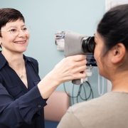
The new partnership will advance pediatric health through innovative discoveries and therapies, with an initial focus on pediatric cancers, including brain tumors.
Children’s National Hospital and Virginia Tech are expanding their research partnership, building on a successful collaboration established in 2019. This partnership will advance pediatric health through innovative discoveries and therapies, with an initial focus on pediatric cancers, including brain tumors.
The partnership brings together Children’s National, ranked among the nation’s top pediatric hospitals by U.S. News & World Report, and Virginia Tech, a leading academic research institution. Together, they aim to deliver transformative advancements to enhance outcomes for children facing devastating diagnoses.
The goals of the research-focused partnership include:
Accelerating the understanding of the biology, improvements in prevention and treatment of pediatric cancers and other childhood diseases.
Developing new diagnostic and therapeutic tools to improve care for children.
Training the next generation of scientists and physician-scientists.
What they’re saying
“Over the years, our partnership with Virginia Tech has demonstrated the power of combining top-tier research expertise with a shared commitment to improving pediatric health,” said Catherine Bollard, MBChB, MD, senior vice president and chief research officer and director of the Center for Cancer and Immunology Research. “This expansion underscores our belief that by working together, we can accelerate discoveries and develop life-changing therapies for children with cancer and other rare diseases.”
“Children’s National Hospital has been an important partner for us in biomedical research and innovation,” said Michael Friedlander, PhD, Virginia Tech vice president for health sciences and technology. “Our collaboration deepened with the launch of Children’s National Research & Innovation Campus in Washington, D.C., and now, as our partnership grows even stronger, we’re poised together to take on some of the biggest challenges in cancer research to contribute to the health of children and adults.”
“Partnering with Children’s National connects us to a world-class clinical trial institute that has been a pioneer in treating brain tumors with focused ultrasound technology, and this presents a unique opportunity to help children and families struggling with cancer,” said Cheng-Chia “Fred” Wu, MD, PhD, a member of the Children’s National Brain Tumor Research Institute and a principal investigator in cancer research and faculty member at the Fralin Biomedical Research Institute in Roanoke and in the Virginia Tech Carilion School of Medicine.“I can’t wait to see where this takes us.”
Big picture
The initial focus of the collaboration is pediatric cancers, including brain tumors — among the most challenging childhood diagnoses. By combining Virginia Tech’s leading-edge technology and research infrastructure with Children’s National’s expertise in pediatric care, the organizations aim to make significant strides in understanding these diseases.
An interdisciplinary approach is at the heart of the ongoing strategy. The collaboration first began with the launch of a 12,000-square-foot Virginia Tech biomedical research facility within the Children’s National Research & Innovation Campus, which opened in 2020. Located on a 12-acre portion of the former Walter Reed Army Medical Center in Washington, D.C., the campus was the nation’s first innovation hub focused exclusively on pediatric research.
https://innovationdistrict.childrensnational.org/wp-content/uploads/2025/02/boy-getting-eye-exam-CNRI.jpg385685Innovation Districthttps://innovationdistrict.childrensnational.org/wp-content/uploads/2023/12/innovationdistrict_logo-1-1030x165.pngInnovation District2025-02-04 13:31:522025-02-04 13:32:49Expanded partnership with Virginia Tech accelerates pediatric cancer research
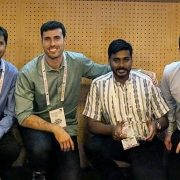
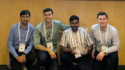
Meet the winners (left to right): Syed M. Anwar, Ph.D., M.S., principal investigator at Children’s National; Daniel Capellan Martin, M.Sc., Polytechnic University of Madrid; Abhijeet Parida, data scientist at Children’s National; and Austin Tapp, Ph.D., postdoctoral research fellow at Children’s National.
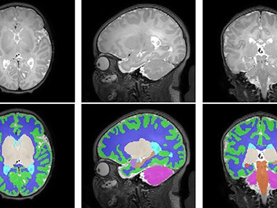
Using an award-winning artificial intelligence (AI) algorithm developed at Children’s National Hospital, researchers ranked first in the world in the Brain Tumor Segmentation-Africa (BraTS-Africa) challenge for their approach to identifying different parts of deadly gliomas. The details of their innovative method were recently published on arXiv, a curated research-sharing platform.
“Technology can bridge the gap in healthcare between high- and low-resource countries,” said Marius George Linguraru, D.Phil., M.A., M.Sc., the Connor Family Professor in Research and Innovation and principal investigator in the Sheikh Zayed Institute for Pediatric Surgical Innovation (SZI). “By tailoring methods we created at our hospital to fit the needs of specific regions, such as sub-Saharan Africa, our research helps improve medical imaging and diagnosis in challenging environments.”
Dr. Linguraru was the program chair of the International Conference on Medical Image Computing and Computer Assisted Intervention (MICCAI) 2024 in Marrakesh, Morocco, the leading global meeting on AI in medical imaging.
Children’s National leads the way
Gliomas are a type of brain tumor with a high death rate and are especially difficult to diagnose in low- and middle-income countries. Given the increased need in Africa, researchers worldwide came together in Morocco to compete over the best way to accurately detect and measure tumors using MRI data and AI.
By applying advanced machine-learning techniques, the researchers adapted tools initially designed for well-resourced settings to work in countries with far fewer.
The study focused on transfer learning, a process in which an AI model is trained in advance on a large number of brain tumor images and then adjusted to work with smaller sets of new data. In this case, the models were adapted to work with local sub-Saharan African data using a strategy to combine different models’ strengths.
When tested, the approach achieved impressive accuracy scores. The Children’s National team, which included a colleague from the Polytechnic University of Madrid, ranked first in the BraTS-Africa 2024 challenge for identifying different parts of gliomas.
“To make the method widely available, the winning algorithm is shared online for others to use and improve upon,” Dr. Linguraru said. “My favorite part of these competitions is how they highlight the way innovation and collaboration can reduce global healthcare inequalities.”
The big picture
Children’s National researchers consistently lead global events using AI and advanced imaging to tackle complex healthcare challenges. In 2023, the team won a global contest to measure pediatric brain tumors at the MICCAI 2023 Conference. This year’s success in the BraTS-Africa challenge builds on this knowledge base and expands its use to adult gliomas.
At the Radiological Society of North America 2024 annual meeting, which drew 50,000 attendees, Zhifan Jiang, Ph.D., a staff scientist in the Precision Medical Imaging Lab at SZI, also won the Cum Laude Award for his scientific poster on applying AI to radiological images to predict severe outcomes for children with brain tumors caused by neurofibromatosis type 1.
“These achievements show how our science is leading the world in using AI for good,” Dr. Linguraru said. “Every day, we’re building on our knowledge of advanced imaging, brain tumors and AI to improve the diagnosis, measurement and treatment of deadly tumors — on a global scale.”
https://innovationdistrict.childrensnational.org/wp-content/uploads/2025/01/Brats2024-Team-CNRI.jpg385685Innovation Districthttps://innovationdistrict.childrensnational.org/wp-content/uploads/2023/12/innovationdistrict_logo-1-1030x165.pngInnovation District2025-01-10 17:12:152025-01-10 17:17:51AI for good: Children’s National wins global competitions for measuring brain tumors
Sepsis is a rapidly progressing, life-threatening condition characterized by organ dysfunction (OD) resulting from an immune response to infection.
Researchers at Children’s National Hospital are investigating pediatric sepsis, a leading cause of in-hospital death, particularly in underserved and minoritized populations.
A new Maximizing Investigators’ Research Award (MIRA) (R35) is granting over $2.2 million to Children’s National to establish a comprehensive sepsis program that will include a diagnostic artificial intelligence (AI) based, biomarker-enhanced platform for early recognition of pediatric sepsis and cutting-edge, extracellular vesicle-based therapeutics that can significantly decrease mortality and long-term disease sequelae.
MIRA provides support for research in an investigator’s laboratory that falls within the mission of The National Institute of General Medical Sciences.
Why it matters
Sepsis, defined as a rapidly progressive, is a life-threatening organ dysfunction (OD) due to an immune dysregulation as a response to infection. In the U.S., it results in over 75,000 pediatric admissions on an annual basis with an associated mortality rate of 5 to 20%.
“Sepsis has been linked to 20% of deaths worldwide and despite some recent advances in diagnostic tools, the lack of accurate definitions and heterogeneity of this clinical syndrome have led to delays in recognition and treatment with deleterious effects on the health of millions of children, especially those from minority groups,” says Ioannis Koutroulis, M.D., Ph.D., M.B.A., research director of Emergency Medicine at Children’s National. “Additionally, current sepsis therapeutic regimens are mostly supportive, lack the necessary personalization and fail to address the underlying physiological processes leading to sepsis-induced life threating organ failure.”
How does it move the field forward
High mortality rates and no significant new treatments in recent years due to sepsis presents a critical opportunity to make a global impact. Advancements in early recognition and intervention could save millions of lives. By utilizing AI and biomarkers for the early detection of sepsis in children, medical professionals could have the potential to greatly improve outcomes by enabling timely treatment.
“There is an urgent need to focus more attention on this condition and develop effective solutions to combat its devastating effects,” Dr. Koutroulis adds.
How we’re leading the way
The high-volume pediatric emergency room will serve as a crucial site for recruiting patients. With access to state-of-the-art laboratories, the research will be conducted in facilities equipped with cutting-edge technology, ensuring accurate and efficient analysis. This combination of a high patient volume and advanced research infrastructure will enable the program to deliver reliable results and make significant strides in the fight against pediatric sepsis.
“This grant will allow for early recognition of pediatric sepsis and treatment with an innovative approach using extracellular vesicle-based therapies that can directly affect immune and metabolic processes,” Dr. Koutroulis said.
https://innovationdistrict.childrensnational.org/wp-content/uploads/2025/01/bacterial-outbreak-CNRI.jpg385685Innovation Districthttps://innovationdistrict.childrensnational.org/wp-content/uploads/2023/12/innovationdistrict_logo-1-1030x165.pngInnovation District2025-01-10 16:40:202025-01-10 16:41:51$2.2m grant to support AI-driven sepsis program for pediatric care
Some students may be exposed to nearly twice the annual dose of natural background radiation, estimated by the U.S. Nuclear Regulatory Commission at 3.1 mSv (310 mrem).
A Children’s National Hospital researcher teamed up with a high school student from Bethlehem, Pa., to shed light on radon, a silent health risk that may be present in some schools.
They examined radon levels in five eastern Pennsylvania school districts. The neighborhoods surrounding all 37 public schools had average radon levels exceeding the federal action level, or 4.0 pCi/L. According to their findings, the same could be true of the school buildings.
In a new research letter published in JAMA Network Open, researchers found some students may be exposed to nearly twice the annual dose of natural background radiation, estimated by the U.S. Nuclear Regulatory Commission at 3.1 mSv (310 mrem).
An odorless and invisible gas, radon is the leading cause of lung cancer among nonsmokers and the second overall cause of lung cancer nationwide. Its greatest danger lies in prolonged exposure, a risk amplified in school settings where children and teachers spend extensive hours. The U.S. Environmental Protection Agency (EPA) estimates more than 70,000 classrooms have high short-term radon levels.
“This study highlights the urgent need for radon testing in schools,” said Beth Tarini, M.D., M.S., M.B.A., co-director of the Center for Translational Research at Children’s National and the manuscript’s senior author. “Unchecked exposure to radon in these settings could have significant short- and long-term health effects, particularly for children.”
The EPA has found that approximately 20% of schools nationwide have done some testing, and only . In the Washington, D.C., region, Dr. Tarini says testing is often done:
The District of Columbia requires public schools to test for radon and publicize the results. If the results are above the federal limit of >4pCi/L, the schools are required to mitigate the risk.
Maryland doesn’t require schools to test for radon, but some schools test. The state’s largest school district — Montgomery County Public Schools — has been testing for radon since the late 1980s and retests facilities every five years.
In Virginia, the commonwealth requires public schools to test for radon, make the results public and report the results to the state.
Brian Yang, the study’s first author, called for action in Pennsylvania and regions with known radon risks.
“This research underscores the need to test radon levels in schools and, if necessary, mitigate,” said Yang, a senior at Moravian Academy in Bethlehem, Pa. “Addressing this invisible and under-recognized threat should be a public health priority.”
https://innovationdistrict.childrensnational.org/wp-content/uploads/2024/12/kids-in-school-CNRI.jpg385685Innovation Districthttps://innovationdistrict.childrensnational.org/wp-content/uploads/2023/12/innovationdistrict_logo-1-1030x165.pngInnovation District2024-12-09 10:58:462024-12-09 11:00:02Radon in school: A hidden worry for eastern Pennsylvania students
Nearly 200 biomedical leaders from Washington, D.C., Maryland, and Virginia gathered at the Children’s National Research & Innovation Campus for the 2nd annual Cell & Gene Therapy Symposium. The event showcased groundbreaking developments in rare disease treatments and underscored the importance of regional collaboration.
“By targeting diseases at the cellular level, we are on the cusp of breakthroughs in cell and gene therapy that will transform medicine,” said Catherine Bollard, M.D., M.B.Ch.B., director of the Center for Cancer and Immunology Research (CCIR) at Children’s National Hospital and a host of the symposium. “Progress will accelerate if we build partnerships beyond our own organizations.”
The big picture
Scientists and clinicians have worked for more than two decades to develop cell and gene therapies aimed at treating diseases on a cellular level. The past few years have been particularly promising as investment in science has led to advancements. Children’s National is at the forefront, as one of the first pediatric hospitals in the world to offer commercial gene therapies for sickle cell disease.
Many more treatments for rare diseases are in development at Children’s National and beyond. Leaders at CCIR are actively building collaborations with companies, academic institutions and enterprises across the mid-Atlantic region to accelerate these efforts.
During the symposium, Eugene Hwang, M.D., chief of Oncology at Children’s National, addressed the urgent need for more effective and less toxic treatments for pediatric brain tumors. He highlighted the potential of combining immunotherapies with innovations like low-intensity focused ultrasound, which can open the blood-brain barrier temporarily to improve drug delivery to tumors.
“With collaboration between the lab and clinic, alongside industry partners and even between hospitals, we can finally make strides I haven’t seen in my entire career,” Dr. Hwang said. “It’s an incredibly inspiring time for all of us.”
Why it matters
Experts from organizations as diverse as MaxCyte, ScaleReady, RoosterBio, PSC Biotech, Qiagen, FujiFilm and the Frederick County Office of Economic Development came together for the daylong conversation.
Michael Friedlander, Ph.D., executive director of the Fralin Biomedical Research Institute at Virginia Tech, emphasized the critical role of regional partnerships in fulfilling the potential of these emerging therapies. He pointed to the collaborative research between Children’s National and Virginia Tech on brain tumors, where bioengineers and cancer researchers are working side-by-side to create new treatments.
“We are now able to begin delivering these leading-edge therapies to patients,” Dr. Friedlander said. “For example, those who live in rural settings often have much less access to such frontline medical innovations. By collaborating with Children’s National and gaining access to urban pediatric populations, as well as patients in our more rural area, we can start to bring these therapies to a much broader audience.”
What’s next
Patrick Hanley, Ph.D., chief and director of the Cellular Therapy Program at Children’s National, observed that other regions in the U.S. are uniting to advance scientific discoveries with the backing of government, academia and industry. He hopes to see similar collaboration across the D.C., Maryland, and Virginia area, known as the DMV. Children’s National is leading an initiative called CHARM – the Capital Health and Mid-Atlantic Regenerative Medicine – to bring regional experts together for webinars, networking events and partnership opportunities.
“There’s significant interest in cell and gene therapy worldwide,” said Dr. Hanley, a symposium host. “I see an even greater interest in creating cell and gene therapy hubs. The time is right for our mid-Atlantic region, and I’m excited to see what unfolds in the next five years.”
https://innovationdistrict.childrensnational.org/wp-content/uploads/2024/11/CGT-Conference-CNRI.jpg385685Innovation Districthttps://innovationdistrict.childrensnational.org/wp-content/uploads/2023/12/innovationdistrict_logo-1-1030x165.pngInnovation District2024-11-26 10:13:282024-11-26 10:15:14Regional powerhouse: Cell and Gene therapy leaders from mid-Atlantic forge connections
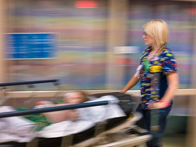
By becoming highly “pediatric ready,” emergency departments could prevent the deaths of 2,143 children each year with an annual cost between $0 and $12 per child resident, depending on the state.
In emergencies, children have distinct needs. Yet 83% of emergency departments (EDs) nationwide are not fully prepared to meet them. A new study has found that bridging that gap, known as becoming highly “pediatric ready,” could prevent the deaths of 2,143 children each year with an annual cost between $0 and $12 per child resident, depending on the state.
“Our country can afford it, and we owe it to our children to do it,” says the study’s senior author Nathan Kuppermann, M.D., chair of Pediatrics and chief academic officer at Children’s National Hospital.
The research team – led by Oregon Health & Science University and Children’s National – analyzed data from 4,840 EDs, focusing on 669,019 children at risk for death upon seeking care. Using predictive models, they assessed how every ED achieving high pediatric readiness – defined as scoring at least 88 out of 100 on the National Pediatric Readiness Project assessment – could impact mortality rates.
“The National Pediatric Readiness Project outlines essential pediatric capabilities for EDs, such as the availability of essential pediatric equipment and pediatric-specific training,” says Dr. Kuppermann, an emergency medicine physician. “While a perfect score of 100 is ideal, past research shows a score of 88 or higher can reduce mortality risk by up to 76% for ill children and 60% for injured children.”
Why it matters
In Maryland, an additional cost of $1.10 per child could save 17 pediatric lives annually, adjusted for population size. In Virginia, $2.42 per child could save 29 lives annually, and $1.59 per child in the District of Columbia could save 16 lives annually. The research team said strategies for implementing the findings would require regulation, incentives and policy-based initiatives.
“This study builds on a growing body of research demonstrating that every hospital can and must be ready for children’s emergencies,” says lead author Craig Newgard, M.D., M.P.H., an emergency physician at Oregon Health & Science University. “For the first time, we have comprehensive national and state-by-state data that emphasizes both the urgency and feasibility of this work.”
The patient benefit
“Our country can afford it, and we owe it to our children to do it,” says the study’s senior author Nathan Kuppermann, M.D., chair of Pediatrics and chief academic officer at Children’s National Hospital.
By applying the potential reduction in mortality associated with high readiness to the number of children at risk of death, the researchers identified the number of lives that could be saved each year. State-specific estimates, adjusted for population size, ranged from 0 preventable deaths in Delaware to 69 in South Dakota.
“Achieving high readiness levels can be challenging for small emergency departments with fewer resources, typically in more rural areas. The result is significant inequity and large healthcare deserts in pediatric emergency care across the United States,” Dr. Kuppermann says. “Yet we found the cost of elevating care to the highest quartile of pediatric readiness is not very high.”
What’s next
The study authors estimate achieving universal high pediatric readiness across the United States would cost approximately $207 million annually. Per-child costs by state to raise ED readiness from current levels ranged from $0 to $12 per year.
“This research emphasizes the urgent need for widespread investment in pediatric readiness,” says Kate Remick, M.D., co-author and emergency physician with the Dell School of Medicine at the University of Texas at Austin. “The National Pediatric Readiness Project has provided a roadmap for improvement. But we need the full engagement of clinicians, healthcare administrators, policymakers and families to make universal pediatric readiness a reality.”
The study outlines several strategies to improve pediatric emergency care, such as integrating high pediatric readiness into hospital accreditation requirements and incentivizing readiness through performance-based reimbursement models.
This study was funded by a Department of Health and Human Services (HHS) Health Resources and Services Administration (HRSA) Emergency Medical Services for Children Targeted Issue grant (H34MC33243-01-01) and an HHS National Institutes of Health (NIH) Eunice Kennedy Shriver National Institute of Child Health and Human Development (NICHD) grant (R24 HD085927). The contents are those of the author(s) and do not necessarily represent the official views of, nor an endorsement, by HHS, HRSA, NIH, or the U.S. Government.
https://innovationdistrict.childrensnational.org/wp-content/uploads/2024/10/ER-nurse-CNRI.jpg385685Innovation Districthttps://innovationdistrict.childrensnational.org/wp-content/uploads/2023/12/innovationdistrict_logo-1-1030x165.pngInnovation District2024-11-01 11:00:312024-10-31 11:04:40Investment in pediatric emergency care could save over 2,100 lives annually
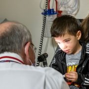
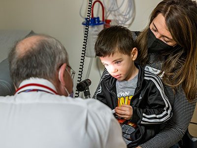
Pain assessment in medicine often relies on imprecise visual rating scales featuring smiling or crying faces, frustrating patients and physicians alike. Children’s National Hospital researchers aim to change this with a new device designed to precisely measure pain, backed by an $8-million award from the Advanced Research Projects Agency for Health (ARPA-H) Sprint for Women’s Health.
The sprint addresses critical unmet challenges in women’s health, champions transformative innovations and tackles health conditions that uniquely or disproportionately affect women. Children’s National researchers – with their collaborators from Johns Hopkins University and Medstar Research Institute – are furthering the development of the AlgometRx Nociometer, a pain measurement device to better understand and treat pain.
Julia Finkel, M.D., pediatric anesthesiologist and director of Pain Medicine Research at the Sheikh Zayed Institute for Pediatric Surgical Innovation (SZI) at Children’s National, said she hopes to improve the understanding of gender differences in pain and better treat patients of all ages with analgesics. Children’s National will receive the $8 million over two years through the Sprint for Women’s Health’s launchpad track for later-stage health solutions.
“As physicians, we often feel helpless to understand how our patients are experiencing pain and whether treatments are working,” Dr. Finkel said. “We use the Visual Analog Scale, which fails to classify the pain’s etiology or help guide a specific intervention, often leaving us to treat by trial and error. The Nociometer will change that.”
The patient benefit
Using a nonpainful stimulus, the portable Nociometer uses noninvasive technology and algorithms to analyze pupil dilation and the body’s response to stimulation along select nerves. In under one minute, the data collected can formulate a patient profile quantifying how the nervous system is processing painful stimuli. This new construct can be used to characterize pain type, intensity and assess the impact of pain relief medications.
Dr. Finkel spent nine years developing the Nociometer at SZI and her spin-out company, AlgometRx, which receives commercialization support from Children’s National Innovation Ventures. She said this transformative platform technology can be applied to any population – including elderly, nonverbal or pediatric patients – and will be instrumental in studying women’s pain experiences.
“Research studies have long shown that pain in women and girls is underestimated and undertreated,” Dr. Finkel said. “Creating a novel technology to quantify pain has tremendous applications in pinpointing and effectively treating pain, potentially altering treatment in nearly any medical setting.”
The big picture
ARPA-H sought solutions within six topics of interest in women’s health and received an unprecedented response of submissions. ARPA-H launched the Sprint for Women’s Health in February, with First Lady Jill Biden announcing the funding as the first major deliverable from the White House Initiative on Women’s Health Research.
The ARPA-H Sprint for Women’s Health is conducted in collaboration with the Investor Catalyst Hub of ARPANET-H, the agency’s nationwide health innovation network that connects people, innovators and institutions to accelerate better health outcomes for everyone. Children’s National is a spoke member of the Investor Catalyst Hub.
Children’s National will work with an ARPA-H program manager and the Investor Catalyst Hub over two years to develop their proposed solution, receiving milestone-based payments aligned to research activities and performance objectives.
The ARPA-H launchpad program accelerates transformative health solutions’ path to impact by providing funding and market transition support. As a launchpad performer, Dr. Finkel will also work with an entrepreneur-in-residence and participate in Launchpad Accelerator, which includes a customized curriculum, virtual events, and in-person workshops to support the transition from research to commercialization.
https://innovationdistrict.childrensnational.org/wp-content/uploads/2024/10/Finkel-pain-assessment-CNRI.jpg385685Innovation Districthttps://innovationdistrict.childrensnational.org/wp-content/uploads/2023/12/innovationdistrict_logo-1-1030x165.pngInnovation District2024-10-23 13:00:172024-10-30 10:20:33ARPA-H awards $8m to advance device for precision pain measurement
“It is an absolute honor and privilege to be elected to the National Academy of Medicine,” Dr. Goyal said. “I am committed to NAM’s mission of using science to inform policy for the achievement of health equity.”
The National Academy of Medicine (NAM) has elected Monika Goyal, M.D., M.S.C.E., to its international body of 2,400 leaders in medical sciences, healthcare and public health. One of the highest honors for physicians, the achievement is reserved for individuals who have demonstrated outstanding professional commitment and service.
Dr. Goyal, emergency medicine physician and co-director of the Center for Translational Research at Children’s National Hospital, was recognized for her national leadership in research focused on pediatric firearm injury prevention. She has spotlighted the burden of firearm violence on child health and advanced pediatric equity science. Her research has led to the development of interventions to address healthcare inequities.
“It is an absolute honor and privilege to be elected to the National Academy of Medicine,” Dr. Goyal said. “I am committed to NAM’s mission of using science to inform policy for the achievement of health equity.”
The big picture
The National Academy of Sciences established NAM in 1970 as the Institute of Medicine. The diverse organization requires that at least one-quarter of its membership be selected from fields outside the health professions, including law, engineering, social sciences and the humanities. NAM elected 90 regular and 10 international members during its recent annual meeting.
NAM works with the National Academy of Sciences and National Academy of Engineering. Upon election, NAM members commit to volunteering their services for National Academies activities.
Why we’re excited
“I am thrilled that Dr. Goyal is joining me in this incredible body that works to provide independent, objective analysis on complex scientific problems and public policy decisions,” said NAM member Nathan Kuppermann, M.D., M.P.H., chief academic officer and chair of Pediatrics at Children’s National. “Dr. Goyal’s contributions to pediatric health will be an incredible asset to the organization.”
https://innovationdistrict.childrensnational.org/wp-content/uploads/2024/03/Monika-Goyal-CNRI.png385685Innovation Districthttps://innovationdistrict.childrensnational.org/wp-content/uploads/2023/12/innovationdistrict_logo-1-1030x165.pngInnovation District2024-10-21 10:50:082024-10-21 10:51:05National Academy of Medicine elects gun violence, health equity expert