Tag Archive for: Duchenne muscular dystrophy

Motor neuron connecting to muscle fiber

FDA approves muscular dystrophy drug built on Children’s National research


Motor neuron connecting to muscle fiber

Duchenne muscular dystrophy (DMD) is the most common hereditary neuromuscular disease.


Boys with Duchenne muscular dystrophy (DMD) have a clinically proven, new treatment option with the Food and Drug Administration’s approval of vamorolone, a steroidal-type, anti-inflammatory drug developed based on research performed at Children’s National Hospital.

Created by ReveraGen BioPharma Inc., vamorolone has a molecular structure similar to traditional corticosteroids, which are currently used to treat DMD. Yet its structure was found to be chemically different enough to reduce unwanted side effects, including brittle bones and reduced stature. Nearly two decades ago, ReveraGen leaders – President and CEO Eric Hoffman, Ph.D., and Vice President for Research Kanneboyina Nagaraju, D.V.M., Ph.D. – launched research efforts into the drug when they led the Center for Genetic Medicine Research at Children’s National. They worked with then-Chief Academic Officer Mark Batshaw, M.D., on the new clinical option.

“Throughout my career, I have treated children with DMD, and I have seen over time how their shorter heights and brittle bones impact them physically and emotionally – in terms of their self-esteem and ability to participate in activities,” Dr. Batshaw said. “This drug should help these boys function more effectively and prevent certain long-term complications.”

The patient benefit

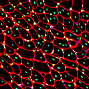
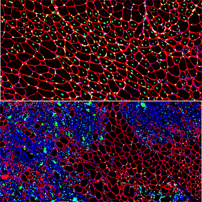
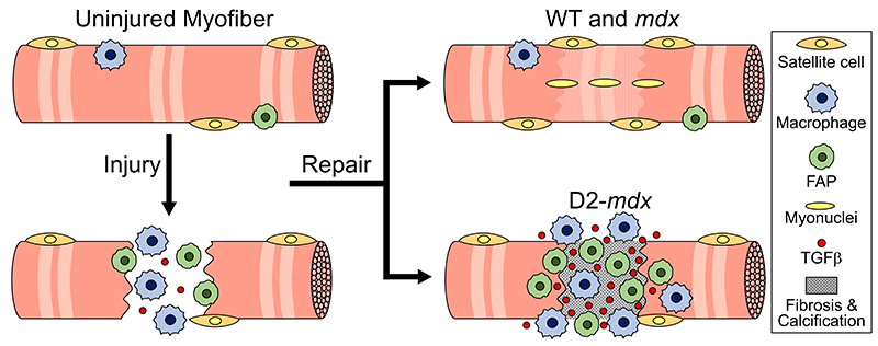
Muscular dystrophy includes a group of degenerative genetically inherited neuro-muscular diseases that strike only boys. DMD is the most common, severe and life-threatening form of muscular dystrophy. ReveraGen studied vamorolone for patients ages two years and up in the hopes of providing a new, FDA-approved treatment option for these children. In clinical trials, daily treatment with vamorolone improved muscle strength and stature with results comparable to prednisolone, but without some of the most impactful side effects of steroids, particularly the stunted growth and weakened bones.

Children’s National Hospital leads the way

Kolaleh Eskandanian, Ph.D., M.B.A., P.M.P., vice president and chief innovation officer for Children’s National, said Drs. Hoffman and Nagaraju’s work on the drug paved the way for entrepreneurship at the hospital, as they were the first faculty members to launch a spin-off company. Since then, more than 130 faculty members have been named as inventors on 132 patents. Children’s National is now home to Innovation Ventures, the hospital’s intellectual property development and commercialization arm, which provides guidance and resources to academic entrepreneurs who introduce a concept for pediatric medical products.

“We cannot wait to see the tremendous effort behind vamorolone in the hands of patients and clinicians treating Duchenne muscular dystrophy,” Eskandanian said. “Today’s FDA approval for ReveraGen shows the importance of supporting clinicians and researchers who are developing solutions to advance healthcare for children.”

Why we’re excited

Hoffman said the drug has been through a series of clinical trials showing advantages over the current treatment options. In 2024, Catalyst Pharma will market vamorolone under the trade name Agamree in the United States.

“Vamorolone was developed using a different business model and drug development approach, including partnerships with the National Institutes of Health, Department of Defense, the European Commission and more than a dozen international nonprofit foundations,” Dr. Hoffman said. “The collaborative, community-engaged approach—including 32 academic clinical sites in 11 countries — and the participation of hundreds of DMD families led to this approval today.”

Hiram receives the first commercial dose of Elevidys

Children’s National gives first commercial dose of new FDA-approved gene therapy for Duchenne muscular dystrophy

Hiram receives the first commercial dose of Elevidys

On the day before his 6th birthday, Hiram, 5, was the first patient ever with DMD to receive the drug after the U.S. Food and Drug Administration (FDA) approved its use last month.

Children’s National Hospital is the first pediatric hospital to administer a commercial dose of Elevidys (delandistrogene moxeparvovec-rokl), the first gene therapy for the treatment of pediatric patients with Duchenne muscular dystrophy (DMD).

On the day before his 6th birthday, Hiram, 5, was the first patient ever with DMD to receive the drug after the U.S. Food and Drug Administration (FDA) approved its use last month.

“The approval of Elevidys opens a new door for young patients with DMD and their families,” says Sarah Wright, D.O., neuromuscular neurologist at Children’s National. “This disease has had limited targeted treatments to date which can help alter the trajectory of disease.”

The background

On June 22, the FDA approved the use of Elevidys for patients 4 through 5 years of age with DMD with a confirmed mutation in the DMD gene who do not have a pre-existing medical reason preventing treatment with this therapy.

DMD is a rare and progressive genetic neuromuscular disease that predominantly affects males. It is caused by genetic changes in the DMD gene that affects the muscles, leading to muscle wasting that gets worse over time. Symptoms include progressive weakness and loss (atrophy) of both skeletal and heart muscle. Muscle weakness is usually noticeable in early childhood when signs like delayed ability to sit, stand or walk, and difficulties learning to speak manifest in a patient.

How it works

Elevidys is a one-time intravenous gene therapy that aims to delay or halt the progression of DMD by delivering a modified, functional version of dystrophin to muscle cells. The dystrophin gene is the largest known human gene.

“Elevidys is a viral vector (the ‘envelope to deliver the gene of interest’) mediated gene therapy that allows for the introduction of a gene that codes for a shortened form of dystrophin protein, or microdystrophin,” Dr. Wright explains. While not a cure for DMD, trials of Elevidys have demonstrated increases in dystrophin expression and improved functional results in young children with the disease.

“We have years of dedicated work on the part of researchers, clinician leaders and advocacy organizations in the field of muscular dystrophy to thank for this ground-breaking moment,” says Dr. Wright. “The approval of Elevidys offers families of patients ages 4-5 with DMD the option to receive this gene therapy that is designed to target the underlying cause of the disease.”

“The time-sensitivity of this medication illustrates the importance of going to a top academic pediatric hospital early on in neurologic care,” adds Elizabeth Wells, M.D., senior vice president of the Center for Neuroscience and Behavioral Medicine at Children’s National.

What’s next

The neuromuscular team at Children’s National is looking forward to offering this therapy to young patients with DMD and to the completion of additional trials/results for therapies in the DMD drug development pipeline.

“The research and approval of novel therapies provides more options for our DMD patients and their families, which is a critical step toward improving the lives of patients with DMD,” Dr. Wright says.

muscle cells

Experimental model mimics early-stage myogenic deficit in boys with DMD

muscle cells

Muscle regeneration marked by incorporation of muscle stem cell nuclei (green) in the myofibers (red) in dystrophic muscles with low TGFβ level (upper image), but not with high TGFβ level (lower image). Inflammatory and other nuclei are labeled blue.

Boys with Duchenne muscular dystrophy (DMD) experience poor muscle regeneration, but the precise reasons for this remain under investigation. An experimental model of severe DMD that experiences a large spike in transforming growth factor-beta (TGFβ) activity after muscle injury shows that high TGFβ activity suppresses muscle regeneration and promotes fibroadipogenic progenitors (FAPs). This leads to replacement of the damaged muscle fibers by calcified and connective tissue, compromising muscle structure and function. While blocking FAP buildup provides a partial solution, a Children’s National Hospital study team identifies correcting the muscle micro-environment caused by high TGFβ as a ripe therapeutic target.

The team’s study was published online March 26, 2020, in JCI Insight.

DMD is a chronic muscle disease that affects 1 in 6,200 young men in the prime of their lives. The disorder, caused by genetic mutations leading to the inability to produce dystrophin protein, leads to ongoing muscle damage, chronic inflammation and poor regeneration of lost muscle tissue. The patients experience progressive muscle wasting, lose the ability to walk by the time they’re teenagers and die prematurely due to cardiorespiratory failure.

The Children’s National team finds for the first time that as early as preadolescence (3 to 4 weeks of age), their experimental model of severe DMD disease showed clear signs of the type of spontaneous muscle damage, regenerative failure and muscle fiber loss seen in preadolescent boys who have DMD.

“In boys, the challenge due to muscle loss exists from early in their lives, but had not been mimicked previously in experimental models,” says Jyoti K. Jaiswal, MSc, Ph.D., principal investigator in the Center for Genetic Medicine Research at Children’s National, and the study’s co-senior author. “TGFβ is widely associated with muscle fibrosis in DMD, when, in fact, our work shows its role in this disease process is far more significant.”

Research teams have searched for experimental models that replicate the sudden onset of symptoms in boys who have DMD as well as its complex progression.

“Our work not only offers insight into the delicate balance needed for regeneration of skeletal muscle, but it also provides quantitative information about muscle stem cell activity when this balanced is disturbed,” says Terence A. Partridge, Ph.D., principal investigator in the Center for Genetic Medicine Research at Children’s National, and the study’s co-senior author.

This schematic depicts the fate of injured myofibers in healthy or dystrophic muscle

This schematic depicts the fate of injured myofibers in healthy or dystrophic muscle (WT or mdx experimental models) that maintain low TGFβ level, compared with D2-mdx experimental models that experience a large increase in TGFβ level. As the legend shows, various cells are involved in this regenerative response.

“The D2-mdx experimental model is a relevant one to use to investigate the interplay between inflammation and muscle degeneration that is seen in humans with DMD,” adds Davi A.G. Mázala, co-lead study author.  “This model faithfully recapitulates many features of the complex disease process seen in humans.”

Between 3 to 4 weeks of age in the experimental models of severe DMD disease, the level of active TGFβ spiked up to 10-fold compared with models with milder disease. Intramuscular injections of an off-the-shelf drug that inhibits TGFβ signaling tamped down the number of FAPs, improving the muscle environment by lowering TGFβ activity.

“This work lays the foundation for studies that could lead to future therapeutic strategies to improve patients’ outcomes and lessen disease severity,” says James S. Novak, Ph.D., principal investigator in Children’s Center for Genetic Medicine Research, and co-lead study author. “Ultimately, our goal is to improve the ability of patients to continue to maintain muscle mass and regenerate muscle.”

In addition to Mázala, Novak, Jaiswal and Partridge, Children’s National study co-authors include Marshall W. Hogarth; Marie Nearing; Prabhat Adusumalli; Christopher B. Tully; Nayab F. Habib; Heather Gordish-Dressman, M.D.; and Yi-Wen Chen, Ph.D.

Financial support for the research described in this post was provided by the National Institutes of Health under award Nos. T32AR056993, R01AR055686 and U54HD090257; Foundation to Eradicate Duchenne; Muscular Dystrophy Association under award Nos. MDA295203, MDA480160 and MDA 477331; Parent Project Muscular Dystrophy; and Duchenne Parent Project – Netherlands.

dystrophin protein

Experimental drug shows promise for slowing cardiac disease and inflammation

dystrophin protein

Duchenne muscular dystrophy (DMD) is caused by mutations in the DMD gene, which provides instructions for making dystrophin, a protein found mostly in skeletal, respiratory and heart muscles.

Vamorolone, an experimental medicine under development, appears to combine the beneficial effects of prednisone and eplerenone – standard treatments for Duchenne muscular dystrophy (DMD) – in the heart and muscles, while also showing improved safety in experimental models. The drug does so by simultaneously targeting two nuclear receptors important in regulating inflammation and cardiomyopathy, indicates a small study published online Feb. 11, 2019, in Life Science Alliance.

DMD is a progressive X-linked disease that occurs mostly in males. It is characterized by muscle weakness that worsens over time, and most kids with DMD will use wheelchairs by the time they’re teenagers. DMD is caused by mutations in the DMD gene, which provides instructions for making dystrophin, a protein found mostly in skeletal, respiratory and heart muscles.

Cardiomyopathy, an umbrella term for diseases that weaken the heart, is a leading cause of death for young adults with DMD, causing up to 50 percent of deaths in patients who lack dystrophin. A collaborative research team co-led by Christopher R. Heier, Ph.D., and Christopher F. Spurney, M.D., of Children’s National Health System, is investigating cardiomyopathy in DMD. They find genetic dystrophin loss provides “a second hit” for a specific pathway that worsens cardiomyopathy in experimental models of DMD.

“Some drugs can interact with both the mineralocorticoid receptor (MR) and glucocorticoid receptor (GR) since these two drug targets evolved from a common ancestor. However, we find these two drug targets can play distinctly different roles in heart and skeletal muscle. The GR regulates muscle inflammation, while the MR plays a key role in heart health,” says Heier, an assistant professor at Children’s National and lead study author. “In our study, the experimental drug vamorolone safely targets both the GR to treat chronic inflammation and the MR to treat the heart.”

After gauging the efficacy of various treatments in test tubes, the study team looked at whether any could mitigate negative impacts of the MR on heart health. Wild type and mdx experimental models were implanted with pumps that activated the MR. These models also received a daily oral MR antagonist (or inhibitor) drug, and either eplerenone, spironolactone or vamorolone. Of note:

  • MR activation increased kidney size and caused elevated blood pressure (hypertension).
  • Treatment with vamorolone maintained normal kidney size and prevented hypertension.
  • MR activation increased mdx heart mass and fibrosis. Vamorolone mitigated these changes.
  • MR activation decreased mdx heart function, while vamorolone prevented declines in function.
  • Daily prednisone caused negative MR- and GR-mediated side effects, such as hyperinsulinemia, whereas vamorolone safely improved heart function without these side effects.

“These findings have the potential to help current and future patients,” Heier says. “Clinicians already prescribe several of these drugs. Our new data support the use of MR antagonists such as eplerenone in protecting DMD hearts, particularly if patients take prednisone. The experimental drug vamorolone is currently in Phase IIb clinical trials and is particularly exciting for its unique potential to simultaneously treat chronic inflammation and heart pathology with improved safety.”

In addition to Heier and senior author Spurney, study co-authors include Qing Yu, Alyson A. Fiorillo, Christopher B. Tully, Asya Tucker and Davi A. Mazala, all of Children’s National; Kitipong Uaesoontrachoon and Sadish Srinivassane, AGADA Biosciences Inc.; and Jesse M. Damsker, Eric P. Hoffman and Kanneboyina Nagaraju, ReveraGen BioPharma.

Financial support for research described in this report was provided by Action Duchenne; the Clark Charitable Foundation; the Department of Defense under award W81XWH-17-1-047; the Foundation to Eradicate Duchenne; the Intellectual and Developmental Disabilities Research Center under award U54HD090257 (through the National Institutes of Health’s (NIH) Eunice Kennedy Shriver National Institute of Child Health and Human Development); and the NIH under awards K99HL130035, R00HL130035, L40AR068727 and T32AR056993.

Financial disclosure:  Co-authors employed by ReveraGen BioPharma were involved in creating this news release.

boy sitting in wheelchair

Long-term glucocorticoids help patients with DMD

boy sitting in wheelchair

Glucocorticoids, a class of steroid hormone medications, have definite long-term benefits for patients with Duchenne muscular dystrophy, including extending muscle strength and function over years and decreasing the risk of death.

There is currently no cure for the devastating, progressive neuromuscular disease known as Duchenne muscular dystrophy (DMD). But clinics that treat patients with this disease have long relied on a class of steroid hormone medications, known as glucocorticoids, to ease its symptoms. Over weeks and months, these drugs help preserve muscle strength and function. Though these short-term benefits have been clear, some physicians have balked at using these medications over the long term – their benefits over years was unknown, making their potential side effects not worth the risk.

Now, a study published online Nov. 22, 2017 in The Lancet suggests that these medicines have definite long-term benefits, including extending muscle strength and function over years and even decreasing the risk of death. These findings support what has become the standard prescribing practice at many clinics and could help sway parents who are on the fence about their children receiving these therapies.

DMD is characterized by loss of muscle function and progressive muscle weakness that begins in the lower limbs and typically affects males due to the location of its causative genetic mutation. Patients with this devastating neuromuscular disease often receive glucocorticoids at some point as the disease progresses. Studies since the late 1980s have confirmed short-term benefits of treating with these drugs, including delaying the loss of muscle strength and function.

However, no prospective study had followed long-term glucocorticoid use in these patients, explains Heather Gordish-Dressman, Ph.D., a statistician at the Center for Genetic Medicine Research at Children’s National Health System and study senior author. The lack of long-term data led some physicians to delay treatment with these drugs since their use can lead to significant side effects, including weight gain, delayed growth and immunosuppression.

“Everyone had the idea that long-term use could be beneficial, but nobody had really rigorously tested that,” Gordish-Dressman says.

Craig McDonald, M.D., a University of California, Davis, professor and lead author of the study adds: “This long-term, follow-up study provides the most definitive evidence that the benefits of glucocorticoid steroid therapy in DMD extend over the entire lifespan. Most importantly, patients with Duchenne using glucocorticoids experienced an overall reduction in risk of death by more than 50 percent.”

To determine whether the short-term benefits of these drugs extend in the long term, Gordish-Dressman and researchers scattered across the country tapped data from the Cooperative International Neuromuscular Research Group’s Duchenne Natural History Study, the largest study to follow patients with DMD over time. They gathered data for 440 males with DMD aged 2 to 8 years old. About 22 percent had never taken glucocorticoids or had taken these medications for less than one year. The remainder had taken them for at least one year or longer.

By analyzing data for up to 10 years for these patients, the long-term benefits became clear, Gordish-Dressman adds. Glucocorticoid treatment for patients who received it for more than one year delayed loss of mobility milestones that affected the lower limbs by 2.1 to 4.4 years, such as going from supine to standing, climbing four stairs, and walking or running 10 meters, compared with boys who received the medications for less than one year. Long-term glucocorticoid therapy also delayed the loss of mobility milestones in upper limbs, such as hand function, performing a full overhead reach and raising the hands to the mouth.

Long-term use of these drugs also was associated with a decreased risk of death over the length of the study. Furthermore, deflazacort – a glucocorticoid recently approved by the Food and Drug Administration specifically for DMD – delayed loss of the ability to move from supine position to standing, walking and hand-to-mouth function significantly better than prednisone, the most popular glucocorticoid prescribed for DMD in the United States.

Gordish-Dressman says that glucocorticoids are currently a standard part of care for most patients with DMD, with some clinics prescribing these medications as soon as patients are diagnosed. However, because long-term data supporting their use was lacking, some physicians hesitate to prescribe glucocorticoids until the disease had progressed, when patients already had lost significant function.

Future studies will examine which medicines in this class of drugs and which regimens might offer the most benefits as well as how benefits differ with longer-term medication use.

Research reported in this news release was supported by the U.S. Department of Education/NIDRR, H133B031118 and H133B090001; the U.S. Department of Defense, W81XWH-12-1-0417; National Institute of Arthritis and Musculoskeletal and Skin Diseases of the National Institutes of Health under award number R01AR061875; and Parent Project Muscular Dystrophy.

macrophage

Improving treatment success for Duchenne muscular dystrophy

macrophage

Macrophages, white blood cells involved in inflammation, readily take up a new medicine for Duchenne muscular dystrophy and promote its sustained delivery to regenerating muscle fibers long after the drug has disappeared from circulation.

Chronic inflammation plays a crucial role in the sustained delivery of a new type of muscular dystrophy drug, according to an experimental model study led by Children’s National Health System.

The study, published online Oct. 16, 2017 in Nature Communications, details the cellular mechanisms of morpholino antisense drug delivery to muscles. Macrophages, white blood cells involved in inflammation, readily take up a new medicine for Duchenne muscular dystrophy (DMD) and promote its sustained delivery to regenerating muscle fibers long after the drug has disappeared from circulation.

Until recently, the only approved medicines for DMD targeted its symptoms, rather than the root genetic cause. However, in 2016 the Food and Drug Administration approved the first exon-skipping medicine to restore dystrophin protein expression in muscle: Eteplirsen, an antisense phosphorodiamidate morpholino oligomer (PMO). The drug had shown promise in preclinical studies but had variable and sporadic results in clinical trials.

The Children’s National study adds to the understanding of how this type of medicine targets muscle tissue and suggests a path to improve treatments for DMD, which is the most common and severe form of muscular dystrophy and currently has no cure, explains study co-leader James S. Novak, Ph.D., a principal investigator in Children’s Center for Genetic Medicine Research.

Because the medication vanishes from the blood circulation within hours after administration, Children’s research efforts have focused on the mechanism of delivery to muscle and on ways to increase its cellular uptake – and, by extension, its effectiveness. However, researchers understand little about how this medication actually gets delivered to muscle fibers or how the disease pathology impacts this process, knowledge that could offer new ways of boosting both its delivery and effectiveness, says Terence Partridge, Ph.D., study co-leader and principal investigator in Children’s Center for Genetic Medicine Research.

To investigate this question, Novak, Partridge and colleagues used an experimental model of DMD that carries a version of the faulty DMD gene that, like its human counterparts, destroys dystrophin expression. To track the route of the PMO into muscle fibers, they labeled it with a fluorescent tag. The medicine traveled to the muscle but only localized to patches of regenerating muscle where it accumulated within the infiltrating macrophages, immune cells involved in the inflammatory response that accompanies this process. While PMO is rapidly cleared from the blood, the medication remained in these immune cells for up to one week and later entered muscle stem cells, allowing direct transport into regenerating muscle fibers. By co-administering the PMO with a traceable DNA nucleotide analog, the research team was able to define the stage during the regeneration process that promotes heightened uptake by muscle stem cells and efficient dystrophin expression in muscle fibers.

“These macrophages appear to extend the period of availability of this medication to the satellite cells and muscle fibers at these sites,” Partridge explains. “Since the macrophages are acting as long-term storage reservoirs for prolonged delivery to muscle fibers, they could possibly represent new therapeutic targets for improving the uptake and delivery of this medicine to muscle.”

Future research for this group will focus on testing whether macrophages might be used as efficient delivery vectors to transport eteplirsen to the muscle, which would avert the rapid clearance currently associated with intravenous delivery.

“Understanding exactly how different classes of exon-skipping drugs are delivered to muscle could open entirely new possibilities for improving future therapeutics and enhancing the clinical benefit for patients,” Novak adds.

mitochondria

Mitochondria key for repairing cell damage in DMD

mitochondria

A research team led by Jyoti K. Jaiswal, M.S.C., Ph.D., found that dysfunctional mitochondria prevent repair of muscle cells in Duchenne muscular dystrophy.

PDF Version

What’s known

Duchenne muscular dystrophy (DMD), one of the most severe forms of muscular dystrophy, is caused by a defect in the dystrophin gene. The protein that this gene encodes is responsible for anchoring muscle cells’ inner frameworks, or cytoskeletons, to proteins and other molecules outside these cells, the extracellular matrix. Without functional dystrophin protein, the cell membranes of muscle cells become damaged, and the cells eventually die. This cell death leads to the progressive muscle loss that characterizes this disease. Why these cells are unable to repair this progressive damage has been unknown.

What’s new

A research team led by Jyoti K. Jaiswal, M.S.C., Ph.D., a principal investigator in the Center for Genetic Medicine Research at Children’s National Health System, investigated this question in two experimental models of DMD that carry different mutations of the dystrophin gene. The researchers monitored the effects of the lack of functional dystrophin protein in these preclinical models on the level and function of muscle cell. They found that mitochondria – organelles that act as powerhouses to supply the chemical energy to drive cellular activities – are among the first to be affected. They found that the decline in mitochondrial level and activity over time in these experimental models preceded the onset of symptoms. The research team also looked at the ability of the experimental models’ muscle cells to repair damage. As the muscle cell mitochondria lost function, the cells’ ability to repair damage also declined. Efforts to increase mitochondrial activity after these organelles became dysfunctional did not improve muscle repair. This suggests that poor muscle repair may not be caused by a deficit in energy production by mitochondria.

Questions for future research

Q: Does similar mitochondrial dysfunction occur in human patients with DMD?
Q: How can the mitochondrial dysfunction be prevented?
Q: Is there a way to reverse mitochondrial dysfunction to better preserve the ability of muscle cells to repair from DMD-related damage?

Source: “Mitochondria mediate cell membrane repair and contribute to Duchenne muscular dystrophy.” Vila, M.C., S. Rayavarapu, M.W. Hogarth, J.H. Van der Meulen, A. Horn, A. Defour, S. Takeda, K.J. Brown, Y. Hathout, K. Nagaraju and J.K. Jaiswal. Published by Cell Death and Differentiation February 2017.

Muscular Dystrophy Association awards grants to two Children’s National scientists

Marshall Hogarth, Ph.D

Marshall Hogarth, Ph.D

James Novak, Ph.D.

James Novak, Ph.D.

Two Children’s National Health System research scientists, Marshall Hogarth, Ph.D. and James Novak, Ph.D., have received Post-Doctoral Development Grants from the Muscular Dystrophy Association (MDA) as part of funding awarded to young, rising researchers who are poised to become independent investigators.

Over the next three years, Hogarth and Novak will be allotted $180,000 each to underwrite their individual research projects.

Hogarth’s research is focused on limb-girdle muscular dystrophy (LGMD), a disease which presents as muscle weakness when patients are in their late teens before rapidly progressing to severe debilitation. The MDA grant will allow Hogarth to continue his research investigating the replacement of muscle with fatty tissue and the role this plays in the late onset and subsequent progression of LGMD in patients.

Novak focuses mainly on researching Duchenne Muscular Dystrophy (DMD), a severely debilitating form of MD, that leads to progressive muscle weakness and respiratory and cardiac failure. Currently, the only Food and  Drug Administration (FDA)  approved treatment for DMD is exon-skipping. The MDA grant will support Novak’s study of the mechanisms that regulate the delivery of exon-skipping drugs in muscle, in order to identify new therapeutic targets and improve drug efficacy for patients with DMD.

While Hogarth and Novak focus on different aspects of neuromuscular disease, both look forward to making significant contributions that lead to overall improvements in the treatment of patients impacted by muscular dystrophy.

Children’s National spin-off ReveraGen announces agreement with actelion for Duchenne Muscular Dystrophy treatment

Earlier this month, ReveraGen BioPharma announced an exclusive option agreement with Actelion Ltd for lead compound vamorolone, a non-hormonal steroid modulator that is primarily used for the treatment of Duchenne Muscular Dystrophy (DMD). ReveraGen, the first Children’s National private spin-off company, is engaged in the discovery and development of proprietary therapeutic products for neuromuscular and inflammatory diseases.

Under the terms of the license agreement, Actelion and ReveraGen will partner to research and co-develop the novel compound vamorolone, which holds the potential to preserve muscle function and prolong ambulation in DMD patients, without some of the side effects that are commonly associated with glucocorticoid therapy. Those commonly associated include growth stunting and immune suppression, which can pose significant challenges for very young patients.

ReveraGen completed Phase I clinical trials for vamorolone in late 2015, and a Phase IIa program is currently underway to investigate the safety and efficiency of vamorolone in male DMD patients between four and seven years of age who have not taken deflazacort or prednisone. A Phase IIb program is also in early planning stages.

ReveraGen Co-Founder and CEO Eric Hoffman, PhD, has worked on DMD since the late 1980’s and has led his own research group for nearly 20 years at Children’s National. He co-founded ReveraGen back in 2008 with John McCall, PhD and Kanneboyina Nagaraju, PhD, DVM, before being named CEO in 2014. Children’s National maintains a 38 percent stake in ReveraGen.

Biomarkers sensitive to daily corticosteroid use

Using a mass spectrometer, Yetrib Hathout, Ph.D., is able to quantify 3,000 to 4,000 proteins from a tissue sample to identify proteins associated with cancer.

Using a Somascan proteomics assay – which simultaneously analyzes 1,129 proteins in a small volume of serum – a team led by Children’s National Health System researchers identified 21 biomarkers that respond to corticosteroids taken daily by children with Duchenne muscular dystrophy (DMD) and inflammatory bowel disease.

Corticosteroids are commonly prescribed to treat inflammatory conditions. High daily doses of corticosteroids are considered the standard of care for DMD, a type of muscular dystrophy characterized by worsening muscle weakness that affects 1 in 3,600 male infants. However, depending on the age of the child and drug dosage, chronic use is associated with such side effects as changes in bone remodeling that can lead to stunted growth, weight gain, facial puffiness caused by fat buildup, mood changes, sleep disturbances, and immune suppression. The research team sought to identify blood biomarkers that could be leveraged to create a fast, reliable way to gauge the safety and efficacy of corticosteroid use by children. The biomarkers also could guide development of a replacement therapy with fewer side effects.

“Ten pro-inflammatory proteins were elevated in untreated patients and suppressed by corticosteroids (MMP12, IL22RA2, CCL22, IGFBP2, FCER2, LY9, ITGa1/b1, LTa1/b2, ANGPT2 and FGG),” Yetrib Hathout, Ph.D., Proteomic Core Director at Children’s National, and colleagues write in the journal Scientific Reports. “These are candidate biomarkers for anti-inflammatory efficacy of corticosteroids.”

The blood biomarkers sensitive to corticosteroids fit into three broad groups, according to the authors. The children taking corticosteroids were matched with children of the same age who had never taken the medicine. Five biomarkers significantly increased in this corticosteroid-naïve group and decreased in kids prescribed corticosteroids. The biomarkers generally were inflammatory proteins and included chemokine, insulin-like growth factor binding protein 2, and integrin alpha-I/beta-1 complex.

The second group of biomarkers included nine proteins associated with macrophage and T-lymphocytes that were significantly reduced in concentration in kids taking corticosteroids. According to the study, this finding hints at corticosteroids blunting the ability of the immune system’s most able fighters to respond to infection.

In the third group were five proteins that were significantly increased by corticosteroid treatment in DMD and included matrix metalloproteinase 3, carnosine dipeptidase 1, angiotensinogen, growth hormone binding protein, insulin, and leptin, a hormone linked to appetite.

What researchers learned with this study will help them more accurately design the next phase of the work, Hathout says.

“We are the first team to report a number of novel discoveries, including that growth hormone binding protein (GHBP) levels increase with corticosteroid use. This represents a candidate biomarker for stunted growth. In order to use that new information effectively in drug development, the next studies must corroborate the role of serum GHBP levels as predictors of diminished stature,” he adds. “The study finding that four adrenal steroid hormones are depressed in kids taking corticosteroids raises additional questions about the broader impact of adrenal insufficiency, including its role in the delay of the onset of puberty.”

This work was supported by National Institutes of Health grants (R01AR062380, R01AR061875, P50AR060836, U54HD071601, K99HL130035, and R44NS095423) and Department of Defense CDMRP program grant W81XWH-15-1-0265. Additional support was provided by AFM-Telethon (18259) and the Muscular Dystrophy Association USA (MDA353094).