Tag Archive for: CHD

Zhe Han, PhD

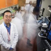
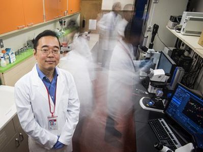
Lab led by Zhe Han, Ph.D., receives $1.75 million from NIH

Zhe Han, PhD

A new four-year NIH grant will enable Zhe Han, Ph.D., to carry out the latest stage in the detective work to determine how histone-modifying genes regulate heart development and the molecular mechanisms of congenital heart disease caused by these genetic mutations.

The National Institutes of Health (NIH) has awarded $1.75 million to a research lab led by Zhe Han, Ph.D., principal investigator and associate professor in the Center for Genetic Medicine Research, in order to build models of congenital heart disease (CHD) that are tailored to the unique genetic sequences of individual patients.

Han was the first researcher to create a Drosophila melanogaster model to efficiently study genes involved in CHD, the No.1 birth defect experienced by newborns, based on sequencing data from patients with the heart condition. While surgery can fix more than 90 percent of such heart defects, an ongoing challenge is how to contend with the remaining cases since mutations of a vast array of genes could trigger any individual CHD case.

In a landmark paper published in 2013 in the journal Nature, five different institutions sequenced the genomes of more than 300 patients with CHD and their families, identifying 200 mutated genes of interest.

“Even though mutations of these genes were identified from patients with CHD, these genes cannot be called ‘CHD genes’ since we had no in vivo evidence to demonstrate these genes are involved in heart development,” Han says. “A key question to be answered: How do we efficiently test a large number of candidate disease genes in an experimental model system?”

In early 2017, Han published a paper in Elife providing the answer to that lingering question. By silencing genes in a fly model of human CHD, the research team confirmed which genes play important roles in development. The largest group of genes that were validated in Han’s study were histone-modifying genes. (DNA winds around the histone protein, like thread wrapped around a spool, to become packed into a higher-level structure.)

The new four-year NIH grant will enable Han to carry out the next stage of the detective work to determine precisely how histone-modifying genes regulate heart development. In order to do so, his group will silence the function of histone-modifying genes one by one, to study their function in the fly heart development and to identify the key histone-modifying genes for heart development. And because patients with CHD can have more than one mutated gene, he will silence multiple genes simultaneously to determine how those genes work in partnership to cause heart development to go awry.

By the end of the four-year research project, Han hopes to be able to identify all of the histone-modified genes that play pivotal roles in development of the heart in order to use those genes to tailor make personalized fly models corresponding to individual patient’s genetic makeup.

Parents with mutations linked to CHD are likely to pass heart disease risk to the next generation. One day, those parents could have an opportunity to sequence their genes to learn the degree of CHD risk their offspring face.

“Funding this type of basic research enables us to understand which genes are important for heart development and how. With this knowledge, in the near future we could predict the chances of a baby being born with CHD, and cure it by using gene-editing approaches to prevent passing disease to the next generation,” Han says.

Spectral data shine light on placenta

preemie baby

A research project led by Subechhya Pradhan, Ph.D., aims to shed light on metabolism of the placenta, a poorly understood organ, and characterize early biomarkers of fetal congenital heart disease.

The placenta serves as an essential intermediary between a pregnant mother and her developing fetus, transporting in life-sustaining oxygen and nutrients, ferrying out waste and serving as interim lungs, kidneys and liver as those vital organs develop in utero.

While the placenta plays a vital role in supporting normal pregnancies, it remains largely a black box to science. A research project led by Subechhya Pradhan, Ph.D., and partially funded by a Clinical and Translational Science Institute Research Award aims to shed light on placenta metabolism and characterize possible early biomarkers of impaired placental function in fetal congenital heart disease (CHD), the most common type of birth defect.

“There is a huge information void,” says Pradhan, a research faculty member of the Developing Brain Research Laboratory at Children’s National Health System. “Right now, we do not have very much information about placenta metabolism in vivo. This would be one of the first steps to understand what is actually going on in the placenta at a biochemical level as pregnancies progress.”

The project Pradhan leads will look at the placentas of 30 women in the second and third trimesters of healthy, uncomplicated pregnancies and will compare them with placentas of 30 pregnant women whose fetuses have been diagnosed with CHD. As volunteers for a different study, the women are already undergoing magnetic resonance imaging, which takes detailed images of the placenta’s structure and architecture. The magnetic resonance spectroscopy scans that Pradhan will review show the unique chemical fingerprints of key metabolites: Choline, lipids and lactate.

Choline, a nutrient the body needs to preserve cellular structural integrity, is a marker of cell membrane turnover. Fetuses with CHD have higher concentrations of lactate in the brain, a telltale sign of a shortage of oxygen. Pradhan’s working hypothesis is that there may be differing lipid profiles and lactate levels in the placenta in pregnancies complicated by CHD.  The research team will extract those metabolite concentrations from the spectral scans to describe how they evolve in both groups of pregnant women.

“While babies born with CHD can undergo surgery as early as the first few days (or sometimes hours) of life to correct their hearts, unfortunately, we still see a high prevalence of neurodevelopmental impairments in infants with CHD. This suggests that neurological dysfunctional may have its origin in fetal life,” Pradhan says.

Having an earlier idea of which fetuses with CHD are most vulnerable has the potential to pinpoint which pregnancies need more oversight and earlier intervention.

Placenta spectral data traditionally have been difficult to acquire because the pregnant mother moves as does the fetus, she adds. During the three-minute scans, the research team will try to limit excess movement using a technique called respiratory gating, which tells the machine to synchronize image acquisition so it occurs in rhythm with the women’s breathing.

Nobuyuki Ishibashi

Congenital heart disease and the brain

Nobuyuki Ishibashi

In a recent review article published in Circulation Research, Nobuyuki Ishibashi, M.D., and his colleagues at Children’s National Health System summarized what is currently known about how congenital heart disease affects brain maturation.

What’s known

Among all known birth defects, congenital heart disease (CHD) is the leading cause of death in infants. Fortunately, advances in surgical techniques and treatments are improving the outlook for these children, and more and more are reaching adulthood. However, because of this increased longevity, it has become increasingly clear that children born with CHD are at risk of developing life-long neurological deficits. Several risk factors for these neurodevelopmental abnormalities have been identified, but direct links between specific factors and neurological defects have yet to be established.

What’s new

In a recent review article published in Circulation Research, a team from Children’s National Health System summarized what is currently known about how CHD affects brain maturation. Drawing from studies conducted at Children’s National as well as other research institutions, Paul D. Morton, Ph.D., Nobuyuki Ishibashi, M.D., and Richard A. Jonas, M.D., write that clinical findings in patients, improvements in imaging analysis, advances in neuromonitoring techniques and the development of animal models have greatly contributed to our understanding of the neurodevelopmental changes that occur with CHD.

Findings from Children’s National include:

  • An assessment of the intraoperative effects of cardiopulmonary bypass surgery on white matter using neonatal piglets.
  • An arterial spin labeling MRI study that showed newborns with complex CHD have a significant reduction in global cerebral blood flow.
  • A rodent study that modeled diffuse white matter brain injury in premature birth and identified the cellular and molecular mechanisms underlying lineage-specific vulnerabilities of oligodendrocytes and their regenerative response after chronic neonatal hypoxia.

The authors conclude that although there is ample clinical evidence of neurological damage associated with CHD, there is limited knowledge of the cellular events associated with these abnormalities. They offer perspectives about what can be done to improve our understanding of neurological deficits in CHD, and emphasize that ultimately, a multidisciplinary approach combining multiple fields and myriad technology will be essential to improve or prevent adverse neurodevelopmental outcomes in individuals with CHD.

Questions for future research

Q: What are the cellular events associated with each factor involved in neurodevelopmental delays?
Q: How does the neurodevelopmental status of a patient with CHD change as they age?
Q: How do the genes involved in structural congenital cardiac anomalies affect brain development and function?

Source: Norton, P.D., Ishibashi, N., Jonas, R.A. Neurodevelopmental Abnormalities and Congenital Heart Disease: Insights Into Altered Brain Maturation,” Circulation Research (2017) 120:960-977.