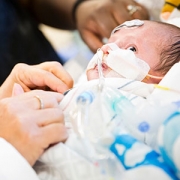
New research maps how opioid exposure may shape the newborn brain
New research from the Developing Brain Institute at Children’s National is helping scientists better understand how prenatal opioid exposure may shape early brain development.
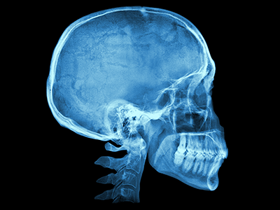
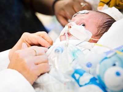
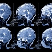
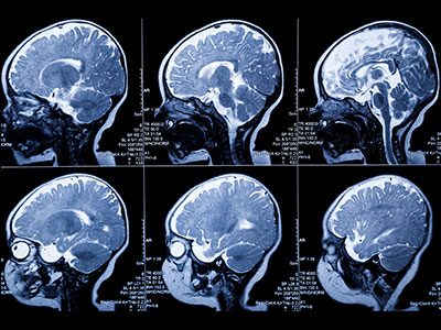
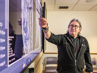
The opioid crisis continues to affect families across the United States. One question researchers are working to answer is how opioid exposure during pregnancy may influence the developing brain. A new study led by scientists at the Developing Brain Institute at Children’s National Hospital offers one of the most comprehensive looks yet. Using advanced brain imaging, the team examined how brain networks are organized in newborns who were exposed to opioids before birth.
The study analyzed brain scans from 248 newborns across four medical centers in the United States. Some infants had prenatal opioid exposure, while others did not. By comparing the two groups, researchers were able to identify subtle differences in how regions of the newborn brain communicate.
What the study found
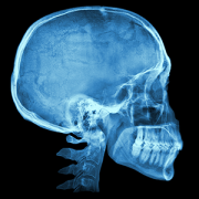
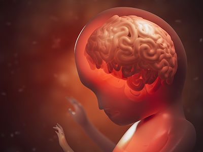
To study early brain development, the researchers used resting-state functional MRI, a technique that measures how different parts of the brain communicate while a person is at rest. Even in newborns, the brain already contains recognizable communication networks that support basic functions such as movement, sensation and early cognitive processes. These networks continue to mature rapidly during infancy and early childhood.
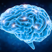
One of the most striking findings from the study was that the overall structure of these major brain networks remained intact in opioid-exposed newborns. But when researchers looked more closely at how specific regions were connected, they found important differences. The study also found that opioid-exposed newborns showed a pattern of both weaker and stronger connections across different brain circuits. Some areas involved in sensorimotor function and frontal brain regulation showed reduced connectivity. These circuits help coordinate movement, attention and self-regulation. At the same time, researchers observed stronger connectivity in several other regions, including areas involved in emotional processing, reward and sensory perception such as the amygdala, putamen and parts of the visual system. Rather than a loss of brain networks, the results point to shifts in how strongly certain regions communicate with one another. Scientists believe these changes may reflect how opioids interact with the developing brain during critical periods of growth.
Why this matters
Many previous imaging studies of opioid exposure in newborns have relied on relatively small groups of infants. Recruiting newborns for advanced brain imaging is challenging, and it is especially difficult to assemble well-matched comparison groups. This study draws on the ACT NOW Outcomes of Babies with Opioid Exposure (OBOE) study, a large multi-site research effort designed to better understand the long-term effects of prenatal opioid exposure.
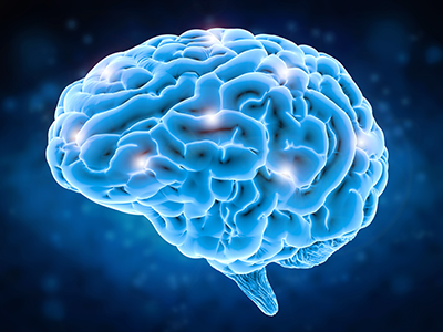
Because the researchers were able to analyze one of the largest cohorts studied to date, they could perform a whole-brain analysis that examined connectivity across 93 brain regions. That broader approach helped reveal widespread but nuanced connectivity differences. The altered brain connectivity observed in the study may represent an early neural signature of prenatal opioid exposure. Researchers believe these changes could reflect a vulnerability that may contribute to developmental or behavioral challenges later in life. Previous studies have shown that children exposed to opioids during pregnancy may face increased risks of motor delays, attention challenges and difficulties with emotional regulation.
However, the authors emphasize that these findings represent an early step in understanding the biology behind those outcomes. Because the OBOE study is designed to follow children over time, future research will explore whether early brain connectivity patterns are linked to later development. If those links are confirmed, brain imaging could eventually help clinicians identify which infants may benefit from closer developmental monitoring or early support.
Leading the way
The Developing Brain Institute at Children’s National serves as the neuroimaging core for the OBOE study, bringing decades of expertise in imaging the developing brain. This work is part of a broader effort to identify early markers of risk and resilience in childhood development. By combining large-scale collaboration with cutting-edge neuroimaging, studies like this one are helping researchers better understand how the earliest stages of life influence brain development and how clinicians can intervene earlier to support healthier futures for children.
This research, Disrupted Brain Connectivity in Newborns Following Antenatal Opioid Exposure, was published in Biological Psychiatry: Cognitive Neuroscience and Neuroimaging. Authors from Children’s National Hospital include Josepheen De Asis-Cruz, MD, PhD; Jung-Hoon Kim, PhD; Kushal Kapse, MS; Yao Wu, PhD; and Catherine Limperopoulos, PhD, in collaboration with colleagues from institutions participating in the multi-site ACT NOW Outcomes of Babies with Opioid Exposure (OBOE) study.


 2024 marked another groundbreaking year for Children’s National Hospital, showcasing remarkable advances across the spectrum of pediatric medicine, research and healthcare innovation. From pioneering surgical procedures to breakthrough artificial intelligence applications, the institution continued to push the boundaries of what’s possible in children’s healthcare. Read on for our list of the most popular articles we published on Innovation District in 2024.