Tag Archive for: brain damage

newborn in incubator

In HIE lower heart rate variability signals stressed newborns

newborn in incubator

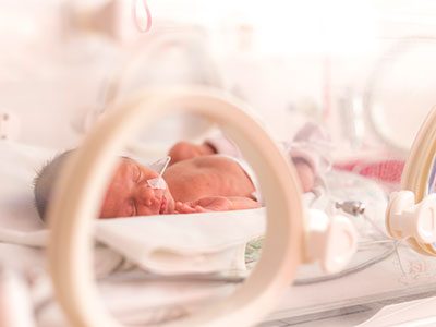
In newborns with hypoxic-ischemic encephalopathy (HIE), lower heart rate variability correlates with autonomic manifestations of stress shortly after birth, underscoring the value of this biomarker, according to Children’s research presented during the Pediatric Academic Societies 2019 Annual Meeting.

Tethered to an array of machines that keep their bodies nourished, warm and alive, newborns with health issues can’t speak. But Children’s research teams are tapping into what the machinery itself says, looking for insights into which vulnerable infants are most in need of earlier intervention.
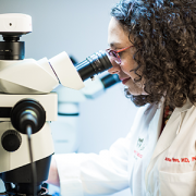
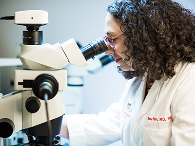
Heart rate variability – or the variation between heartbeats – is a sign of health. Our autonomic nervous system constantly sends signals to adjust our heart rate under normal conditions. We can measure heart rate variability non-invasively, providing a way to detect potential problems with the autonomic nervous system as a sensitive marker of health in critically ill newborns,” says An N. Massaro, M.D., co-Director of Research for the Division of Neonatology at Children’s National, and the study’s senior author. “We’re looking for validated markers of brain injury in babies with HIE, and our study helps to support heart rate variability as one such valuable physiological biomarker.”

In most newborns, the autonomic nervous system reliably and automatically receives information about the body and the outside world and, in response, controls essential functions like blood pressure, body temperature, how quickly the baby breathes and how rapidly the newborn’s heart beats. The sympathetic part stimulates body processes, while the parasympathetic part inhibits body processes. When the nervous system’s internal auto-pilot falters, babies can suffer.

The Children’s team enrolled infants with HIE in the prospective, observational study. (HIE is brain damage that occurs with full-term babies who experience insufficient blood and oxygen flow to the brain around the time they are born.) Fifteen percent had severe encephalopathy. Mean age of babies in the observational study was 38.9 weeks gestation. Their median Apgar score at five minutes was 3; the 0-9 Apgar range indicates how ready newborns are for the rigors of life outside the womb.

The team analyzed heart rate variability metrics for three time periods:

  • The first 24 to 27 hours of life
  • The first three hours after babies undergoing therapeutic cooling were rewarmed and
  • The first three hours after babies’ body temperature had returned to normal.

They correlated the relationship between heart rate variability for 68 infants during at least one of these time periods with the stress z-score from the NICU Network Neurobehavioral Scale. The scale is a standardized assessment of newborn’s neurobehavioral integrity. The stress summary score indicates a newborn’s overall stress response, and six test items specifically relate to autonomic function.

“Alpha exponent and root mean square in short timescales, root mean square in long timescales, as well as low and high frequency powers positively correlated with stress scores and, even after adjusting for covariates, remained independently associated at 24 hours,” says Allie Townsend, the study’s lead author.

Pediatric Academic Societies 2019 Annual Meeting presentation

  • “Heart rate variability (HRV) measures of autonomic nervous system (ANS) function relates to neonatal neurobehavioral manifestations of stress in newborn with hypoxic-ischemic encephalopathy (HIE).”
    • Monday, April 29, 2019, 5:45 p.m. (EST)

Allie Townsend, lead author; Rathinaswamy B. Govindan, Ph.D., staff scientist, Advanced Physiological Signals Processing Lab and co-author; Penny Glass, Ph.D., director, Child Development Program and co-author; Judy Brown, co-author; Tareq Al-Shargabi, M.S., co-author; Taeun Chang, M.D., director, Neonatal Neurology and Neonatal Neurocritical Care Program and co-author; Adré J. du Plessis, M.B.Ch.B., MPH, chief of the Division of Fetal and Transitional Medicine and co-author; An N. Massaro, M.D., co-Director of Research for the Division of Neonatology and senior author, all of Children’s National.

Dr. Anna Penn uses a microscope

New model mimics persistent interneuron loss seen in prematurity

Dr. Anna Penn uses a microscope

Children’s research-clinicians created a novel preclinical model that mimics the persistent interneuron loss seen in preterm human infants, identifying interneuron subtypes that could become future therapeutic targets to prevent or lessen neurodevelopmental risks.

Research-clinicians at Children’s National Health System have created a novel preclinical model that mimics the persistent interneuron loss seen in preterm human infants, identifying interneuron subtypes that could become future therapeutic targets to prevent or lessen neurodevelopmental risks, the team reports Jan. 31, 2019, in eNeuro. The open access journal for Society for Neuroscience recognized the team’s paper as its “featured” article.

In the prefrontal cortex (PFC) of infants born preterm, there are decreased somatostatin and calbindin interneurons seen in upper cortical layers in infants who survived for a few months after preterm birth. This neuronal damage was mimicked in an experimental model of preterm brain injury in the PFC, but only when the newborn experimental models had first experienced a combination of prenatal maternal immune activation and postnatal chronic sublethal hypoxia. Neither neuronal insult on its own produced the pattern of interneuron loss in the upper cortical layers observed in humans, the research team finds.

“These combined insults lead to long-term neurobehavioral deficits that mimic what we see in human infants who are born extremely preterm,” says Anna Penn, M.D., Ph.D., a neonatologist in the Division of Neonatology and the Fetal Medicine Institute and a developmental neuroscientist at Children’s National Health System, and senior study author. “Future success in preventing neuronal damage in newborns relies on having accurate experimental models of preterm brain injury and well-defined outcome measures that can be examined in young infants and experimental models of the same developmental stage.”

According to the Centers for Disease Control and Prevention 1 in 10 infants is born preterm, before the 37th week of pregnancy. Many of these preterm births result from infection or inflammation in utero. After delivery, many infants experience other health challenges, like respiratory failure. These multi-hits can exacerbate brain damage.

Prematurity is associated with significantly increased risk of neurobehavioral pathologies, including autism spectrum disorder and schizophrenia. In both psychiatric disorders, the prefrontal cortex inhibitory circuit is disrupted due to alterations of gamma-aminobutyric acid (GABA) interneurons in a brain region involved in working memory and social cognition.

Cortical interneurons are created and migrate late in pregnancy and early infancy. That timing leaves them particularly vulnerable to insults, such as preterm birth.

In order to investigate the effects of perinatal insults on GABAergic interneuron development, the Children’s research team, led by Helene Lacaille, Ph.D., in Dr. Penn’s laboratory, subjected the new preterm encephalopathy experimental model to a battery of neurobehavioral tests, including working memory, cognitive flexibility and social cognition.

“This translational study, which examined the prefrontal cortex in age-matched term and preterm babies supports our hypothesis that specific cellular alterations seen in preterm encephalopathy can be linked with a heightened risk of children experiencing neuropsychiatric disorders later in life,” Dr. Penn adds. “Specific interneuron subtypes may provide specific therapeutic targets for medicines that hold the promise of preventing or lessening these neurodevelopmental risks.”

In addition to Dr. Penn and Lead Author Lacaille, Children’s co-authors include Claire-Marie Vacher; Dana Bakalar, Jiaqi J. O’Reilly and Jacquelyn Salzbank, all of Children’s Center for Neuroscience Research.

Financial support for research described in this post was provided by the National Institutes of Health under award R01HD092593, District of Columbia Intellectual Developmental Disabilities Research Center under award U54HD090257, Cerebral Palsy Alliance Research Foundation, Children’s National Board of Visitors, Children’s Research Institute and Fetal Medicine Institute.

An-Massaro

Keeping an eye on autonomic function for infants with HIE

An-Massaro

“By including heart rate variability measurements and other markers of autonomic function in our current predictive armamentarium,” says An Massaro, M.D., “we may be able to offer new hope for infants with HIE.”

In about two to three in every 1,000 full-term births, babies develop a neurological condition called hypoxic ischemic encephalopathy (HIE) when their brains receive insufficient oxygen. HIE can be a devastating condition, leading to severe developmental or cognitive delays or motor impairments that become more evident as the child grows older. Despite improvements in care – including therapeutic hypothermia, a whole-body cooling method administered shortly after birth that can slow brain damage – about half of children with this condition die from neurological complications by age 2.

Finding ways to identify children with the most severe HIE could help researchers focus their efforts and provide even more intense neuroprotective care, explains An Massaro, M.D., a neonatologist at Children’s National Health System. But thus far, it’s been unclear which symptoms reflect the extent of HIE-induced brain damage.

That’s why Dr. Massaro and colleagues embarked on a study published in the May 2018 issue of Journal of Pediatrics. The team sought to determine whether dysfunction of the autonomic nervous system (ANS) – the auto-pilot part of the nervous system responsible for unconscious bodily functions, such as breathing and digestion – reflected in routine care events can be used as a marker for brain injury severity.

The researchers collected data from 25 infants who were treated for HIE with therapeutic hypothermia at Children’s National. Thanks to multi-modal monitoring, these babies’ medical records hold a treasure trove of information, explains Rathinaswamy B. Govindan, Ph.D., a staff scientist in Children’s Advanced Physiological Signals Processing Lab.

In addition to including continuous heart rate tracings and blood pressure readings that are standard for many infants in the neonatal intensive care unit (NICU), they also recorded cerebral near infrared spectroscopy, a monitor that measures brain tissue oxygen levels. The investigators performed detailed analyses to evaluate how these monitor readings change in response to a variety of routine care events, such as diaper changes, heel sticks, endotracheal tube manipulations and pupil examinations.

The researchers stratified these infants based on how dysfunctional their ANS behaved by using heart rate variability as a marker: The fewer natural fluctuations in heart rate, the more damaged their ANS was thought to be. And they also used non-invasive brain magnetic resonance imaging (MRI) to determine brain damage. They then compared this information with the babies’ physiological responses during each care event.

Their findings show that infants with impaired ANS, based on depressed heart rate variability before the care event, had significantly different responses to these care events compared with babies with intact ANS.

  • For stimulating interventions, such as diaper changes and heel sticks, both heart rate and blood pressure increased in babies with intact ANS but decreased in babies with impaired ones.
  • Shining a light in their pupils led to an expected decreased heart rate with stable blood pressure in ANS-intact infants, but in ANS-impaired infants, there was no responsive change in heart rate and, additionally, a decrease in blood pressure was observed.
  • Responses were similar between the two groups during breathing tube manipulations, except for a slight increase in heart rate a few minutes later in the ANS-impaired group.

These results, Govindan explains, suggest that a real-time, continuous way to assess ANS function may offer insights into the expected physiological response for a given infant during routine NICU care.

“This is exactly the type of additional information that intensivists need to pinpoint infants who may benefit from additional neuroprotective support,” he says. “Right now, it is standard practice to monitor brain activity continuously using electroencephalogram and to check the status of the brain using MRI to assess the response to therapeutic cooling. Neither of these assessments can be readily used by neonatologists at the bedside in real-time to make clinical decisions.”

Assessing ANS function in real-time can help guide neuroprotective care in high-risk newborns by providing insight into the evolving nature of brain damage in these infants, Dr. Massaro adds.

Beyond simply serving as a biomarker into brain injury, poor ANS function also could contribute to the development of secondary injury in newborns with HIE by stymieing the normal changes in heart rate and blood pressure that help oxygenate and heal injured brains. The researchers found that the cumulative duration of autonomic impairment was significantly correlated with the severity of brain injury visible by MRI in this group of infants.

“By including heart rate variability measurements and other markers of autonomic function in our current predictive armamentarium,” says Dr. Massaro, “we may be able to offer new hope for infants with HIE.”

In addition to Dr. Massaro, the Senior Author, study co-authors include Lead Author, Heather Campbell, M.D.; Rathinaswamy B. Govindan, Ph.D., Children’s Advanced Physiological Signals Processing Lab; Srinivas Kota, Ph.D.; Tareq Al-Shargabi, M.S.; Marina Metzler, B.S.; Nickie Andescavage, M.D., Children’s neonatalogist; Taeun Chang, M.D., Children’s neonatal and fetal neurologist; L. Gilbert Vezina, M.D., attending in Children’s Division of Diagnostic Imaging and Radiology; and Adré J. du Plessis, M.B.Ch.B., M.P.H., chief of Children’s Division of Fetal and Transitional Medicine.

This research was supported by the Clinical and Translational Science Institute at Children’s National under awards UL1TR000075 and 1KL2RR031987-01 and the Intellectual and Developmental Disabilities Research Consortium within the National Institutes of Health under award P30HD040677.