Tag Archive for: biomarker

Julia Finkel

Novel pupillary response biomarker for BBD discovered

Julia Finkel

“To our knowledge, this unique pupillary signature has not been previously seen or described in other patient populations and we have not seen it in any of our other studies,” says Julia Finkel, M.D. “It may represent a distinctive and readily-identifiable physiologic marker of disease.”

Researchers at Children’s National have discovered a potential biomarker in the pupillary response of some pediatric patients with Bowel and Bladder Dysfunction (BBD) that could improve the speed and accuracy of diagnosis and treatment, according to a recent study published in the Journal of Pediatric Urology.

BBD describes a range of lower urinary tract symptoms accompanied by bowel complaints such as enuresis (bedwetting), urgency and urinary retention, often accompanied by constipation. While these symptoms represent 40% of pediatric urology visits, BBD is considered an underdiagnosed pediatric ailment.

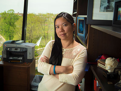
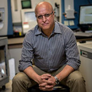
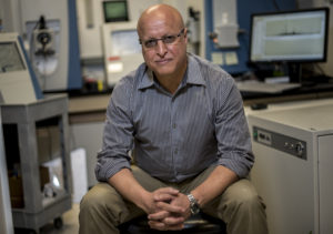
Julia Finkel, M.D., pediatric anesthesiologist and director of Research and Development for Pain Medicine at the Sheikh Zayed Institute for Pediatric Surgical Innovation at Children’s National, led the pilot study to explore whether BBD could be detected from a patient’s pupillary light reflex response. Using a novel application of pupillometry, Dr. Finkel and her research team recorded and analyzed the pupillary light reflex responses of 28 patients with BBD, ages 7 – 21, from the Wetting, Infections and Stooling Help (WISH) clinic at Children’s National Health System. The study included baseline static and dynamic pupillometry assessments obtained from each patient before and after voiding. Pupillary measurements were also taken after five minutes of lying down by the patient, and again after five minutes of standing.

In reviewing the patient’s graphed data, the researchers noted a distinct “notch” shape repeated in the pupillary response graph of 11 of 28 patients with BBD symptoms. In those 11 patients, the graph notch appears to indicate a brief repeat constriction of the patient’s pupil before returning to its resting size.

Considering that bowel and bladder functions are controlled in part by the autonomic nervous system, researchers surmised that the notch on the graph is likely to reflect a characteristic disturbance in the regulation of the autonomic nervous system of those 11 patients, which would indicate a physiological cause for their BBD, either alone or in combination with a behavioral cause.

“To our knowledge, this unique pupillary signature has not been previously seen or described in other patient populations and we have not seen it in any of our other studies,” says Dr. Finkel. “It may represent a distinctive and readily-identifiable physiologic marker of disease.”

Causes of BBD can be physiological, such as anomalies in the synapsis of the nervous system, and can be related to behavioral health issues such as anxiety. Early diagnosis and treatment of BBD is important in avoiding secondary complications that can adversely impact a child’s kidney and bladder function as well as psychosocial well-being.

Dr. Finkel says that, while the results of this study are broadly consistent with other studies that examined the autonomic nervous system activity of BBD patients, this small study is preliminary. She notes that further research is needed and would include assessing abnormalities in pupillary response stemming from the parasympathetic and sympathetic functions of the autonomic nervous system.

Her hope is that further study will lead to more effective diagnostic and monitoring tools for clinicians treating BBD patients.

Dr. Finkel’s research focuses on the diagnostic potential of various pupillary reflexes. She says that pupillometry makes an ideal point-of-care diagnostic tool because it is noninvasive, easy to use, portable and provides real-time data for diagnosis and monitoring of therapeutic effects.

In addition to Dr. Finkel, study co-authors include Kevin G. Jackson, Nadia B. Kalloo, M.D., and Emily Blum, M.D., of Children’s National Health System; and Elizabeth L. Malphrus, MS-III, George Washington University.

newborn in incubator

In HIE lower heart rate variability signals stressed newborns

newborn in incubator

In newborns with hypoxic-ischemic encephalopathy (HIE), lower heart rate variability correlates with autonomic manifestations of stress shortly after birth, underscoring the value of this biomarker, according to Children’s research presented during the Pediatric Academic Societies 2019 Annual Meeting.

Tethered to an array of machines that keep their bodies nourished, warm and alive, newborns with health issues can’t speak. But Children’s research teams are tapping into what the machinery itself says, looking for insights into which vulnerable infants are most in need of earlier intervention.

Heart rate variability – or the variation between heartbeats – is a sign of health. Our autonomic nervous system constantly sends signals to adjust our heart rate under normal conditions. We can measure heart rate variability non-invasively, providing a way to detect potential problems with the autonomic nervous system as a sensitive marker of health in critically ill newborns,” says An N. Massaro, M.D., co-Director of Research for the Division of Neonatology at Children’s National, and the study’s senior author. “We’re looking for validated markers of brain injury in babies with HIE, and our study helps to support heart rate variability as one such valuable physiological biomarker.”

In most newborns, the autonomic nervous system reliably and automatically receives information about the body and the outside world and, in response, controls essential functions like blood pressure, body temperature, how quickly the baby breathes and how rapidly the newborn’s heart beats. The sympathetic part stimulates body processes, while the parasympathetic part inhibits body processes. When the nervous system’s internal auto-pilot falters, babies can suffer.

The Children’s team enrolled infants with HIE in the prospective, observational study. (HIE is brain damage that occurs with full-term babies who experience insufficient blood and oxygen flow to the brain around the time they are born.) Fifteen percent had severe encephalopathy. Mean age of babies in the observational study was 38.9 weeks gestation. Their median Apgar score at five minutes was 3; the 0-9 Apgar range indicates how ready newborns are for the rigors of life outside the womb.

The team analyzed heart rate variability metrics for three time periods:

  • The first 24 to 27 hours of life
  • The first three hours after babies undergoing therapeutic cooling were rewarmed and
  • The first three hours after babies’ body temperature had returned to normal.

They correlated the relationship between heart rate variability for 68 infants during at least one of these time periods with the stress z-score from the NICU Network Neurobehavioral Scale. The scale is a standardized assessment of newborn’s neurobehavioral integrity. The stress summary score indicates a newborn’s overall stress response, and six test items specifically relate to autonomic function.

“Alpha exponent and root mean square in short timescales, root mean square in long timescales, as well as low and high frequency powers positively correlated with stress scores and, even after adjusting for covariates, remained independently associated at 24 hours,” says Allie Townsend, the study’s lead author.

Pediatric Academic Societies 2019 Annual Meeting presentation

  • “Heart rate variability (HRV) measures of autonomic nervous system (ANS) function relates to neonatal neurobehavioral manifestations of stress in newborn with hypoxic-ischemic encephalopathy (HIE).”
    • Monday, April 29, 2019, 5:45 p.m. (EST)

Allie Townsend, lead author; Rathinaswamy B. Govindan, Ph.D., staff scientist, Advanced Physiological Signals Processing Lab and co-author; Penny Glass, Ph.D., director, Child Development Program and co-author; Judy Brown, co-author; Tareq Al-Shargabi, M.S., co-author; Taeun Chang, M.D., director, Neonatal Neurology and Neonatal Neurocritical Care Program and co-author; Adré J. du Plessis, M.B.Ch.B., MPH, chief of the Division of Fetal and Transitional Medicine and co-author; An N. Massaro, M.D., co-Director of Research for the Division of Neonatology and senior author, all of Children’s National.

DNA moleucle

PAC1R mutation may be linked to severity of social deficits in autism

DNA moleucle

A mutation of the gene PAC1R may be linked to the severity of social deficits experienced by kids with autism spectrum disorder (ASD), finds a study from a multi-institutional research team led by Children’s National faculty. If the pilot findings are corroborated in larger, multi-center studies, the research published online Dec. 17, 2018, in Autism Research represents the first step toward identifying a potential novel biomarker to guide interventions and better predict outcomes for children with autism.

As many as 1 in 40 children are affected by ASD. Symptoms of the disorder – such as not making eye contact, not responding to one’s name when called, an inability to follow a conversation of more than one speaker or incessantly repeating certain words or phrases – usually crop up by the time a child turns 3.

The developmental disorder is believed to be linked, in part, to disrupted circuitry within the amygdala, a brain structure integral for processing social-emotional information. This study reveals that PAC1R is expressed during key periods of brain development when the amygdala – an almond-shaped cluster of neurons – develops and matures. A properly functioning amygdala, along with brain structures like the prefrontal cortex and cerebellum, are crucial to neurotypical social-emotional processing.

“Our study suggests that an individual with autism who is carrying a mutation in PAC1R may have a greater chance of more severe social problems and disrupted functional brain connectivity with the amygdala,” says Joshua G. Corbin, Ph.D., interim director of the Center for Neuroscience Research at Children’s National Health System and the study’s co-senior author. “Our study is one important step along the pathway to developing new biomarkers for autism spectrum disorder and, hopefully, predicting patients’ outcomes.”

The research team’s insights came through investigating multiple lines of evidence:

  • They looked at gene expression in the brains of an experimental model at days 13.5 and 18.5 of fetal development and day 7 of life, dates that correspond with early, mid and late amygdala development. They confirmed that Pac1r is expressed in the experimental model at a critical time frame for brain development that coincides with the timing for altered brain trajectories with ASD.
  • They looked at gene expression in the human brain by mining publicly available genome-wide transcriptome data, plotting median PAC1R expression values for key brain regions. They found high levels of PAC1R expression at multiple ages with higher PAC1R expression in male brains during the fetal period and higher PAC1R expression in female brains during childhood and early adulthood.
  • One hundred twenty-nine patients with ASD aged 6 to 14 were recruited for behavioral assessment. Of the 48 patients who also participated in neuroimaging, 20 were able to stay awake for five minutes without too much movement as the resting state functional magnetic resonance images were captured. Children who were carriers of the high-risk genotype had higher resting-state connectivity between the amygdala and right posterior temporal gyrus. Connectivity alterations in a region of the brain involved in processing visual motion may influence how kids with ASD perceive socially meaningful information, the authors write.
  • Each child also submitted a saliva sample for DNA genotyping. Previously published research finds that a G to C single nucleotide polymorphism, a single swap in the nucleotides that make up DNA, in PAC1R is associated with higher risk for post traumatic stress disorder in girls. In this behavioral assessment, the research team found children with autism who carried the homozygous CC genotype had higher scores as measured through a validated tool, meaning they had greater social deficits than kids with the heterozygous genotype.

All told, the project is the fruit of six years of painstaking research and data collection, say the researchers. That includes banking patients’ saliva samples collected during clinical visits for future retrospective analyses to determine which genetic mutations were correlated with behavioral and functional brain deficits, Corbin adds.

Lauren Kenworthy, who directs our Center for Autism Spectrum Disorders, and I have been talking over the years about how we could bring our programs together. We homed in on this project to look at about a dozen genes to assess correlations and brought in experts from genetics and genomics at Children’s National to sequence genes of interest,” he adds. “Linking the bench to bedside is especially difficult in neuroscience. It takes a huge amount of effort and dozens of discussions, and it’s very rare. It’s an exemplar of what we strive for.”

In addition to Corbin, study co-authors include Lead Author Meredith Goodrich and Maria Jesus Herrero, post-doctoral fellow, Children’s Center for Neuroscience Research; Anna Chelsea Armour and co-Senior Author Lauren Kenworthy, Ph.D., Children’s Center for Autism Spectrum Disorders; Karuna Panchapakesan, Joseph Devaney and Susan Knoblach, Ph.D., Children’s Center for Genetic Medicine Research; Xiaozhen You and Chandan J. Vaidya, Georgetown University; and Catherine A.W. Sullivan and Abha R. Gupta, Yale School of Medicine.

Financial support for the research described in this report was provided by DC-IDDRC under awards HD040677-07 and 1U54HD090257, the Clinical and Translational Science Institute at Children’s National, The Isidore and Bertha Gudelsky Family Foundation and the National Institutes of Health under awards MH083053-01A2 and MH084961.

An-Massaro

Keeping an eye on autonomic function for infants with HIE

An-Massaro

“By including heart rate variability measurements and other markers of autonomic function in our current predictive armamentarium,” says An Massaro, M.D., “we may be able to offer new hope for infants with HIE.”

In about two to three in every 1,000 full-term births, babies develop a neurological condition called hypoxic ischemic encephalopathy (HIE) when their brains receive insufficient oxygen. HIE can be a devastating condition, leading to severe developmental or cognitive delays or motor impairments that become more evident as the child grows older. Despite improvements in care – including therapeutic hypothermia, a whole-body cooling method administered shortly after birth that can slow brain damage – about half of children with this condition die from neurological complications by age 2.

Finding ways to identify children with the most severe HIE could help researchers focus their efforts and provide even more intense neuroprotective care, explains An Massaro, M.D., a neonatologist at Children’s National Health System. But thus far, it’s been unclear which symptoms reflect the extent of HIE-induced brain damage.

That’s why Dr. Massaro and colleagues embarked on a study published in the May 2018 issue of Journal of Pediatrics. The team sought to determine whether dysfunction of the autonomic nervous system (ANS) – the auto-pilot part of the nervous system responsible for unconscious bodily functions, such as breathing and digestion – reflected in routine care events can be used as a marker for brain injury severity.

The researchers collected data from 25 infants who were treated for HIE with therapeutic hypothermia at Children’s National. Thanks to multi-modal monitoring, these babies’ medical records hold a treasure trove of information, explains Rathinaswamy B. Govindan, Ph.D., a staff scientist in Children’s Advanced Physiological Signals Processing Lab.

In addition to including continuous heart rate tracings and blood pressure readings that are standard for many infants in the neonatal intensive care unit (NICU), they also recorded cerebral near infrared spectroscopy, a monitor that measures brain tissue oxygen levels. The investigators performed detailed analyses to evaluate how these monitor readings change in response to a variety of routine care events, such as diaper changes, heel sticks, endotracheal tube manipulations and pupil examinations.

The researchers stratified these infants based on how dysfunctional their ANS behaved by using heart rate variability as a marker: The fewer natural fluctuations in heart rate, the more damaged their ANS was thought to be. And they also used non-invasive brain magnetic resonance imaging (MRI) to determine brain damage. They then compared this information with the babies’ physiological responses during each care event.

Their findings show that infants with impaired ANS, based on depressed heart rate variability before the care event, had significantly different responses to these care events compared with babies with intact ANS.

  • For stimulating interventions, such as diaper changes and heel sticks, both heart rate and blood pressure increased in babies with intact ANS but decreased in babies with impaired ones.
  • Shining a light in their pupils led to an expected decreased heart rate with stable blood pressure in ANS-intact infants, but in ANS-impaired infants, there was no responsive change in heart rate and, additionally, a decrease in blood pressure was observed.
  • Responses were similar between the two groups during breathing tube manipulations, except for a slight increase in heart rate a few minutes later in the ANS-impaired group.

These results, Govindan explains, suggest that a real-time, continuous way to assess ANS function may offer insights into the expected physiological response for a given infant during routine NICU care.

“This is exactly the type of additional information that intensivists need to pinpoint infants who may benefit from additional neuroprotective support,” he says. “Right now, it is standard practice to monitor brain activity continuously using electroencephalogram and to check the status of the brain using MRI to assess the response to therapeutic cooling. Neither of these assessments can be readily used by neonatologists at the bedside in real-time to make clinical decisions.”

Assessing ANS function in real-time can help guide neuroprotective care in high-risk newborns by providing insight into the evolving nature of brain damage in these infants, Dr. Massaro adds.

Beyond simply serving as a biomarker into brain injury, poor ANS function also could contribute to the development of secondary injury in newborns with HIE by stymieing the normal changes in heart rate and blood pressure that help oxygenate and heal injured brains. The researchers found that the cumulative duration of autonomic impairment was significantly correlated with the severity of brain injury visible by MRI in this group of infants.

“By including heart rate variability measurements and other markers of autonomic function in our current predictive armamentarium,” says Dr. Massaro, “we may be able to offer new hope for infants with HIE.”

In addition to Dr. Massaro, the Senior Author, study co-authors include Lead Author, Heather Campbell, M.D.; Rathinaswamy B. Govindan, Ph.D., Children’s Advanced Physiological Signals Processing Lab; Srinivas Kota, Ph.D.; Tareq Al-Shargabi, M.S.; Marina Metzler, B.S.; Nickie Andescavage, M.D., Children’s neonatalogist; Taeun Chang, M.D., Children’s neonatal and fetal neurologist; L. Gilbert Vezina, M.D., attending in Children’s Division of Diagnostic Imaging and Radiology; and Adré J. du Plessis, M.B.Ch.B., M.P.H., chief of Children’s Division of Fetal and Transitional Medicine.

This research was supported by the Clinical and Translational Science Institute at Children’s National under awards UL1TR000075 and 1KL2RR031987-01 and the Intellectual and Developmental Disabilities Research Consortium within the National Institutes of Health under award P30HD040677.

effects of cardiopulmonary bypass surgery on the white matter of piglets.

The effects of cardiopulmonary bypass on white matter development

 cardiopulmonary bypass

Nobuyuki Ishibashi, M.D., and a team of researchers looked the effects of cardiopulmonary bypass surgery on the white matter of an animal model.

Mortality rates for infants born with congenital heart disease (CHD) have dramatically decreased over the past two decades, with more and more children reaching adulthood. However, many survivors are at risk for neurodevelopmental abnormalities  associated with cardiopulmonary bypass surgery (CPB), including long-term injuries to the brain’s white matter and neural connectivity impairments that can lead to neurological dysfunction.

“Clinical studies have found a connection between abnormal neurological outcomes and surgery, but we don’t know what’s happening at the cellular level,” explains Nobuyuki Ishibashi, M.D., Director of the Cardiac Surgery Research Laboratory at Children’s National. To help shed light on this matter, Ishibashi and a team of researchers looked at the effects of CPB on the white matter of an animal model.

The research team randomly assigned models to receive one of three CPB-induced insults: a sham surgery (control group); full-flow bypass for 60 minutes; and 25°C circulatory arrest for 60 minutes. The team then used fractional anisotropy — a technique that measures the directionality of axon mylenation — to determine white matter organization in the models’ brains. They also used immunohistology techniques to assess the integrity of white matter oligodendrocytes, astrocytes and microglia.

The results, published in the Journal of the American Heart Association, show that white matter experiences region-specific vulnerability to insults associated with CPB, with fibers within the frontal cortex appearing the most susceptible. The team also found that fractional anisotropy changes after CPB were insult dependent and that regions most resilient to CPB-induced fractional anisotropy reduction were those that maintained mature oligodendrocytes.

From these findings, Ishibashi and his co-authors conclude that reducing alterations of oligodendrocyte development in the frontal cortex can be both a metric and a goal to improve neurodevelopmental impairment in the congenital heart disease population. “Because we are seeing cellular damage in these regions, we can target them for future therapies,” explains Ishibashi.

The study also demonstrates the dynamic relationship between fractional anisotropy and cellular events after pediatric cardiac surgery, and indicates that the technique is a clinically relevant biomarker in white matter injury after cardiac surgery.

Biomarkers sensitive to daily corticosteroid use

Using a mass spectrometer, Yetrib Hathout, Ph.D., is able to quantify 3,000 to 4,000 proteins from a tissue sample to identify proteins associated with cancer.

Using a Somascan proteomics assay – which simultaneously analyzes 1,129 proteins in a small volume of serum – a team led by Children’s National Health System researchers identified 21 biomarkers that respond to corticosteroids taken daily by children with Duchenne muscular dystrophy (DMD) and inflammatory bowel disease.

Corticosteroids are commonly prescribed to treat inflammatory conditions. High daily doses of corticosteroids are considered the standard of care for DMD, a type of muscular dystrophy characterized by worsening muscle weakness that affects 1 in 3,600 male infants. However, depending on the age of the child and drug dosage, chronic use is associated with such side effects as changes in bone remodeling that can lead to stunted growth, weight gain, facial puffiness caused by fat buildup, mood changes, sleep disturbances, and immune suppression. The research team sought to identify blood biomarkers that could be leveraged to create a fast, reliable way to gauge the safety and efficacy of corticosteroid use by children. The biomarkers also could guide development of a replacement therapy with fewer side effects.

“Ten pro-inflammatory proteins were elevated in untreated patients and suppressed by corticosteroids (MMP12, IL22RA2, CCL22, IGFBP2, FCER2, LY9, ITGa1/b1, LTa1/b2, ANGPT2 and FGG),” Yetrib Hathout, Ph.D., Proteomic Core Director at Children’s National, and colleagues write in the journal Scientific Reports. “These are candidate biomarkers for anti-inflammatory efficacy of corticosteroids.”

The blood biomarkers sensitive to corticosteroids fit into three broad groups, according to the authors. The children taking corticosteroids were matched with children of the same age who had never taken the medicine. Five biomarkers significantly increased in this corticosteroid-naïve group and decreased in kids prescribed corticosteroids. The biomarkers generally were inflammatory proteins and included chemokine, insulin-like growth factor binding protein 2, and integrin alpha-I/beta-1 complex.

The second group of biomarkers included nine proteins associated with macrophage and T-lymphocytes that were significantly reduced in concentration in kids taking corticosteroids. According to the study, this finding hints at corticosteroids blunting the ability of the immune system’s most able fighters to respond to infection.

In the third group were five proteins that were significantly increased by corticosteroid treatment in DMD and included matrix metalloproteinase 3, carnosine dipeptidase 1, angiotensinogen, growth hormone binding protein, insulin, and leptin, a hormone linked to appetite.

What researchers learned with this study will help them more accurately design the next phase of the work, Hathout says.

“We are the first team to report a number of novel discoveries, including that growth hormone binding protein (GHBP) levels increase with corticosteroid use. This represents a candidate biomarker for stunted growth. In order to use that new information effectively in drug development, the next studies must corroborate the role of serum GHBP levels as predictors of diminished stature,” he adds. “The study finding that four adrenal steroid hormones are depressed in kids taking corticosteroids raises additional questions about the broader impact of adrenal insufficiency, including its role in the delay of the onset of puberty.”

This work was supported by National Institutes of Health grants (R01AR062380, R01AR061875, P50AR060836, U54HD071601, K99HL130035, and R44NS095423) and Department of Defense CDMRP program grant W81XWH-15-1-0265. Additional support was provided by AFM-Telethon (18259) and the Muscular Dystrophy Association USA (MDA353094).

The search for precise blood biomarkers of neonatal brain injury

Bloodbiomarkers

PDF Version

What’s Known:
Hypoxic-ischemic encephalopathy (HIE) is characterized by reduced blood and oxygen flow to a baby’s brain around birth and may cause neurologic disability or death. It occurs most commonly after intrauterine asphyxia brought on by such difficulties as circulatory problems, placental abruption, or inflammatory processes. Newborns with HIE may suffer seizures, difficulty feeding, and disturbed control of heart rate and breathing. Cooling therapy, which is the standard of care, offers some protection to the developing brain, but up to 50 percent of HIE-affected infants still have poor outcomes.

What’s New:
Research scientists at Children’s National Health System are involved in a multi-center clinical trial to determine if erythropoietin (EPO), a hormone naturally secreted by the kidneys and commonly used to treat anemia, helps to prevent brain injury in these infants. The trial, called the HEAL Study (High Dose Erythropoietin for Asphyxia and Encephalopathy), is exploring whether EPO, given in addition to hypothermia, further lowers the risk of brain injury in HIE-affected babies. As a part of this study, researchers at Children’s National are leading the investigation to identify biomarkers of brain injury. Biomarkers are telltale chemicals in the blood and are used in tests that evaluate whether patients have suffered a heart attack. While available biomarkers warn when the heart, kidney, or liver is in trouble, there is no blood biomarker that signals ongoing brain injury. Such blood biomarkers could help to determine which infants are responding to treatment as well as to precisely identify which HIE-affected infants are still struggling and require additional treatments, such as EPO, to protect the brain and improve outcomes.

Questions for Future Research: 

  • Does EPO, in tandem with hypothermia, improve long- term neurodevelopmental outcomes in newborns with HIE?
  • Which biomarkers, or panel of biomarkers, best reflect the timing and severity of neonatal brain injury?
  • Can biomarkers direct which types of treatments are best for specific patients and when they should be used?

Source: Plasma Biomarkers of Brain Injury in Neonatal HIE (Hypoxic-Ischemic Encephalopathy).” A.N. Massaro, Y. Wu, T.K. Bammler, A. Mathur, R.C. McKinstry, T. Chang, D.E. Mayock, S. Mulkey, K. Van Meurs, L. Dong, R. Ballard, and S. Juul. Presented during the 2016 Pediatric Academic Societies Annual Meeting, Baltimore, MD. May 3, 2016.