Tag Archive for: autonomic nervous system

Autonomic nervous system appears to function well regardless of mode of childbirth

Late in pregnancy, the human body carefully prepares fetuses for the rigors of life outside the protection of the womb. Levels of cortisol, a stress hormone, ramp up and spike during labor. Catecholamines, another stress hormone, also rise at birth, helping to kick start the necessary functions that the baby will need to regulate breathing, heartbeat, blood pressure and energy metabolism levels at delivery. Oxytocin surges, promoting contractions for the mother during labor and stimulating milk production after the infant is born.

These processes also can play a role in preparing the fetal brain during the transition to life outside the womb by readying the autonomic nervous system and adapting its cerebral connections. The autonomic nervous system acts like the body’s autopilot, taking in information it needs to ensure that internal organs run steadily without willful action, such as ensuring the heart beats and eyelids blink at steady intervals. Its yin, the sympathetic division, stimulates body processes while its yang, the parasympathetic division, inhibits them.

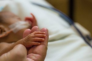
Infants born preterm have reduced autonomic function compared with their full-term peers and also face possible serious neurodevelopmental impairment later in life. But is there a difference in autonomic nervous system function for full-term babies after undergoing labor compared with infants delivered via cesarean section (C-section)?

A team from the Children’s National Inova Collaborative Research Program (CNICA) – a research collaboration between Children’s National in Washington, D.C., and Inova Women’s and Children’s Hospital in Virginia – set out to answer that question in a paper published online July 30, 2019, in Scientific Reports.

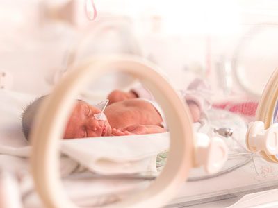
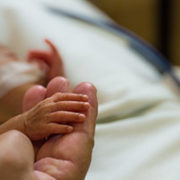
They enrolled newborns who had experienced normal, full-term pregnancies and recorded their brain function and heart performance when they were about 2 days old. Infants whose conditions were fragile enough to require observation in the neonatal intensive care unit were excluded from the study. Of 167 infants recruited for the prospective cohort study, 118 newborns had sufficiently robust data to include them in the research.  Of these newborns:

  • 62 (52.5%) were born by vaginal delivery
  • 22 (18.6%) started out with vaginal delivery but ultimately switched to C-section based on failure to progress, failed labor induction or fetal intolerance to labor
  • And 34 (28.8%) were born by elective C-section.

The CNICA research team swaddled infants for comfort and slipped electrode nets over their tiny heads to simultaneously measure heart rate variability and electrocortical function through non-invasive techniques. The team hypothesized that infants who had been exposed to labor would have enhanced autonomic tone and higher cortical electroencephalogram (EEG) power than babies born via C-section.

“In a low-risk group of babies born full-term, the autonomic nervous system and cortical systems appear to function well regardless of whether infants were exposed to labor prior to birth,” says Sarah B. Mulkey, M.D., Ph.D., a fetalneonatal neurologist in the Division of Fetal and Transitional Medicine at Children’s National and the study’s lead author.

However, infants born by C-section following a period of labor had significantly increased accelerations in their heart rates. And the infants born by C-section during labor had significantly lower relative gamma frequency EEG at 25.2 hours old compared with the other two groups studied.

“Together these findings point to a possible increased stress response and arousal difference in infants who started with vaginal delivery and finished delivery with C-section,” Dr. Mulkey says. “There is so little published research about the neurologic impacts of the mode of delivery, so our work helps to provide a normal reference point for future studies looking at high-risk infants, including babies born preterm.”

Because the research team saw little differences in autonomic tone or other EEG frequencies when the infants were 1 day old, future research will explore these measures at different points in the newborns’ early life as well as the role of the sleep-wake cycle on heart rate variability.

In addition to Dr. Mulkey, study co-authors include Srinivas Kota, Ph.D., Rathinaswamy B. Govindan, Ph.D., Tareq Al-Shargabi, MSc, Christopher B. Swisher, BS, Laura Hitchings, BScM, Stephanie Russo, BS, Nicole Herrera, MPH, Robert McCarter, ScD, and Senior Author Adré  J. du Plessis, M.B.Ch.B., MPH, all of Children’s National; and Augustine Eze Jr., MS, G. Larry Maxwell, M.D., and Robin Baker, M.D., all of Inova Women’s and Children’s Hospital.

Financial support for research described in this post was provided by the National Institutes of Health National Center for Advancing Translational Sciences under award numbers UL1TR001876 and KL2TR001877.

newborn in incubator

In HIE lower heart rate variability signals stressed newborns

newborn in incubator

In newborns with hypoxic-ischemic encephalopathy (HIE), lower heart rate variability correlates with autonomic manifestations of stress shortly after birth, underscoring the value of this biomarker, according to Children’s research presented during the Pediatric Academic Societies 2019 Annual Meeting.

Tethered to an array of machines that keep their bodies nourished, warm and alive, newborns with health issues can’t speak. But Children’s research teams are tapping into what the machinery itself says, looking for insights into which vulnerable infants are most in need of earlier intervention.

Heart rate variability – or the variation between heartbeats – is a sign of health. Our autonomic nervous system constantly sends signals to adjust our heart rate under normal conditions. We can measure heart rate variability non-invasively, providing a way to detect potential problems with the autonomic nervous system as a sensitive marker of health in critically ill newborns,” says An N. Massaro, M.D., co-Director of Research for the Division of Neonatology at Children’s National, and the study’s senior author. “We’re looking for validated markers of brain injury in babies with HIE, and our study helps to support heart rate variability as one such valuable physiological biomarker.”

In most newborns, the autonomic nervous system reliably and automatically receives information about the body and the outside world and, in response, controls essential functions like blood pressure, body temperature, how quickly the baby breathes and how rapidly the newborn’s heart beats. The sympathetic part stimulates body processes, while the parasympathetic part inhibits body processes. When the nervous system’s internal auto-pilot falters, babies can suffer.

The Children’s team enrolled infants with HIE in the prospective, observational study. (HIE is brain damage that occurs with full-term babies who experience insufficient blood and oxygen flow to the brain around the time they are born.) Fifteen percent had severe encephalopathy. Mean age of babies in the observational study was 38.9 weeks gestation. Their median Apgar score at five minutes was 3; the 0-9 Apgar range indicates how ready newborns are for the rigors of life outside the womb.

The team analyzed heart rate variability metrics for three time periods:

  • The first 24 to 27 hours of life
  • The first three hours after babies undergoing therapeutic cooling were rewarmed and
  • The first three hours after babies’ body temperature had returned to normal.

They correlated the relationship between heart rate variability for 68 infants during at least one of these time periods with the stress z-score from the NICU Network Neurobehavioral Scale. The scale is a standardized assessment of newborn’s neurobehavioral integrity. The stress summary score indicates a newborn’s overall stress response, and six test items specifically relate to autonomic function.

“Alpha exponent and root mean square in short timescales, root mean square in long timescales, as well as low and high frequency powers positively correlated with stress scores and, even after adjusting for covariates, remained independently associated at 24 hours,” says Allie Townsend, the study’s lead author.

Pediatric Academic Societies 2019 Annual Meeting presentation

  • “Heart rate variability (HRV) measures of autonomic nervous system (ANS) function relates to neonatal neurobehavioral manifestations of stress in newborn with hypoxic-ischemic encephalopathy (HIE).”
    • Monday, April 29, 2019, 5:45 p.m. (EST)

Allie Townsend, lead author; Rathinaswamy B. Govindan, Ph.D., staff scientist, Advanced Physiological Signals Processing Lab and co-author; Penny Glass, Ph.D., director, Child Development Program and co-author; Judy Brown, co-author; Tareq Al-Shargabi, M.S., co-author; Taeun Chang, M.D., director, Neonatal Neurology and Neonatal Neurocritical Care Program and co-author; Adré J. du Plessis, M.B.Ch.B., MPH, chief of the Division of Fetal and Transitional Medicine and co-author; An N. Massaro, M.D., co-Director of Research for the Division of Neonatology and senior author, all of Children’s National.

An-Massaro

Keeping an eye on autonomic function for infants with HIE

An-Massaro

“By including heart rate variability measurements and other markers of autonomic function in our current predictive armamentarium,” says An Massaro, M.D., “we may be able to offer new hope for infants with HIE.”

In about two to three in every 1,000 full-term births, babies develop a neurological condition called hypoxic ischemic encephalopathy (HIE) when their brains receive insufficient oxygen. HIE can be a devastating condition, leading to severe developmental or cognitive delays or motor impairments that become more evident as the child grows older. Despite improvements in care – including therapeutic hypothermia, a whole-body cooling method administered shortly after birth that can slow brain damage – about half of children with this condition die from neurological complications by age 2.

Finding ways to identify children with the most severe HIE could help researchers focus their efforts and provide even more intense neuroprotective care, explains An Massaro, M.D., a neonatologist at Children’s National Health System. But thus far, it’s been unclear which symptoms reflect the extent of HIE-induced brain damage.

That’s why Dr. Massaro and colleagues embarked on a study published in the May 2018 issue of Journal of Pediatrics. The team sought to determine whether dysfunction of the autonomic nervous system (ANS) – the auto-pilot part of the nervous system responsible for unconscious bodily functions, such as breathing and digestion – reflected in routine care events can be used as a marker for brain injury severity.

The researchers collected data from 25 infants who were treated for HIE with therapeutic hypothermia at Children’s National. Thanks to multi-modal monitoring, these babies’ medical records hold a treasure trove of information, explains Rathinaswamy B. Govindan, Ph.D., a staff scientist in Children’s Advanced Physiological Signals Processing Lab.

In addition to including continuous heart rate tracings and blood pressure readings that are standard for many infants in the neonatal intensive care unit (NICU), they also recorded cerebral near infrared spectroscopy, a monitor that measures brain tissue oxygen levels. The investigators performed detailed analyses to evaluate how these monitor readings change in response to a variety of routine care events, such as diaper changes, heel sticks, endotracheal tube manipulations and pupil examinations.

The researchers stratified these infants based on how dysfunctional their ANS behaved by using heart rate variability as a marker: The fewer natural fluctuations in heart rate, the more damaged their ANS was thought to be. And they also used non-invasive brain magnetic resonance imaging (MRI) to determine brain damage. They then compared this information with the babies’ physiological responses during each care event.

Their findings show that infants with impaired ANS, based on depressed heart rate variability before the care event, had significantly different responses to these care events compared with babies with intact ANS.

  • For stimulating interventions, such as diaper changes and heel sticks, both heart rate and blood pressure increased in babies with intact ANS but decreased in babies with impaired ones.
  • Shining a light in their pupils led to an expected decreased heart rate with stable blood pressure in ANS-intact infants, but in ANS-impaired infants, there was no responsive change in heart rate and, additionally, a decrease in blood pressure was observed.
  • Responses were similar between the two groups during breathing tube manipulations, except for a slight increase in heart rate a few minutes later in the ANS-impaired group.

These results, Govindan explains, suggest that a real-time, continuous way to assess ANS function may offer insights into the expected physiological response for a given infant during routine NICU care.

“This is exactly the type of additional information that intensivists need to pinpoint infants who may benefit from additional neuroprotective support,” he says. “Right now, it is standard practice to monitor brain activity continuously using electroencephalogram and to check the status of the brain using MRI to assess the response to therapeutic cooling. Neither of these assessments can be readily used by neonatologists at the bedside in real-time to make clinical decisions.”

Assessing ANS function in real-time can help guide neuroprotective care in high-risk newborns by providing insight into the evolving nature of brain damage in these infants, Dr. Massaro adds.

Beyond simply serving as a biomarker into brain injury, poor ANS function also could contribute to the development of secondary injury in newborns with HIE by stymieing the normal changes in heart rate and blood pressure that help oxygenate and heal injured brains. The researchers found that the cumulative duration of autonomic impairment was significantly correlated with the severity of brain injury visible by MRI in this group of infants.

“By including heart rate variability measurements and other markers of autonomic function in our current predictive armamentarium,” says Dr. Massaro, “we may be able to offer new hope for infants with HIE.”

In addition to Dr. Massaro, the Senior Author, study co-authors include Lead Author, Heather Campbell, M.D.; Rathinaswamy B. Govindan, Ph.D., Children’s Advanced Physiological Signals Processing Lab; Srinivas Kota, Ph.D.; Tareq Al-Shargabi, M.S.; Marina Metzler, B.S.; Nickie Andescavage, M.D., Children’s neonatalogist; Taeun Chang, M.D., Children’s neonatal and fetal neurologist; L. Gilbert Vezina, M.D., attending in Children’s Division of Diagnostic Imaging and Radiology; and Adré J. du Plessis, M.B.Ch.B., M.P.H., chief of Children’s Division of Fetal and Transitional Medicine.

This research was supported by the Clinical and Translational Science Institute at Children’s National under awards UL1TR000075 and 1KL2RR031987-01 and the Intellectual and Developmental Disabilities Research Consortium within the National Institutes of Health under award P30HD040677.

NPosnack-Heart-image

NIH funding to improve devices and safeguard cardiovascular health

Nearly 15 million blood transfusions are performed each year in the U.S., and pediatric patients alone receive roughly 425,000 transfused units. Endocrine-disrupting chemicals, such as bisphenol A and di-2-ethylhexyl-phthalate (DEHP), can leach from some plastic devices used in such transfusions. However, it remains unclear how many complications following a transfusion can be attributed to the interplay between local and systemic reactions to these chemical contaminants.

NPosnack-Heart-image

Top: Live, excised heart that is being perfused with a crystalloid buffer via the aorta. The heart is stained with a voltage-sensitive fluorescent dye, which is excited by an LED light source. Bottom, right: Cardiac action potentials are optically mapped across the epicardial surface in real-time by monitoring changes in the fluorescence signal that are proportional to changes in transmembrane voltage. Bottom, left: An activation map (middle) depicts the speed of electrical conduction across the heart surface. Credit: Rafael Jaimes, Ph.D.; Luther Swift, Ph.D.; Manelle Ramadan, B.S.; Bryan Siegel, M.D.; James Hiebert, B.S., all of Children’s National Health System; and Daniel McInerney, student at The George Washington University.

The National Heart, Lung and Blood Institute within the National Institutes of Health has awarded a $3.4 million, five-year grant to Nikki Gillum Posnack, Ph.D., assistant professor at the Children’s National Heart Institute within the Sheikh Zayed Institute for Pediatric Surgical Innovation (SZI) at Children’s National Health System, to answer that question and to provide insights that could accelerate development of safer biomaterials.

According to the Food and Drug Administration, patients who are undergoing IV therapy, blood transfusion, cardiopulmonary bypass or extracorporeal membrane oxygenation or who receive nutrition through feeding support tubes have the potential to be exposed to DEHP.

Posnack led a recent study that found that an experimental model exposed to DEHP experienced altered autonomic regulation, heart rate variability and cardiovascular reactivity and reported the findings Nov. 6, 2017, in the American Journal of Physiology. The pre-clinical model study is the first to show such an association between phthalate chemicals used in everyday medical devices like IV tubing and cardiovascular health.

In the follow-on research, Posnack and colleagues will:

  • Use in vivo and whole heart models to define the extent to which biomaterial leaching and chemical exposure alters cardiovascular and autonomic function in experimental models
  • Determine whether biocompatibility and incidental chemical exposure are linked to cardiovascular and autonomic abnormalities experienced by pediatric patients post transfusion
  • Compare and contrast alternative biomaterials, chemicals and manufacturing techniques to identify safer transfusion device options.

“Ultimately, we hope to strengthen the evidence base used to inform decisions by the scientific, medical and regulatory communities about whether chemical additives that have endocrine-disrupting properties should be used to manufacture medical devices,” Posnack says. “Our findings also will highlight incentives that could accelerate development of alternative biomaterials, additives and fabrication techniques to improve safety for patients undergoing transfusion.”

Nikki Gillum Posnack

Experimental model study links phthalates and cardiovascular health

Nikki Gillum Posnack

“Because phthalate chemicals are known to migrate out of plastic products, our study highlights the importance of adopting safer materials, chemical additives and/or surface coatings for use in medical devices to reduce the risk of inadvertent exposure,” explains study senior author Nikki Gillum Posnack, Ph.D.

An experimental model exposed to di-2-ethylhexyl-phthalate (DEHP), a chemical that can leach from plastic-based medical devices, experienced altered autonomic regulation, heart rate variability and cardiovascular reactivity, according to a study published online Nov. 6, 2017 by the American Journal of Physiology. The pre-clinical model study is the first to show such an association between phthalate chemicals used in everyday medical devices like IV tubing and cardiovascular health.

“Plastics have revolutionized medical devices, transformed how we treat blood-based diseases and helped to make innovative cardiology procedures possible,” says Nikki Gillum Posnack, Ph.D., study senior author and assistant professor at the Children’s National Heart Institute within the Sheikh Zayed Institute for Pediatric Surgical Innovation (SZI) at Children’s National Health System. “Because phthalate chemicals are known to migrate out of plastic products, our study highlights the importance of adopting safer materials, chemical additives and/or surface coatings for use in medical devices to reduce the risk of inadvertent exposure.”

According to the Food and Drug Administration, patients who are undergoing IV therapy, blood transfusion, cardiopulmonary bypass or extracorporeal membrane oxygenation or who receive nutrition through feeding support tubes have the potential to be exposed to DEHP.

Patients undergoing extensive interventions to save their lives may be exposed to multiple plastic-based devices that supply oxygen and nutrition or that pump newly oxygenated blood to oxygen-starved organs.

“These interventions keep very fragile kids alive. What’s most important is getting patients the care they need when they need it,” Posnack says. “In the biomaterials field, our ultimate goal is to reduce inadvertent risks to patients that can result from contact with plastic products by identifying replacement materials or safer coatings to lower overall risk.”

In order to assess the safety of phthalate chemicals used in such medical devices, the Children’s-led research team implanted adult experimental models with radiofrequency transmitters that monitored their heart rate variability, blood pressure and autonomic regulation. Then, they exposed the experimental models to DEHP, a softener used in making the plastic polymer, polyvinyl chloride, flexible.

DEHP-treated pre-clinical models had decreased heart rate variability with lower-than-normal variation in the intervals between heart beats. The experimental models also showed an exaggerated mean arterial pressure response to ganglionic blockade. And in response to a stressor, the experimental models in the treatment group displayed enhanced cardiovascular reactivity as well as prolonged blood pressure recovery, according to the study team.

“The autonomic nervous system is a part of the nervous system that automatically regulates such essential functions as blood pressure and breathing rate without any conscious effort by the individual,” Posnack adds. “Because alterations in the autonomic balance provide an early warning sign of trouble – before symptoms of hypertension or atherosclerosis manifest – our findings underscore the importance of additional studies to explore the potential impact of phthalate chemicals on organ function.”

Billie Lou Short, M.D., chief of Children’s Division of Neonatology, called the paper an “important study” that builds on a foundation laid in the late 199os by Children’s researchers who were the first to show that plasticizers migrated from tubing in the extracorporeal membrane oxygenation (ECMO) circuit. Children’s researchers also led a study published in 2004 that evaluated the effect of plasticizers on the human reproductive system. A small number of adolescents who had undergone ECMO as newborns did not experience the complications that had been seen in in experimental models, Dr. Short says.

Posnack’s study co-authors include Rafael Jaimes III, Ph.D., SZI staff scientist; Meredith Sherman, SZI research technician; and Adam Swiercz, Narine Muselimyan and Paul J. Marvar, all of The George Washington University.

Sarah Mulkey receives NIH career development grant

Sarah Mulkey

Sarah B. Mulkey, M.D., Ph.D., a fetal-neonatal neurologist in the Division of Fetal and Translational Medicine at Children’s National Health System, has received a KL2 award from the Clinical and Translational Science Institute at Children’s National, which is funded through the National Institutes of Health. This grant, totaling $135,000 over two years, will allow Dr. Mulkey to reserve dedicated research time — apart from her clinical duties — to pursue a research project studying the autonomic nervous system in newborns.

Dr. Mulkey’s project will focus on developing a better understanding of this part of the nervous system — responsible for unconscious control of basic bodily functions, such as heart rate and breathing — in healthy, full-term babies, and how this system integrates with other brain regions responsible for mood and stress responses. Dr. Mulkey and colleagues then will compare these findings to those from babies whose autonomic nervous systems might have abnormal development, such as infants born pre-term or those with congenital heart defects or intrauterine growth restriction. The findings could help researchers develop new interventions to optimize autonomic nervous system development in vulnerable patients and improve long-term neurologic and psychological health in children.

“This award is an incredible opportunity for a young investigator since it provides protected time both for research and career development,” Dr. Mulkey says. “We need more clinicians in pediatric research to improve medical care and outcomes for children. This award makes it possible for me to devote significant time to research in order to contribute to new knowledge about babies throughout my career.”

To that end, NIH’s National Center for Advancing Translational Sciences has created a new LinkedIn page to highlight the innovative work of KL2 scholars.

Unlocking the ‘black box’ of NICU monitors to protect vulnerable preemies

MiningdatafromNICUmonitors

What’s Known
Around the world, some 15 million infants are born prematurely each year. Babies born prematurely can spend their first weeks to months of life in the neonatal intensive care unit (NICU) tethered to machines that closely monitor vital signs, such as breathing and heart rate.

After discharge, preemies have a very high risk of returning to the NICU, often due to breathing difficulties, such as experiencing excessively long pauses between breaths. Such acute life-threatening events are a major cause of preemies’ hospital readmission and may result in death.

What’s New
During infants’ NICU stays, cardiorespiratory monitors amass a mountain of data about each child. Through the unprecedented collaboration of researchers working in various divisions of Children’s National Health System, the team was able to unlock that black box of information by creating algorithms to extract data and by using retrospective analyses to tease out new insights. This multidisciplinary team has been able to predict with a greater degree of precision which babies are at higher risk of returning to the NICU after discharge. What these most vulnerable preemies have in common is the degree of maturation of their autonomic nervous system, which controls such involuntary actions as heart rate and breathing. The sympathetic nervous system, which the body leverages as it copes with the stress of life-threatening events (ALTE), also plays a role in these infants’ heightened vulnerability. Being able to identify these newborns earlier has the potential to lower readmissions and save lives.

Questions for Future Research
Q: How can further computer-based analyses of NICU monitor data be used to determine how preemies respond to routine activities, such as feeding to predict which infants have compromised cardiorespiratory systems?
Q: How can we develop a test to assess all premature infants for physiologic readiness for safe NICU discharge and, thus, prevent ALTE and sudden death in this vulnerable population?

Source: Vagal Hypersensitivity in Premature Infants and Risk of Hospital Readmission Due to Acute Life-Threatening Events (ALTE).” G. Nino, R. Govindan, T. AlShargabi, M. Metzler, R. Joshi, G. Perez, A.N. Massaro, R. McCarter, and A. du Plessis. Presented during the 2016 Pediatric Academic Societies Annual Meeting, Baltimore, MD. May 2, 2016.