Novel camera + machine learning = hope for more precise neurosurgery
Researchers at Children’s National Hospital developed a compact imaging camera capable of seeing beyond the human visual spectrum to help segment healthy brain tissue from tumors during surgery. The groundbreaking technology will allow neurosurgeons to make more precise, real-time decisions in the operating room, rather than sending samples to pathology labs for biopsies.
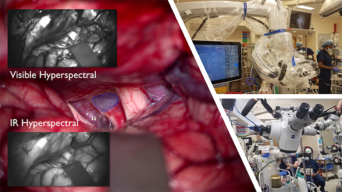
In a manuscript published in Bioengineering, the team of engineers and neurosurgeons details how its snapshot hyperspectral imaging (sHSI) camera can be used to capture and process images of brain tissue, using the wide spectrum of light between visible and infrared wavelengths. That additional information — beyond the human eye — has the potential to allow for more accurate and complete tumor removal.
“In the hands of a neurosurgeon, this camera, when combined with machine learning, could dramatically improve outcomes for some of our most vulnerable brain tumor patients,” said Richard Jaepyeong Cha, Ph.D., an optical engineer and principal investigator at the Sheikh Zayed Institute of Pediatric Surgical Innovation. “We are able to attach the camera to a surgical microscope and process a significant amount of information from the patient while in the operating room. Not only could this lead to more complete tumor resection, it will also allow the surgeon to save as much healthy brain tissue as possible and reduce lifelong neurological complications.”
Why we’re excited
Brain tumors are the most common solid tumors in children, accounting for the highest number of pediatric cancer deaths globally each year. To develop a treatment plan, neurosurgeons need to understand the tumor’s features, including its type, grade of malignancy, location and its categorization as a primary or metastatic cancer. This information leads to decisions about how to remove or biopsy a tumor.
Under the current protocols, surgeons evaluate tumor margins in the operating room by examining the appearance of the brain tissue and sending out small samples to the pathology department for biopsies. This can lead to longer surgeries and difficult real-time surgical decisions. For instance, some low-grade tumors are visually indistinguishable from healthy brain tissue.
In four investigational cases approved by the hospital’s institutional research board, the sHSI camera was used in the operating room to help segment healthy pediatric brain tissue from tumors. Unlike the conventional red-green-blue (RGB) imaging cameras, which use only those three colors, HSI captures spectral data at each pixel of the image — a task too complex for the human eye — and sends it instantly for processing by an algorithm designed to assist in tumor segmentation.
What’s ahead
Despite the small dataset, the researchers were able to successfully segment healthy brain tissue from lesions with a high specificity during pediatric brain tumor resection procedures. Significant work remains to refine the technology and the machine learning behind it. Researchers also plan to integrate the sHSI camera into a laparoscope to visualize tumors that are not on the brain’s surface and collect data from more angles.
“As we develop these groundbreaking tools, we plan to continue to expand the dataset and refine the algorithm to make pediatric neurosurgery continually more precise,” said Naomi Kifle, M.S., research and development engineer at Children’s National and first author on the paper. “As our dataset grows, we hope to create a model that can distinguish healthy brain tissue, tumor and skull. This groundbreaking surgical tool shows significant promise.”