Microscopic visual of a diseased muscle section

Gene therapy offers potential long-term treatment for limb-girdle muscular dystrophy 2B

Microscopic visual of a diseased muscle section

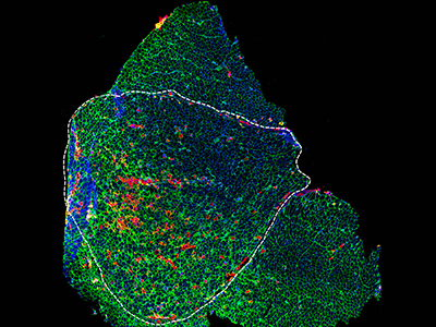
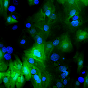
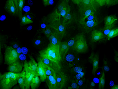
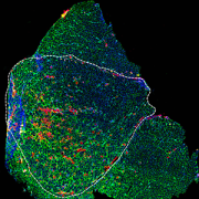
Microscopic visual of a diseased muscle section. Credit: Daniel Bittel.

Children’s National Hospital experts developed a new pre-clinical gene therapy for a rare disorder, known as limb-girdle muscular dystrophy (LGMD) 2B, that addresses the primary cellular deficit associated with this disease. Using a single injection of a low dose gene therapy vector, researchers restored the ability of injured muscle fibers to repair in a way that reduced muscle degeneration and enhanced the functioning of the diseased muscle. The treatment was safe, attenuated fibro-fatty muscle degeneration, and restored myofiber size and muscle strength, according to the study published in the Journal of Clinical Investigation.

With an incidence of less than 1 in 100,000, LGMD2B is a rare disorder caused by a genetic mutation in a large gene called dysferlin. This faulty gene leads to muscle weakness in the arms, legs, shoulder and pelvic girdle. Affected children and adults face trouble walking, climbing stairs and getting out of chairs. Individuals typically lose the ability to walk within years after the onset of symptoms, and often need assistance with everyday tasks such as showering, dressing and transferring.

This study described a new approach that avoids the need for packaging a large gene, like dysferlin, or giving a large vector dose to target the muscles, which are bottlenecks faced in ongoing gene therapy efforts aimed at muscular dystrophies.

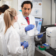
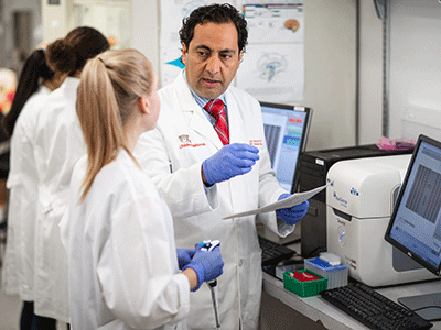
“Currently, patients with LGMD2B have no gene or drug-based therapies available to them, and we are amongst the few centers developing therapeutic approaches for this disease,” said Jyoti K. Jaiswal, M.Sc. Ph.D., senior investigator of the Center for Genetic Medicine Research at Children’s National. “We are working to further enhance the efficacy of this approach and perform a longer-term safety and efficacy study to enable the clinical translation of this therapy.”

The genetic defect in dysferlin that is associated with LGMD2B causes the encoded protein to be truncated or degraded. This hinders the muscle fiber’s ability to heal, which is required for healthy muscles. In recessive genetic disorders, like LGMD2B, common pre-clinical gene therapy approaches usually target the mutated gene in the muscle, making them capable of producing the missing proteins.

“The large size of the gene mutated in this disease, and impediments in body-wide delivery of gene therapy vectors to reach all the muscles, pose significant challenges for developing gene therapies to treat this disease,” said Jaiswal.

To overcome these challenges, the researchers found another way to slow down the disease’s progression. The authors built upon their previous discovery that acid sphingomyelinase (hASM) protein is required to repair injured muscle cells. In this current work, the research team administered a single in vivo dose of an Adeno-associated virus (AAV) vector that produces a secreted version of hASM in the liver, which then was delivered to the muscles via blood circulation at a level determined to be efficacious in repairing LGMD2B patient’s injured muscle cells.

“Increased muscle degeneration necessitates greater muscle regeneration, and we found that improved repair of dysferlin-deficient myofibers by hASM-AAV reduces the need for regeneration, causing a 2-fold decrease in the number of regenerated myofibers,” said Daniel Bittel, D.P.T., PhD., research postdoctoral fellow of the Center for Genetic Medicine Research at Children’s National and a lead author of this study.

Sreetama Sen Chandra, Ph.D., who was a research postdoctoral fellow at Children’s National at the time of this study and served as co-lead author, also added that “these findings are also of interest to patients with Niemann-Pick disease type A since the pre-clinical model for this disease also manifests poor sarcolemma repair.”

Children’s National researchers of the Center for Genetic Medicine Research and the Rare Disease Institute (RDI) are constantly pursuing high-impact opportunities in pediatric genomic and precision medicine. Both centers combine its strengths with public and private partners, including industry, universities, federal agencies, start-up companies and academic medical centers. They also serve as an international referral site for rare disorders.

Gene therapy Schematic

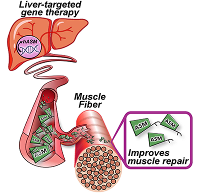
Gene therapy Schematic. Credit: Daniel Bittel.

computer circuit board

Integrating clinical parameters with lung imaging to predict respiratory outcomes in premature babies

computer circuit board

The team will develop an objective framework to predict the risk and assess the severity of respiratory disease in premature babies using non-invasive low-radiation X-ray imaging biomarkers and clinical parameters from the patient bedside.

Children’s National Hospital received a $1.7M award from the National Institutes of Health (NIH) National Heart, Lung, and Blood Institute (NHLBI) to develop computational tools that integrate continuous clinical parameters with lung imaging to predict respiratory outcomes for babies born severely premature in newborn intensive care unit (NICU) settings.

The multi-disciplinary team of internationally recognized experts in quantitative imaging, machine learning and neonatal respiratory research believes they can improve clinical practice. To get there, they will develop an objective framework to predict the risk and assess the severity of respiratory disease in premature babies using non-invasive low-radiation X-ray imaging biomarkers and clinical parameters from the patient bedside.

“This computational tool will assist clinicians in making critical decisions about the course of therapy and other necessary follow-ups,” said Gustavo Nino, M.D., M.S.H.S., D’A.B.S.M., principal investigator in the Center for Genetic Medicine at Children’s National. “An objective informed decision about the severity of lung disease in prematurity will result in fewer rehospitalizations, better long-term outcomes and life-saving benefits.”

Prematurity is the largest single cause of death in children under five in the world. Lower respiratory tract infections (LRTI) are the top cause of hospitalization and mortality in premature infants. Clinical tools to predict the risk and assess the severity of LRTI in premature babies are needed to allow early interventions that can decrease the high morbidity and mortality in this patient group.

“Our new technology will provide clinicians an accurate, fast and comprehensive summary of the respiratory status of premature babies,” said Dr. Nino. “The data analysis along with the software technology will help determine if a premature baby seen in the NICU can be safely discharged or will require further monitoring and treatment.”

Predictive analytics could help in many ways. For example, there are instances where newborns in the NICU are on the right path with no risks in the future, but there are babies who will come back with severe infections.

“In the first scenario, if we can predict earlier that they’re fine, this could reduce the number of chest X-rays and extra tests, so we assess that this child can be safely sent home,” said Marius George Linguraru, D.Phil., M.A., M.Sc., principal investigator in the Sheikh Zayed Institute for Pediatric Surgical Innovation at Children’s National. “On the other hand, for kids that may come back to the hospital in the near future, we could predict earlier that they are not that well by looking at images and other continuous measurements such as supplemental oxygen.”

This approach, in essence, is a collection of continuous data from the NICU, which is very complex itself because it needs to be collected every day and fed into a machine learning model that digests the data to identify risk patterns for the health of the lung.

“If we find that there is still a risk, it does not necessarily mean that the child has to stay in the NICU any longer, but they might continue treatment, and we will have to define how this integrates into the clinical management of these patients,” said Linguraru. “If there is something in the data that we can put our finger on, we will know which kids require timely attention, hopefully reducing future adverse situations with potential comorbidities and financial burdens.”

cystic kidney disease

American Heart Association grant funds study of vascular complications in ADPKD

cystic kidney disease

Ashima Gulati, M.D., Ph.D., pediatric nephrologist at Children’s National Hospital recently was awarded a grant from the American Heart Association. Dr. Gulati’s work will aim to identify the genetic determinants of vascular complications in autosomal dominant polycystic kidney disease (ADPKD).

Cerebrovascular complications such as vascular aneurysms and anomalies are an important cause of morbidity in ADPKD that need to be studied. The goal of Dr. Gulati’s research is to contribute to knowledge towards using molecular medicine to inform individual genetic risk of clinically significant vascular complications in ADPKD.

This work addresses a clinically significant vascular complication in ADPKD, the most common inherited form of kidney failure world-wide.

Hans Pohl

Q&A with Hans Pohl, M.D., on the future of pediatric urology

Hans Pohl

Hans Pohl, Division Chief, Urology

The Urology team at Children’s National Hospital is led by Hans Pohl, M.D., and includes seven other fellowship-trained pediatric urologists and three nurse practitioners. Dr. Pohl has experience in treating patients with spina bifida and bladder exstrophy, in addition to the other more common diagnoses and in using laparoscopy to perform surgery through minimally invasive techniques.

Here, Dr. Pohl tells us more about the department he is leading and what it means for the future of pediatric urology patients at Children’s National.

What excites you most about current research in the field of urology?

The most cutting-edge research these days pertains to understanding the complex and diverse role played by bacteria in the urinary tract. We previously thought the urinary tract is sterile but that turns out to not be the case. Just like the friendly bacteria living in our bodies, there are bacteria that live within our urinary tracts. It is thought that when the normal function of the urinary tract is disrupted, the balance of healthy to unhealthy bacteria is disrupted. Our faculty at the Children’s National Urology Division are looking at urinary tract infection (UTI) from various aspects. Michael Hsieh, M.D., is investigating the role of bacteria in causing inflammation; Daniel Casella, M.D., has investigated how a drug called varenicline might reduce inflammation in infected kidneys; and Briony Varda, M.D., will be investigating the socioeconomic factors affecting how people living with spina bifida manage their urinary tracts and UTIs. By looking at UTIs at all levels from bacteria factors to host factors to treatment we will understand better how to reduce the impact of a very common problem on human beings.

What are some of the most valuable changes or advancements for the program you hope to see in the next couple of years?

I believe that our next step in program evolution will focus on improving the quality of life of children with urination abnormalities and UTIs. Everyone has experienced that their child has urinary symptoms at some point during childhood, some also have UTIs. Pediatric urologists have learned that normal lower urinary tract function is critically important in reducing a child’s risk for a UTI. Even if a child does not have UTIs, there can be significant social stigma amongst a child’s peers when loss of urine control happens.

What makes the Urology Division at Children’s National unique from other programs in the country?

We have grown considerably over the past several years, adding to our faculty surgeons with complimentary skills outside of the operating room. For instance, we have pediatric urologists who are also basic scientists, translational scientists, systems scientists, a clinical informaticist and minimally invasive surgeons. These varied qualities create a dynamic group of people who bring diverse perspectives to treating patients’ problems and generating creative solutions. We believe that our democratic process of complex care management where all surgeons can openly think about how to optimize patient management is unique. Patients don’t get one opinion from their surgeon, they get ten.

Blood sample tube for anti-Müllerian hormone

A look at the clinical utility of anti-Mullerian hormone

Blood sample tube for anti-Müllerian hormone

Anti-Mullerian hormone (AMH), also known as Mullerian inhibiting substance (MIS), is a hormone produced exclusively in the gonads. It was originally described in the context of sexual differentiation in the male fetus but has gained prominence now as a marker of ovarian reserve and fertility in females.

In a mini-review published in The Journal of Clinical Endocrinology and Metabolism, Roopa Kanakatti Shankar, M.D., pediatric endocrinologist at Children’s National Hospital and an associate professor of pediatrics at the George Washington University School of Medicine and Health Sciences, and co-authors offer an updated synopsis on AMH and its clinical utility in pediatric patients.

The authors performed a systematic search for studies related to the physiology of AMH, normative data and clinical role in pediatrics. After reviewing 70 clinical studies and systematic reviews, they conclude that, “AMH has widespread clinical diagnostic utility in pediatrics but interpretation is often challenging and should be undertaken in the context of not only age and sex but also developmental and pubertal stage of the child.”

Other authors from Children’s National Hospital include Andrew Dauber, M.D., MMSc, Tazim Dowlut-Mcelroy, M.D., and Veronica Gomez-Lobo, M.D.

Read the full review in The Journal of Clinical Endocrinology and Metabolism.

Rare Diseases Institute sign

Children’s National Rare Disease Institute named a Center of Excellence

Rare Diseases Institute sign

RDI, which includes the largest clinical group of pediatric geneticists in the nation, focuses on developing the clinical care field of more than 8,000 rare diseases currently recognized and advancing the best possible treatments for children with these diseases.

The Rare Disease Institute (RDI) at Children’s National Hospital announced its designation as a NORD Rare Disease Center of Excellence, joining a highly select group of 31 medical centers nationwide. This new, innovative network seeks to expand access and advance care and research for rare disease patients in the United States. The program is being led by the National Organization for Rare Disorders (NORD), with a goal to foster knowledge sharing between experts across the country, connect patients to appropriate specialists regardless of disease or geography, and to improve the pace of progress in rare disease diagnosis, treatment and research.

“Children’s National has worked closely with NORD to move this program forward and is very proud to be amongst the first group of recognized centers,” said Marshall Summar, M.D., chief of the Division of Genetics and Metabolism and the director of RDI at Children’s National. “This is a recognition of the institutional efforts, as we take care of patients with the rare disease and help set the standard for the field.”

RDI, which includes the largest clinical group of pediatric geneticists in the nation, focuses on developing the clinical care field of more than 8,000 rare diseases currently recognized and advancing the best possible treatments for children with these diseases.

In February 2021, RDI became the first occupant of the new Children’s National Research & Innovation Campus, a first-of-its-kind pediatric research and innovation hub. The campus now also houses the Center for Genetic Medicine Research, and together researchers are constantly pursuing high-impact opportunities in pediatric genomic and precision medicine. Both centers combine its strengths with public and private partners, including industry, universities, federal agencies, start-up companies and academic medical centers. They also serve as an international referral site for rare disorders.

People living with rare diseases frequently face many challenges in finding a diagnosis and quality clinical care. In establishing the Centers of Excellence program, NORD has designated clinical centers across the U.S. that provide exceptional rare disease care and have demonstrated a deep commitment to serving rare disease patients and their families using a holistic, state of the art approach.

“Right now, far too many rare diseases are without an established standard of care. The Centers of Excellence program will help set that standard – for patients, clinicians, and medical centers alike,” said Ed Neilan, chief scientific and medical officer of NORD. “We are proud to announce Children’s National as a NORD Rare Disease Center of Excellence and look forward to their many further contributions as we collectively seek to improve health equity, care and research to support all individuals with rare diseases.”

Each center was selected by NORD in a competitive application process requiring evidence of staffing with experts across multiple specialties to meet the needs of rare disease patients and significant contributions to rare disease patient education, physician training and research.

Dr. Javad Nazarian

Q&A with Dr. Javad Nazarian on his upcoming work on low-grade gliomas

Dr. Javad Nazarian

Supported by the Gilbert Family Foundation, Dr. Nazarian’s return is part of a special research program within the Gilbert Family Neurofibromatosis Institute that focuses on NF1 research.

Javad Nazarian, Ph.D., M.Sc., associate professor of Pediatrics at George Washington University and professor at the University of Zurich, has expanded his research group at Children’s National to focus on Neurofibromatosis type 1 (NF1) transformed low-grade gliomas (LGGs). Dr. Nazarian will apply his expertise from establishing a successful DIPG (diffuse intrinsic pontine glioma) and DMG (diffuse midline glioma) program in Zurich Switzerland and previously at Children’s National.

In addition to his continued research in Zurich, as a principal investigator at the Department of Genomics and Precision Medicine at Children’s National Dr. Nazarian plans on aggregating his knowledge to the new research and work spearheaded at Children’s National. As one of the first research teams to move to the Children’s National Research & Innovation Campus, Dr. Nazarian’s group is excited to use the opportunity to establish cutting-edge and clinically translational platforms.

Supported by the Gilbert Family Foundation, Dr. Nazarian’s return is part of a special research program within the Gilbert Family Neurofibromatosis Institute that focuses on NF1 research. This research includes associated gliomas with a special emphasis on NF1-associated transformed anaplastic LGGs. His team will develop new avenues of research into childhood and young adult NF-associated LGGs with a special emphasis on transformed high-grade gliomas.

Dr. Nazarian is excited for what’s to come and his goals are clear and set. Here, Dr. Nazarian tells us more about his main objectives and what it means for the future of pediatric neuro-oncology care at Children’s National.

  1. What excites you most about being back at Children’s National?

I have received most of my training at Children’s National, so this is home for me. Being one of the nation’s top children’s hospitals gives a unique advantage and ability to advocate for childhood diseases and cancers. It is always exciting to play a part in the vision of Children’s National.

  1. What are some of the lessons learned during your time working in Zurich? And how do you think these will compliment your work at Children’s National?

We developed a focused group with basic research activities intertwined with clinical needs.  The result was the launch of two clinical trials. I also helped in developing the Diffuse Midline Glioma-Adaptive Combinatory Trial (DMG-ACT) working group that spans across the world with over 18-member institutions that will help to design the next generation clinical trials. I will continue leading the research component of these efforts, which will have a positive impact on our research activities at Children’s National.

  1. How does your work focusing on low-grade gliomas formulating into high-grade gliomas expand and place Children’s National as a leader in the field?

Scientifically speaking, transformed LLGs are very intriguing. I became interested in the field because these tumors share molecular signatures similar to high-grade gliomas (HGGs). Our team has done a great job at Children’s National to develop tools – including biorepositories, avatar models, drug screening platforms, focused working groups, etc. – for HGGs. We will apply the same model to transformed LGGs with the goal of developing biology-derived clinical therapeutics for this patient population.

  1. How will this work support families and patients seeking specific neuro-oncology care?

We will develop new and high thruput tools so that we can better study cancer formation or transformation. These tools and platforms will allow us to screen candidate drugs that will be clinically effective. The main focus is to accelerate discovery, push drugs to the clinic, feed information back to the lab from clinical and subsequently design better therapies.

  1. You are one of the first scientists to move to the Children’s National Research & Innovation Campus. What are some of the valuable changes or advancements you hope to see as a result of the move?

The campus will provide high-end facilities, including cutting-edge preclinical space, and allow for team expansion. The close proximity to Virginia Tech will also provide an environment for cross-discipline interactions.

  1. Anything else you think peers in your field should know about you, the field or our program?

The team at Children’s National includes Drs. Roger Packer and Miriam Bornhorst. Both have provided constant clinical support, innovation and clinical translation of our findings. I look forward to working with them.

smiling baby sleeping

Link between early lower respiratory tract infections and obstructive sleep apnea

smiling baby sleeping

For the first time, researchers at Children’s National Hospital have identified the association between early LRTI and the development of OSA in children.

Several birth cohorts have defined the pivotal role of early lower respiratory tract infections (LRTI) in the inception of pediatric respiratory conditions. However, the association between early LRTI and the development of obstructive sleep apnea (OSA) in children had not previously been made.

Now, for the first time, researchers at Children’s National Hospital have identified the association between early LRTI and the development of OSA in children, according to a study published in the journal SLEEP.

“These results suggest that respiratory syncytial virus LRTI may contribute to the pathophysiology of OSA in children,” said Gustavo Nino, M.D., director of sleep medicine at Children’s National.

The study also demonstrated that children with a history of severe respiratory syncytial virus (RSV) bronchiolitis during early infancy had more than double the odds of developing OSA during the first five years of life independently of other risk factors.

“The results suggest that RSV LRTI may contribute to the pathophysiology of OSA in children, raising concern for the possibility that primary prevention strategies can hinder the initial establishment of OSA following early viral LRTIs,” said Dr. Nino. “Primary prevention of OSA in children would have a dramatic effect in reducing the increasing incidence of this condition and in preventing its detrimental effects on childhood health and beyond.”

The novel findings also raise the possibility that anticipatory strategies and interventions can be developed to identify and prevent the initial establishment of OSA following viral respiratory infections during early infancy. This could provide a dramatic effect in reducing the increasing incidence of this condition and its multiple detrimental effects on childhood health and beyond.

“Our study offers a new paradigm for investigating mechanisms implicated in the early pathogenesis of OSA in the pediatric population,” said Dr. Nino.

Marishka Brown, Ph.D., director of the National Center on Sleep Disorders Research at the National Heart, Lung, and Blood Institute (NHLBI), part of the National Institutes of Health (NIH), agreed.

“The findings from this study suggest that viral lower respiratory tract infections could predispose to the development of sleep-disordered breathing in later childhood,” Brown said. “More research to determine how these infections affect airway function could lead to a better understanding of how sleep apnea develops in pediatric patients.”

This study includes funding support from the NIH, including the NHLBI.

The Pulmonary Division at Children’s National has been ranked as one of the top ten programs in the nation by U.S. News & World Report.

RSV infected infant cells

$2.13M grant accelerates treatments for kids with Down syndrome experiencing respiratory viruses

RSV infected infant cells

Children’s National Hospital received a combined $2.13 million award from the National Institutes of Health’s (NIH) National Heart, Lung and Blood Institute to better understand the mechanisms of severe viral respiratory infections in patients with Down syndrome and to develop new diagnostic tools and innovative precision medicine approaches for this vulnerable population.

“We have a unique opportunity to discover novel targets that can treat severe viral respiratory infections, including SARS-CoV-2,” said Gustavo Nino, M.D., M.S.H.S., D’A.B.S.M., principal investigator in the Center for Genetic Medicine at Children’s National. “Part of the award will help us accelerate the development of these novel approaches to prevent severe respiratory infections caused by SARS-CoV-2 and other viruses like respiratory syncytial virus infection (RSV) in children and adults with Down syndrome.”

Lower respiratory tract infections are a leading cause of hospitalization and death in children with Down syndrome. Those children have a nine times higher risk for hospitalization and mortality due to respiratory viruses that cause lower respiratory tract infections.

Chromosome 21, which is an extra chromosome copy found in patients with Down syndrome, encodes four of the six known interferon receptors, leading to hyperactivation of interferon response in Down syndrome. With the central role of interferons focused on antiviral defense, it remains puzzling how interferon hyperactivation contributes to severe viral lower respiratory tract infections in children with Down syndrome. This is an area that the researchers will explore to better manage and treat viral lower respiratory tract infections in these patients, with the support of NIH’s INCLUDE initiative. INCLUDE provides institutions with grants to help clinical research and therapeutics to understand and diminish risk factors that influence the overall health, longevity, and quality of life for people with Down syndrome related to respiratory viruses.

“While many of the other studies focus on intellectual and other disabilities, we are exploring a novel viral respiratory infectious disease mechanism and are doing so by working directly with patients and patient-derived samples,” said Jyoti Jaiswal, M.Sc., Ph.D., senior investigator in the Center for Genetic Medicine Research at Children’s National.

Children with Down syndrome have historically been excluded in research related to airway antiviral immunity, which is a focus of this human-based transformative study to improve the health and survival of patients with Down syndrome. There is a critical need for studies that define targetable molecular and cellular mechanisms to address dysregulated antiviral responses in this patient population.

“The clinical expertise at Children’s National in studying Down syndrome and the work of our team in caring for these patients with respiratory and sleep disorders positions us well to pursue this work,” said Jaiswal. “This is further supplemented by our initial studies that have identified a novel mechanism of impaired airway antiviral responses in these patients.”

Congresswoman Eleanor Holmes Norton (D-DC) also celebrated Children’s National and its NIH research funding benefitting people with Down syndrome.

“I am pleased to congratulate Dr. Nino and staff on being the recipients of the National Heart, Lung, & Blood Institute grant. You were chosen from a competitive group of applicants and should be proud of this notable achievement,” said Norton in a letter. “By receiving this grant, you have demonstrated outstanding promise in your field. It is my hope that this grant will enable you to better the local and global community.”

Dr. Matthew Bramble, Vincent Kambale, and Neerja Vashist

Gut microbiome may impact susceptibility to konzo

Dr. Matthew Bramble, Vincent Kambale, and Neerja Vashist

From left to right: Dr. Matthew Bramble, Vincent Kambale, and Neerja Vashist. Here, the team is processing samples in the field collected from the study cohort prior to storage in liquid nitrogen. Bramble et al. Nature Communications (2021).

Differences between gut flora and genes from konzo-prone regions of the Democratic Republic of Congo (DRC) may affect the release of cyanide after poorly processed cassava is consumed, according to a study with 180 children. Cassava is a food security crop for over half a billion people in the developing world. Children living in high-risk konzo areas have high glucosidase (linamarase) microbes and low rhodanese microbes in their gut, which could mean more susceptibility and less protection against the disease, suggest Children’s National Hospital researchers who led the study published in Nature Communications.

Konzo is a severe, irreversible neurologic disease that results in paralysis. It occurs after consuming poorly processed cassava — a manioc root and essential crop for DRC and other low-income nations. Poorly processed cassava contains linamarin, a cyanogenic compound. While enzymes with glucosidase activity convert starch to simple sugars, they also break down linamarin, which then releases cyanide into the body.

Neerja Vashist learning how to make fufu

Neerja Vashist is learning how to make fufu. Fufu is a traditional food made from cassava flour, and the cassava flour used in the making of the fufu here has gone through the wetting method to further remove toxins from the cassava flour prior to consumption. Bramble et al. Nature Communications (2021).

“Knowing who is more at risk could result in targeted interventions to process cassava better or try to diversify the diet,” said Eric Vilain, M.D., Ph.D., director of the Center for Genetic Medicine Research at Children’s National. “An alternative intervention is to modify the microbiome to increase the level of protection. This is, however, a difficult task which may have unintended consequences and other side effects.”

The exact biological mechanisms underlying konzo disease susceptibility and severity remained poorly understood until now. This is the first study to shed light on the gut microbiome of populations that rely on toxic cassava as their primary food source.

“While the gut microbiome is not the sole cause of disease given that environment and malnourishment play a role, it is a required modulator,” said Matthew S. Bramble, Ph.D., staff scientist at Children’s National. “Simply stated, without gut microbes, linamarin and other cyanogenic glucosides would pose little to no risk to humans.”

To understand the influence of a detrimental subsistence on the gut flora and its relationship to this debilitating multifactorial neurological disease, the researchers compared the gut microbiome profiles in 180 children from the DRC using shotgun metagenomic sequencing. This approach evaluates bacterial diversity and detects the abundance of microbes and microbial genes in various environments.

The samples were collected in Kinshasa, an urban area with diversified diet and without konzo; Masi-Manimba, a rural area with predominant cassava diet and low prevalence of konzo; and Kahemba, a region with predominant cassava diet and high prevalence of konzo.

Dr. Nicole Mashukano and Dr. Matthew Bramble wetting cassava flour

From left to right: Dr. Nicole Mashukano and Dr. Matthew Bramble. Dr. Mashukano leads the efforts in Kahemba to teach the wetting method to individuals in different health zones. The wetting method is used as an additional step to further detoxify toxins from cassava flour prior to consumption. Here, Dr. Mashukano and Dr. Bramble are spreading out the wet mixture of cassava flour and water into a thin layer on a tarp for drying in the sun, which allows cyanogen breakdown and release in the form of hydrogen cyanide gas. Bramble et al. Nature Communications (2021).

“This study overcame many challenges of doing research in low-resource settings,” said Desire Tshala-Katumbay, M.D., M.P.H., Ph.D., FANA, co-senior author and expert scientist at Institut National de Recherche Biomédicale in Kinshasa, DRC, and professor of neurology at Oregon Health & Science University. “It will open novel avenues to prevent konzo, a devastating disease for many children in Sub-Saharan Africa.”

For next steps, the researchers will study sibling pairs from konzo-prone regions of Kahemba where only one sibling is affected with the disease.

“Studying siblings will help us control for factors that cannot be controlled otherwise, such as the cassava preparation in the household,” said Neerja Vashist, Ph.D. candidate and research trainee at Children’s National. “In this work, each sample had approximately 5 million DNA reads each, so for our follow-up, we plan to increase that to greater than 40 million reads per sample and the overall study cohort size. This study design will allow us to confirm that the trends we observed hold on a larger scale, while enhancing our ability to comprehensively characterize the gut microbiome.”

Could whole-exome sequencing become a standard part of state newborn screening?

smiling baby boy

There are concerns about implementing whole-exome sequencing since it takes away the child’s right to decide if they want to know — or not — about their specific inherited disease.

It is still premature to standardize an innovative methodology known as whole-exome sequencing (WES) as part of state newborn screening programs, argues Beth A. Tarini, M.D., M.S., associate director for the Center of Translational Research at Children’s National Hospital, in a new editorial published in JAMA Pediatrics.

About 4 million infants are born annually in the United States. Newborn screening is a mandatory state-run public health program that screens infants for inherited diseases in the first days of life so they can receive treatment before irreversible damage occurs. Several of these screening tests are done on blood drawn from an infant’s heel.

WES holds the potential to screen infants for thousands of disorders and traits, including those that appear in adulthood. But there are concerns about implementing WES since it takes away the child’s right to decide if they want to know — or not — about their specific inherited disease. There is also the unknown effect that it could have on their ability to obtain health insurance.

“As caretakers for their children, parents have the challenge of deciding what kind of information, including genetic, will be valuable for their child,” says Dr. Tarini. “As a society, we have the responsibility of deciding where the healthcare dollars get the best return – especially when it comes to children. We need to start that conversation for universal genomic sequencing of newborns sooner rather than later.”

The Pereira et al. study, appearing in the new edition of JAMA Pediatrics and referenced in Dr. Tarini’s editorial, is the first to demonstrate no significant harm in the initial 10 months of life after performing WES under the best conditions of access to resources and a controlled environment.

While the Pereira et al. study has limited data on the effects of WES on families from underrepresented backgrounds, Dr. Tarini notes that it does provide a critical first step in this area of pediatric genomic research and for policy decision-making about the widespread implementation of WES in newborns.

“Moving forward, the U.S. will have to make a collective decision about the value of WES for newborns,” says Dr. Tarini. That value calculus cannot be made without consideration of the general state of healthcare for infants. As she points out, “This is not an easy question to answer in a country whose infant mortality ranks 34th according to the Organization for Economic Co-operation and Development (OECD).”

Dr. Tarini’s research identifies ways to optimize the delivery of genetic services to families and children, particularly newborn screening. She has also chaired state newborn screening committees and served on several federal newborn screening committees.

Drs. Packer and van den Acker at the Pediatric Device Innovators Forum

Pediatric Device Innovators Forum explores state of focused ultrasound

For children living with pediatric tumors, less invasive and less painful treatment with no radiation exposure was not always possible. In recent years, the development of technologies like Magnetic resonance guided high intensity focused ultrasound (MR-HIFU) and Low intensity transcranial focused ultrasound (LIFU) is helping to reverse that trend.

This topic was the focus of the recent Pediatric Device Innovators Forum (PDIF) hosted by the National Capital Consortium for Pediatric Device Innovation (NCC-PDI) in partnership with the U.S. Food and Drug Administration’s (FDA) Pediatric Device Consortia (PDC) grant program. A collaboration between Children’s National Hospital and University of Maryland Fischell Institute for Biomedical Devices, NCC-PDI is one of five PDCs funded by the FDA to support pediatric device innovators in bringing more medical devices to market for children.

The discussion, moderated by Kolaleh Eskandanian, Ph.D., MBA, PMP, vice president and chief innovation officer at Children’s National and principal investigator of NCC-PDI, explored the use of focused ultrasound’s noninvasive therapeutic technology for two pediatric indications, Osteoid Osteoma (OO) and Diffuse Intrinsic Pontine Glioma (DIPG), and the ways it can increase the quality of life for pediatric patients while also decreasing the cost of care.

The discussion also examined the most common barriers preventing more widespread implementation of focused ultrasound technology, specifically small sample size for evidence generation, lack of funding opportunities and reimbursement issues that can make or break a technology’s chances at reaching the patients that need it.

Karun Sharma, M.D., director of Interventional Radiology at Children’s National, emphasized the potential for focused ultrasound to treat localized pain relief and treat other diseases that, like OO, do not have any other therapeutic alternative

“At Children’s National, we use MR-HIFU to focus an ultrasound beam into lesions, usually tumors of the bone and soft tissues, to heat and destroy the harmful tissue in that region, eliminating the need for incisions,” says Sharma. “In 2015, Children’s National doctors became the first in the U.S. to use MR-HIFU to treat pediatric osteoid osteoma (OO), a painful, but benign, bone tumor that commonly occurs in children and young adults. The trial demonstrated early success in establishing the safety and feasibility of noninvasive MR-HIFU in children as an alternative to current, more invasive approaches to treat these tumors.”

In November 2020, the FDA approved this MR-HIFU system to treat OO in pediatric patients.

Roger Packer, M.D., senior vice president of the Center for Neuroscience and Behavioral Medicine at Children’s National, also discussed how focused ultrasound, specifically LIFU, has also proven to be an attractive modality for its ability to non-invasively, focally and temporarily disrupt the blood brain barrier (BBB) to allow therapies to reach tumors that, until recently, would have been considered unreachable without severe intervention.

“This presents an opportunity in pediatric care to treat conditions like Diffuse Intrinsic Pontine Glioma (DIPG), a highly aggressive brain tumor that typically causes death and morbidity,” says Packer.

Packer is planning a clinical trial protocol to investigate the safety and efficacy of LIFU for this pediatric indication.

The forum also featured insight from Jessica Foley, M.D., chief scientific officer, Focused Ultrasound Foundation; Arjun Desai, M.D., chief strategic innovation officer, Insighttec; Arun Menawat, M.D., chairman and CEO, Profound Medical; Francesca Joseph, M.D., Children’s National; Johannes N. van den Anker, M.D., Ph.D., vice chair of Experimental Therapeutics, Children’s National; Gordon Schatz, president, Schatz Reimbursement Strategies; Mary Daymont, vice president of Revenue Cycle and Care Management, Children’s National; and Michael Anderson, MD, MBA, FAAP, FCCM, FAARC, senior advisor to US Department of Health and Human Services (HHS/ASPR) and Children’s National.

Anthony Sandler, M.D., senior vice president and surgeon-in-chief of the Joseph E. Robert Jr. Center for Surgical Care and director of the Sheikh Zayed Institute for Pediatric Surgical Innovation at Children’s National Hospital, and Sally Allain, regional head of Johnson & Johnson Innovation, JLABS @ Washington, DC, opened the forum by reinforcing both organizations’ commitment to improving pediatric health.

In September 2020, the Focused Ultrasound Foundation designated Children’s National Hospital as the first global pediatric Center of Excellence for using this technology to help patients with specific types of childhood tumors. As a designated COE, Children’s National has the necessary infrastructure to support the ongoing use of this technology, especially for carrying out future pediatric clinical trials. This infrastructure includes an ethics committee familiar with focused ultrasound, a robust clinical trials research support team, a data review committee for ongoing safety monitoring and annual safety reviews, and a scientific review committee for protocol evaluation.

The Pediatric Device Innovators Forum is a recurring collaborative educational experience designed by the FDA-supported pediatric device consortia to connect and foster synergy among innovators across the technology development ecosystem interested in pediatric medical device development. Each forum is hosted by one of the five consortia. This hybrid event took place at the new Children’s National Research and Innovation Campus, the first-of-its-kind focused on pediatric health care innovation, on the former Walter Reed Army Medical Center campus in Washington, D.C.

To view the latest edition of the forum, visit the NCC-PDI website.

Panelists at the Pediatric Device Innovators Forum

The recent Pediatric Device Innovators Forum (PDIF) exploring the state of focused ultrasound was held at the new Children’s National Research and Innovation Campus, a first-of-its-kind focused on pediatric health care innovation.

morphine vial and needle

Replacing morphine with methadone in the NICU

morphine vial and needle

A synthetic analgesic drug, known as methadone, may serve as a better alternative for newborns in the neonatal intensive care unit (NICU) suffering from opioid withdrawal syndrome, according to a commentary published in Pediatric Research.

A synthetic analgesic drug, known as methadone, may serve as a better alternative for newborns in the neonatal intensive care unit (NICU) suffering from opioid withdrawal syndrome, according to a commentary published in Pediatric Research. Some existing literature suggests that methadone may also address painful stimuli that hinders neurodevelopment throughout adulthood, added Johannes van den Anker, M.D., Ph.D., division chief of Clinical Pharmacology at Children’s National Hospital.

The commentary was selected as the Editor’s Focus in Pediatric Research for the June editionsignaling the scientific community as noteworthy to further explore methadone’s potential as an alternative for pharmacologic treatments instead of morphine.

“It is important to define the pharmacokinetics and pharmacodynamics of methadone to treat pain in neonates in intensive care before replacing morphine with methadone. Pre-clinical research shows that the use of methadone might have fewer side effects than morphine,” said Dr. van den Anker. “If this is also the case in the human neonate, then a shift from morphine to methadone might be beneficial. However, first, we need to define what the safe and effective dose of methadone will be for this purpose.”

While there is a need to better understand how newborns and preemies metabolize methadone, there is existing knowledge that this drug minimizes pain. The commentary, too, raises the question for clinicians to possibly consider methadone as a better option to avoid long-term negative neurodevelopmental consequences — such as hypersensitivity to re-injury in later life — usually associated with pain.

The current but limited data out there still provides “exciting and stimulating” information about the possible use of methadone for the treatment of neonatal pain in the NICU, according to Dr. van den Anker. He believes that, in the future, methadone could also serve as mechanism-based analgesia in newborns experiencing pain.

“There needs to be a collaboration between neonatal medicine specialists, pharmacometricians and developmental pharmacologists to assure not only the generation of evidence-based data to determine these optimal dosing regimens, but also to facilitate the implementation of this new knowledge into daily clinical care in neonatal intensive care units across the globe,” added Dr. van den Anker.