Orthopedics

Scoliosis X-ray image

Improved procedures, reduced harm: Moving the needle on spinal fusion

Scoliosis X-ray image

In many cases of pediatric scoliosis, a surgical posterior spinal fusion – a life-changing yet complicated process – is needed to straighten the spine.

As part of its ongoing transition to value-based care, Children’s National is constantly reevaluating systems and processes across specialties and proactively seeking ways to deliver the highest quality care. This includes treatments for everything from the rarest of diseases to more frequent conditions, such as pediatric scoliosis.

In many cases of pediatric scoliosis, a surgical posterior spinal fusion – a life-changing yet complicated process – is needed to straighten the spine. The procedure involves permanently fusing bones over the curved part of the spine and requires expert coordination among physicians, nurses and therapists. To improve the procedure and make it as safe and efficient as possible, experts at Children’s National developed a first-of-its-kind pediatric spinal fusion surgical home, an innovative, family-centered approach that is making a real impact.

Prior to this initiative, patients who underwent posterior spinal fusion to treat scoliosis spent multiple days across multiple units in the hospital. Thanks to a comprehensive care pathway with input from all care providers treating these patients, overall recovery time has been reduced as well as days in the hospital. This in turn decreased the costs to both the families and Children’s National.

In the first six months of implementation, changes included decreasing the average length of stay from approximately five days to three and a half days, decreased blood transfusion rate and less use of opioid pain medications. Each of these pieces directly contributes to the safety of a child and decreased costs across the board. Ultimately, implementing cutting-edge practices like these brings the organization closer to zero harm and helps move the needle on patient care across the industry.

Femoral fracture

POSNA grant addresses variations in femoral fracture treatment

Femoral fracture

While there are plenty of options for treating pediatric femoral diaphyseal fractures, doctors don’t have a lot of specific guidance on the optimal regimen for each patient age, fracture location and fracture pattern.

Pediatric femoral diaphyseal fractures are some of the most common types of long bone fractures. There are many effective ways to treat these injuries, but unfortunately this assortment of options also leads to variations in cost and clinical outcome for patients and makes it difficult to develop clinical trials exploring the treatment of pediatric femur fractures.

To address this issue, a Children’s National research team led by Matthew Oetgen, M.D., M.B.A., Division Chief of Orthopaedic Surgery and Sports Medicine, received a $30,000 grant from the Pediatric Orthopaedic Society of North America (POSNA) to design a multi-centered, randomized, controlled clinical trial for the treatment of pediatric diaphyseal femur fractures. The team’s ultimate goal is to submit the resulting trial design to an extramural agency for study funding.

While there are plenty of options for treating pediatric femoral diaphyseal fractures, doctors don’t have a lot of specific guidance on the optimal regimen for each patient age, fracture location and fracture pattern. As a result, many treatment decisions are based on surgeon preference, regional variation in care and previous training or experience.

Another issue that arises in the treatment of diaphyseal femur fractures is the impact on the patient’s family. In general, femur fractures are caused by significant trauma that affects both the patient and the family members. On top of this, families are faced with issues such as extended hospitalization, the need for wheelchairs and walkers, pain control, missed school and secondary surgeries for removal of implants. Often, families are left to their own devices to resolve these issues, many of which are more impactful than the injury itself.

Dr. Oetgen believes that a well-planned and well-structured randomized clinical trial guided by patient and family concerns as well as expert surgical opinion has the potential to improve both treatment and care of femoral diaphyseal fracture patients.

“It is no longer good enough to design studies that only look at healing time for femur fractures,” explains Dr. Oetgen. “These injuries have such significant secondary impacts for the families of these patients, we need to determine which treatment is optimal for both fracture healing and is easiest for the families to tolerate. This grant will allow us to consider all of these outcomes in designing a study to find the best treatment for these injuries.”

To aid in the design of their clinical trial, Dr. Oetgen and his team will:

  1. Conduct an extensive literature review on the impact and treatment of pediatric femur fractures.
  2. Survey a diverse group of pediatric orthopaedic surgeons to establish areas of agreement, opposition and equipoise on the surgical treatment of pediatric femur fractures, and use that information to form a consensus opinion on the optimal design of the clinical trial.
  3. Solicit input from non-physician stakeholders (families, parents, payers, state Medicaid representatives, patient advocacy groups, professional organizations) on the important aspects of care in pediatric femur fracture treatment.

The team expects to have the study design competed by February 2019.

Clubfoot

Assessing clubfoot recurrence rates and causes

Clubfoot

A Children’s National research team performed a structured literature review to determine the reported rates of clubfoot correction and recurrence after the Ponseti technique and to identify factors that contribute to these issues.

Clubfoot, or talipes equinovarus, is a congenital foot deformity that affects the bones, muscles, tendons and blood vessels in the feet. It occurs in approximately 1 to 3 of every 1,000 births and is traditionally treated with the nonsurgical Ponseti technique, which uses manipulation and casting to correct the condition. Unfortunately, recurrence of clubfoot after treatment is somewhat common.

A Children’s National research team led by Matthew Oetgen, M.D., Division Chief of Orthopaedic Surgery and Sports Medicine, recently performed a structured literature review to determine the reported rates of clubfoot correction and recurrence after the Ponseti technique and to identify factors that contribute to these issues.

Ponseti treatment is generally administered during the first few weeks of life in order to take advantage of the elasticity of tissues forming the ligaments, joint capsules and tendons. These structures are stretched with weekly, gentle manipulations, and a plaster cast is applied after each session to retain the degree of correction obtained and to soften the ligaments. Over the course of six to eight weeks, the displaced bones are brought into the correct alignment. In order to maintain this alignment, braces are then worn for 23 hours a day for up to three months, and then at night for two to four years.

Matthew Oetgen

The team, led by Matthew Oetgen, M.D., determined that the average rate for correction of clubfoot via the Ponseti technique is 95 percent, with a recurrence rate of 23 percent.

The team from Children’s National, which included Princeton intern Michelle Richardson and Allison Matthews, focused on 81 articles found in the PubMed, Cumulative Index to Nursing and Allied Health Literature (CINAHL), and Scopus databases. From this data, they determined that the average rate for correction of clubfoot via the Ponseti technique is 95 percent, with a recurrence rate of 23 percent.

The researchers also found that 78 percent of recurrence was due to five factors: brace non-compliance, lack of parent education, functional brace issues, casting issues and poor patient tolerance.

Looking further into non-compliance of bracing, the team discovered that the average non-compliance rate was 27 percent, and that factors affecting non-compliance in about half the cases included parent education, financial difficulties, practical brace issues, social difficulties and cultural issues.

The team’s findings should be helpful in establishing programs aimed at decreasing recurrence rates and improving compliance with bracing in children treated for clubfoot.

Karun Sharma

Osteoid osteoma successfully treated with MR-HIFU

Karun Sharma

Doctors from the Sheik Zayed Institute for Pediatric Surgical Innovation at Children’s National Health System have completed a clinical trial that demonstrates how osteoid osteoma, a benign but painful bone tumor that commonly occurs in children and young adults, can be safely and successfully treated using an incisionless surgery method called magnetic resonance-guided high-intensity focused ultrasound (MR-HIFU).

Published in The Journal of Pediatrics on Aug. 17, 2017, the study compares nine patients, ages 6 to 16 years old, who were treated for osteoid osteoma using MR-HIFU with a nine-patient historical control group, ages 6 to 10 years old, who were treated at Children’s National using radiofrequency ablation (RFA) surgery, the standard treatment at most U.S. hospitals. The study results show that treatment using MR-HIFU is feasible and safe for patients, eliminating the incisions or exposure to ionizing radiation that is associated with the RFA procedure. Children’s National is the first U.S. children’s hospital to successfully use MR-HIFU to treat osteoid osteoma.

CT-guided RFA, the most commonly used osteoid osteoma treatment, requires drilling through muscle and soft tissue into bone and also exposes the patient and operator to radiation from the imaging necessary to guide the probe that is inserted to heat and destroy tumor tissue.

“Our objective is to provide a noninvasive treatment option for children with osteoid osteoma and we’re very pleased with the results of this clinical trial,” says Karun Sharma, M.D., Ph.D., director of Interventional Radiology at Children’s National and principal investigator for the osteoid osteoma trial. “We have now shown that MR-HIFU can be performed safely with clinical improvement that is comparable to RFA, but without any incisions or ionizing radiation exposure to children.”

High-intensity focused ultrasound therapy uses focused sound wave energy to heat and destroy the targeted tumor under MRI guidance. This precise and controlled method does not require a scalpel or needle, greatly reducing the risk of complications like infections and bone fractures. It is also a faster treatment option, with expected total procedure time of 90 minutes or less. In the U.S., MR-HIFU is used to treat uterine fibroids and painful bone metastases from several types of cancer in adults, but has not previously been used in children.

This breakthrough is the latest from the Image-Guided Non-Invasive Therapeutic Energy (IGNITE) program, a collaboration of the Sheikh Zayed Institute and the departments of RadiologyOncologySurgery, and Anesthesiology at Children’s National. The goal of the IGNITE program is to improve the quality of life and outcomes for pediatric patients through the development and clinical introduction of novel minimally invasive and noninvasive surgery technologies and combination therapy approaches. The team is led by Peter Kim, M.D., Ph.D., vice president of the Sheikh Zayed Institute.

“The use of MR-HIFU ablation of osteoid osteoma is a perfect example of our mission in the Sheikh Zayed Institute to make pediatric surgery more precise, less invasive and pain-free,” says Dr. Kim. “Our leading team of experts are also exploring the use of MR-HIFU as a noninvasive technique of ablating growth plates and pediatric solid tumors. We also have another clinical trial open for children and young adults with refractory soft tissue tumors, which is being performed in collaboration with Dr. Bradford Wood’s team at the National Institutes of Health, and if successful, it would be the first in the world.”

In addition to Drs. Sharma and Kim, the Children’s National team for the ablation of osteoid osteoma clinical trial included: AeRang Kim, M.D., Ph.D., pediatric oncologist; Matthew Oetgen, M.D., division chief of Orthopaedic Surgery and Sports Medicine; Anilawan Smitthimedhin, M.D., radiology research fellow; Pavel Yarmolenko, Ph.D., Haydar Celik, Ph.D., and Avinash Eranki, engineers; and Janish Patel, M.D., and Domiciano Santos, M.D., pediatric anesthesiologists. Ari Partanen, Ph.D., a senior clinical scientist from Philips, was also a member.

$250K awarded to six winners presenting innovative pediatric medical devices

SZI Symposium Winners

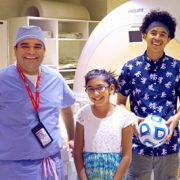
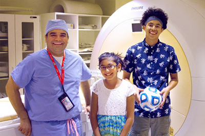
Six companies presenting innovative medical device solutions that address significant unmet needs in pediatric health were awarded a total of $250,000 in grant money yesterday in San Jose, Calif. at the Fifth Annual Pediatric Device Innovation Symposium, organized by the Sheikh Zayed Institute for Pediatric Surgical Innovation at Children’s National Health System.

The “Make Your Medical Device Pitch for Kids!” competition is sponsored by the National Capital Consortium for Pediatric Device Innovation (NCC-PDI), an FDA-funded consortium led by Children’s National and the A. James Clark School of Engineering at the University of Maryland. Four companies were awarded $50,000 each and two were awarded $25,000. The six winners were selected from a field of twelve finalists. A record 98 total submissions from five countries were received for the competition this year.

“To improve care for children, it is imperative that we recognize and encourage relevant new solutions in pediatric medical devices, especially in light of the challenges innovators face in addressing this specialized market,” said Kurt Newman, M.D., president and CEO of Children’s National. “Children’s National is committed to fostering collaboration among innovators, clinicians, policy makers and investors to advance pediatric device development for the benefit of children everywhere.”

This year’s winning innovations receiving $50,000 awards are:

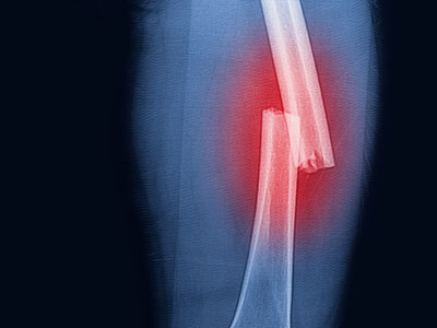
  • CorInnova, Houston, Texas – soft robotic, non-blood-contacting biventricular cardiac assist device for the treatment of heart failure in children
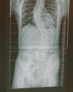
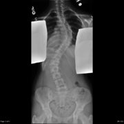
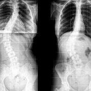
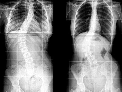
  • Green Sun Medical, Fort Collins, Colo. – novel device that provides necessary pressure for the correction of spinal deformity while providing real-time feedback to clinicians
  • Hub Hygiene and Georgia Institute of Technology, Atlanta, Ga. – low-cost, single-use cleaning technology to prevent central line-associated blood stream infections (CLABSI), a hospital-acquired infection by pediatric ICU patients
  • NAVi Medical Technologies, Houston, Texas – device to provide accurate information about the localization of an umbilical venous catheter (UVC) used in critically-ill newborns to reduce the risk of catheter malposition

Winning innovations receiving $25,000 awards are:

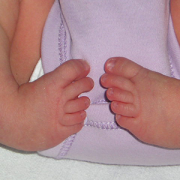
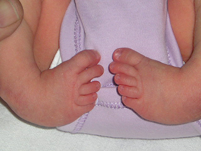
  • Prapela, LLC, Boston, Mass. – novel “baby box” that will allow for a non-pharmacological approach to help drug-exposed infants relax and sleep during withdrawal and post-withdrawal care
  • X-Biomedical, Inc., Philadelphia, Pa. – portable surgical microscope for use in surgeries for treatable causes of blindness in low-income countries and under-resourced setting

“We are honored to recognize these outstanding innovations with this funding,” said Kolaleh Eskandanian, Ph.D., executive director of the Sheikh Zayed Institute and NCC-PDI. “We are even more excited about welcoming this new cohort of companies to our family of pediatric device startups and entrepreneurs. Together we can move the needle a bit faster and safer to bring pediatric products to market.”

She added that in addition to the financial support and consultation services through NCC-PDI, the awardees can leverage the validation received through this highly competitive process to raise the additional capital needed for commercialization. Since inception in 2013, NCC-PDI has supported 67 pediatric devices and the companies and research labs owning these devices have collectively raised $55 million in additional funding.

The twelve finalists each made five-minute presentations to the symposium audience and then responded to judges’ questions. Finalists also included Anecare, LLC, Salt Lake City, Utah; ApnoSystems, Buenos Aires, Argentina; Deton Corp., Pasadena, Calif.; Kite Medical, Dublin, Ireland; Moyarta 2, LLC, The Plains, Va.; and Oculogica, Inc., New York, N.Y.

Serving on the distinguished panel of judges were Susan Alpert, M.D., of SFA Consulting, a former director of the FDA Office of Device Evaluation and former senior vice president and chief regulatory officer of Medtronic; Charles Berul, M.D., co-director, Children’s National Heart Institute; Andrew Elbardissi, M.D., of Deerfield Management; Rick Greenwald, Ph.D., of the New England Pediatric Device Consortium (NEPDC); James Love, J.D., of Oblon; Josh Makower, M.D., of NEA; Jennifer McCaney, Ph.D., of MedTech Innovator; Jackie Phillips, M.D., of Johnson & Johnson; and Tracy Warren of Astarte Ventures.

The pitch competition is a highlight of the annual symposium organized by the Sheikh Zayed Institute at Children’s National, designed to foster innovation that will advance pediatric healthcare and address the unmet surgical and medical device needs for children. New this year, the symposium co-located in a joint effort with The MedTech Conference powered by AdvaMed, the premier gathering of medtech professionals in North America.

Keynote speakers at the event included Daniel Kraft, M.D., faculty chair of Medicine & Neuroscience, Singularity University and executive director, Exponential Medicine; Vasum Peiris, M.D., chief medical officer, Pediatrics and Special Populations, FDA;  and Alan Flake, M.D., director of Center for Fetal Research, Children’s Hospital of Philadelphia.

Panel discussions focused on gap funding for pediatric innovation, the journey from ideation to commercialization, and the pediatric device needs assessment in the future regulatory environment.

Laura Tosi

Giving voice to adult osteogenesis imperfecta patients

Laura Tosi

“I have a number of OI patients moving into adulthood who cannot get care in the adult world, because my colleagues who care for adults have less experience with the disease and because caring for OI adult patients is largely uncharted territory,” says Laura L. Tosi, M.D.

With the influx of increasingly effective technology at our fingertips, the landscape of patient care for complex diseases has changed for the better in recent years. Doctors and researchers can accelerate new discoveries and improvements in patient care by querying and utilizing patient data gathered from all over the world.

For Laura L. Tosi, M.D., director of the Bone Health Program at Children’s National Health System, these changes have galvanized years of research into patients with osteogenesis imperfecta (OI), a population that is particularly difficult to trace into adulthood.

OI is a rare genetic disorder characterized by excessively fragile bones with a high susceptibility to recurrent fractures. Commonly known as “brittle bone disease,” OI is mostly caused by mutations in type I collagen genes. The severity of the disease varies widely and a cure for OI still remains to be found. Currently, treatment methods include medications, physical and occupational therapy, as well as surgery – all of which aim to reduce risk of further fracture, help patients manage pain and promote a healthy lifestyle.

Two of the most critical challenges that accompany the treatment of rare diseases, however, are the paucity of data on the adult patient experience and the challenge of transitioning patients safely from the multispecialty clinics frequently available in childhood to adult care givers who may have never seen the disorder in their career.

Through her research, Dr. Tosi aims to fill these critical knowledge gaps, and has found the Patient-Reported Outcome Measurement Information System (PROMIS®), the patient-reported outcome platform funded by the National Institutes of Health, to be particularly advantageous.

PROMIS® harnesses a set of measurement tools that uses computer adaptive technology and person-centered measurements to evaluate and monitor physical, mental and social health in adults and children. These tools quickly tailor themselves to individual responses and, because of their user-friendly design, provide a level of convenience and easy accessibility that other platforms lack.

“I have a number of OI patients moving into adulthood who cannot get care in the adult world, because my colleagues who care for adults have less experience with the disease and because caring for OI adult patients is largely uncharted territory,” says Dr. Tosi. Realizing the importance of giving voice to adults with OI, Dr. Tosi has harnessed a diverse range of standardized PROMIS® tools to attempt to capture a more complete understanding of the patient experience, ranging from the quality of social participation and peer relationships to physical and emotional distress.

When her first PROMIS®-based mailing to patients received an overwhelming response of more than 1,100 respondents in just 90 days, Dr. Tosi knew that pushing this research forward and out into the community was imperative. The results from that first survey, published in 2015 in the Orphanet Journal of Rare Diseases, demonstrated that adults with OI generally reported lower physical health status and were more likely to struggle with auditory and musculoskeletal problems.

Continued research in this area will not only generate much-needed knowledge about long-term healthcare issues and needs for OI patients, but also help clinicians improve their current treatment methodologies to anticipate these concerns ahead of time, if possible.

“The number of responses to our first survey demonstrated that patients really want to be heard. When you give them tools and ask them to tell you about themselves in ways that they hope will change how you practice, they want to help,” says Dr. Tosi, “because everyone wants to grow old well.”

At the end of August, Dr. Tosi will present her research at the 13th International Conference on Osteogenesis Imperfecta in Oslo, Norway. She also presented her research at the 8th International Conference on Children’s Bone Health as well as at the 17th Annual OI Foundation Scientific Meeting.

Now taking part in designing and executing a national natural history study of patients with OI, Dr. Tosi plans to lead the charge for incorporating and implementing PROMIS® tools into the study. “Once we improve our tools, we will have the ability to query individuals from Alaska to Timbuktu, and provide a far more comprehensive understanding of this very complex and multi-faceted disorder. Harnessing the power of the internet and engaging the patient in delineating their disorder as well as their response to treatment offer a giant step forward in caring for individuals with rare diseases,” she says.

Roberta DeBiasi

Panel: Significant Zika risks linger for pregnant women and developing fetuses in US

Roberta DeBiasi

The threat from Zika “is not over. It is just beginning for the families who are affected by this,” says Roberta L. DeBiasi, M.D., M.S., chief of the Division of Pediatric Infectious Diseases and co-director of the Congenital Zika Virus Program at Children’s National Health System.

The Zika virus epidemic may have fallen off the radar for many media outlets, but significant risks continue to linger for pregnant women and developing fetuses, a panel of experts told staff working for U.S. Congressional leaders.

“The threat of this virus is real, and the threat continues,” Margaret Honein, Ph.D., M.P.H., of the Centers for Disease Control and Prevention’s (CDC) pregnancy and birth defects task force, said during the July 13 briefing held in the Russell Senate Office Building.

Dr. Honein told about 100 attendees that more than 200 Zika-affected babies have been born in the United States suffering from serious birth defects, such as rigid joints, inconsolable distress that causes them to cry continuously and difficulties swallowing. Some of these infants experience seizures that cause further brain damage.

Predicting what Zika will do next in the United States is very difficult, Dr. Honein said, adding that local outbreaks could occur “at any time.” A map she displayed showed Zika’s impact in shades of blue, with Zika infections documented in nearly every state and the highest number of infections – and deepest shade of blue­ – for California, Florida and Texas.

The threat from Zika “is not over. It is just beginning for the families who are affected by this,” agreed Roberta L. DeBiasi, M.D., M.S., chief of the Division of Pediatric Infectious Diseases and co-director of the Congenital Zika Virus Program at Children’s National Health System.

Since Children’s National launched its Zika program in May 2016, the multidisciplinary team has consulted on 65 mother-fetus/infant pairs, Dr. DeBiasi said. Because in utero Zika infection can result in a wide range of side effects, the Children’s team includes pediatric infectious diseases experts, fetal/neonatal neurologists to consult on seizures, audiologists to assess hearing, physical therapists and orthopaedists to contend with limb contractures, pulmonologists to relieve breathing problems and ophthalmologists to diagnose and treat vision disorders – among other specialists.

“You really need a program that has all of these areas of expertise available for a family,” Dr. DeBiasi told attendees. “It is not possible for a family to organize 27 different appointments if you have a child with these needs.”

Children’s Zika experts also collaborate with researchers in Colombia to gauge the ability of magnetic resonance imaging to produce earlier Zika diagnoses, to assess the role of viral load as biomarkers and to document Zika’s long-term impact on children’s neurodevelopment. The Colombia study has enrolled an additional 85 women/infant pairs.

In one presentation slide, Dr. DeBiasi showed sharp magnetic resonance imaging scans from their research study of a fetal brain at 18 and 22 weeks gestation that indicated clear abnormalities, including abnormal cortical folding. Ultrasound images taken at the exact same time points did not detect these abnormalities, she said.

Asked for advice by an attendee whose clinic treats women who regularly travel between California and Mexico, Dr. DeBiasi underscored the fact that Zika infection poses a risk to developing fetuses even if the pregnant woman has no symptoms of infection. “Whether or not they’re symptomatic, the risk is the same. It’s hard for people to understand that. That is No. 1,” she said.

Another challenge is for women who scrupulously follow the CDC’s guidance on lowering their infection risk while traveling. Upon return, those women may be unaware that they could still be exposed to Zika through unprotected sex with their partner who also has travelled, for as long as six months after travel.

US News Honor Roll 2017-18

Children’s National is #1 in Neonatology and Top 10 overall in U.S. News & World Report Survey

US News Honor Roll 2017-18Children’s National is proud to be named #1 in Neonatology in the U.S. News & World Report 2017-18 Best Children’s Hospitals survey. Also, Children’s National was once again named to the coveted Honor Roll, placing them among the Top 10 children’s hospitals in the country.

Being the #1 ranked Neonatology program reflects the quality of care throughout Children’s National because it requires the support and partnership of many other specialties, including cardiology, neurology and surgery. In addition to this honor, Children’s National ranked in the Top 10 in four additional services: Cancer (#7), Neurology and Neurosurgery (#9), Orthopedics (#9) and Nephrology (#10).  For the seventh year in a row, Children’s National has ranked in all ten services, a testament to the pediatric care experts across the organization and their commitment to children and families.

“This recognition is a great achievement for Children’s National, affirming our place as a premier destination for pediatric care, and the commitment of our people, partners and supporters to helping every child grow up stronger,” said Kurt Newman, M.D., President and CEO of Children’s National. “I’m particularly proud of our #1 ranking in Neonatology as, in many ways it reflects the quality of care across our hospital. Treating these tiny patients often encompasses many other specialties, including our Fetal Medicine Institute.”

Children’s National is dedicated to improving the lives of children through innovative research, expert care and advocacy on behalf of children’s needs. In addition to being recognized among the “best of the best” by U.S. News & World Report, Children’s National is a Magnet® designated hospital for excellence in nursing and is a Leapfrog Group Top Hospital. As a top NIH-funded pediatric health system, Children’s National marries cutting-edge research with the highest quality care, to deliver the best possible outcomes for children today and in the future.

POSNA awards clinical trials planning grant to Children’s National

Children’s National Health System has received a prestigious grant from the Pediatric Orthopaedic Society of North America (POSNA) to investigate pediatric femur fractures. The Clinical Trials Planning Grant in the amount of $30,000 will propel work to develop a clinical trial focused on femur fractures, with the ultimate goal of designing a multicenter trial for surgical treatment of pediatric femur fractures.

Karun Sharma, M.D., poses with two patients

Treating osteoid osteoma with MR-HIFU

Karun Sharma, M.D., poses with two patients

Karun Sharma, M.D., poses with two patients who participated in the MR-HIFU trial for pediatric osteoid osteoma.

Doctors from the Sheikh Zayed Institute for Pediatric Surgical Innovation and surgeons from Children’s National are the first in the U.S. to use Magnetic Resonance-Guided High-intensity Focused Ultrasound (MR-HIFU) to treat pediatric osteoid osteoma.

The trial, led by Principal Investigator Karun Sharma, M.D., Ph.D., Director of Interventional Radiology at Children’s National, began in 2015 and is demonstrating early success in establishing the safety and feasibility of noninvasive MR-HIFU as an alternative to the current, more invasive approaches to remove tumor tissue.

Osteoid osteoma is a painful, but benign, bone tumor that commonly occurs in children and young adults. Removal generally requires orthopaedic surgery to scrape the tumor from the bone or CT (computerized tomography) image-guided radiofrequency ablation (RFA), which is less invasive than surgery but is associated with ionizing radiation exposure and requires drilling through muscle and soft tissue into bone.

MR-HIFU, on the other hand, is a precise and controlled method that does not require a scalpel or needle, greatly reducing the risk of complications, including infections and bone fractures. Even better, it promises reduced procedure time, typically an hour or less.

“Our team set out to provide a noninvasive and radiation free treatment option for children with osteoid osteoma and our pilot feasibility and safety trial is almost completed. We have treated 9 patients and we’re very pleased with the success of the treatments so far. Although follow up will continue for another year, results to date that show that MR-HIFU may be a completely non-invasive and radiation free treatment for osteoid osteoma,” Dr. Sharma says. “Several of the children we treated were very active prior to the onset of their tumor, one a soccer player and the other a swimmer, but because of pain from the tumor, they were unable to enjoy their favorite activities, until now.”

“The use of MR-HIFU ablation of osteoid osteoma is a perfect example of our mission in the Sheikh Zayed Institute to make pediatric surgery more precise and less invasive,” adds Peter Kim, M.D., C.M., Ph.D., Vice President of the Sheikh Zayed Institute, who leads the Image Guided Non-Invasive Therapeutic Energy (IGNITE) program.

IGNITE is a joint clinical and research collaboration between the Sheikh Zayed Institute and the Divisions of Radiology, Oncology, Surgery, and Anesthesiology at Children’s National. MR-HIFU is also being used to treat pediatric refractory soft tissue tumors, a first-in-the-world clinical trial that is a collaboration between Children’s National and the NIH Center for Interventional Oncology directed by Bradford Wood, MD. Additionally, the IGNITE team has started preliminary work to explore applications of MR-HIFU for noninvasive ablation of growth plates and pediatric solid tumors.

In addition to Drs. Sharma and Kim, the team for the ablation of osteoid osteoma clinical trial includes: AeRang Kim, MD, PhD, pediatric oncologist; Matthew Oetgen, M.D., Division Chief of Orthopaedic Surgery and Sports Medicine; Kaleb Friend, M.D., pediatric orthopedic surgeon; Pavel Yarmolenko, Ph.D., Haydar Celik, Ph.D., and Avinash Eranki, biomedical engineers; Viktoriya Beskin, MR technologist; and Janish Patel, M.D., and Domiciano Santos, M.D., pediatric anesthesiologists.

MAGEC Rod Tool

MAGEC growing rod improves orthopaedics

MAGEC Spinal Growing Rod Inside Boy

After implanting a MAGEC Spinal Growing Rod, doctors use an external remote control to lengthen the magnetically controlled rod as the child grows.

Children’s National Health System was among the first in the country to offer a novel spinal growing rod for children with scoliosis after it was approved by the FDA just three years ago – and has now treated 30 patients with this innovative technique. The MAGEC™ (MAGnetic Expansion Control) Spinal Growing Rod is a non-invasive treatment for children with early onset scoliosis.

After the initial procedure to implant the rod, doctors use an external remote control outside of the body to lengthen the magnetically controlled rod as the child grows. The adjustments are non-invasive, reducing the number of surgeries required during the course of treatment.

Growing rods have become effective tools for children whose spinal curvature is too significant to control with bracing or casting. The rods—which are surgically attached to the spine above and below the curve and then traditionally lengthened during follow-up surgical procedures—allow the spine to continue growing while managing the curve until the child is old enough for spinal fusion.

The problem: Children must bear the physical and psychological burden of undergoing lengthening procedures every six to 12 months until they are skeletally mature enough to have spinal fusion—typically around age 10 for girls and age 12 or 13 for boys.

Now, instead of returning to the hospital for a major surgery to adjust growth rods twice a year, children with the MAGEC rod have adjustments in just a few minutes four times a year – minus invasive surgery and recovery time, says Matthew Oetgen, M.D., Division Chief of Orthopaedic Surgery and Sports Medicine and Director of Orthopaedic Research at Children’s National.

“Traditional growing rods work, but they require multiple surgeries that increase complication rates and time spent in the hospital,” he says. “We treat many children each year who have or are candidates for growing rods, so it’s important for us to embrace new technology to make the lengthening process easier and less painful for children while decreasing morbidity.”

Children’s National orthopaedic surgeons lengthen the MAGEC rod every three to four months in the office using the electronic remote control. They then monitor the scoliosis and treatment progress with radiographs. Like traditional growing rods, MAGEC is a means, not an end—the system provides a bridge treatment spanning the years between the initial lengthening surgery and spinal fusion.

Dr. Oetgen says this game-changing technology may not be the right solution for every patient, but is the preferred choice because the patients can avoid some additional surgeries down the line. Patients in the 5-7 year age range at the time the rod is in place would potentially face 10 years of surgeries every six months with traditional growing rods.

“We’ve eliminated these regularly scheduled procedures, which is great if you’re a healthy kid,” Oetgen says. “But if you are a kid with other health challenges, such as a neuromuscular disease – it’s really life changing not to have to go into surgery every six months. It saves them a tremendous amount of intervention.”

Following MAGEC’s approval by the U.S. Food and Drug Administration in February 2014, surgeons at Children’s National performed two of the first 15 MAGEC implantations in the country, and the first in the greater Washington, DC, area. MAGEC rods are approved for children with scoliosis greater than 50 degrees in magnitude and under 10 years of age.

On the horizon for this new technology are some improvements Oetgen says would allow physicians using MAGEC rods to improve the patient experience even more. “Smarter” devices could potentially tell doctors how much lengthening has actually occurred after they’ve pushed the buttons on the remote control – instead of having to follow up the procedure with an x-ray to see how the rod interacted with soft tissue around the spine, he says.

And the next generations of MAGEC rods may be smaller devices, allowing younger, smaller kids to reap the benefits.

“In the future these improvements will allow us to treat more patients, and allow us to know what we’re doing and what kind of feedback we’re getting,” Oetgen says.

Spinal fusion surgical home helps kids go home sooner

scoli-1

The first of its kind for pediatric patients, the Children’s National Spinal Fusion Surgical Home implements a newly developed model of care to streamline and optimize the spinal fusion process for adolescent idiopathic scoliosis patients.

Using frameworks of care used in adult models, along with best practices and literature reviews, a multidisciplinary team developed the first Spinal Fusion Surgical Home for pediatric patients. It standardizes the infection-control process, pain-management pathway, and physical-therapy program for patients undergoing spinal fusion.

“This model eliminates variability in the care process and increases the quality of care for pediatric patients,” said Matthew Oetgen, MD, MBA, Chief of Orthopaedic Surgery and Sports Medicine. “It’s just the start—by developing this model specifically for our young patients with adolescent idiopathic scoliosis, we are paving the way for a number of other kids that require different kinds of surgeries.”

Hallmarks of the spinal fusion surgical home
From pre-operative care through recovery, the Spinal Fusion Surgical Home streamlines care with an emphasis on increasing quality outcomes for patients. Children’s National provides an informational website and a single point of contact for scheduling procedures and pre-operative laboratory exams. Before surgery, patients and families attend an evening education class that features presentations from orthopaedic nurse practitioners, physical therapists, and anesthesiologists.

After surgery, a nurse follows up by phone to assess how the patient is handling pain and healing.

Increasing the quality of care
By implementing these standardized protocols, Children’s National has seen a decrease in the average length of stay for spinal fusion patients from about five days to three and a half days. The surgical home also has reduced the transfusion rate from 30 to 12 percent, and patient pain scores have decreased.  “Patients are getting better faster with less pain, and are getting to leave the hospital sooner,” says Karen Thomson, MD.

Children’s National also is creating surgical homes for sickle cell disease patients, who need a variety of different types of surgery, as well as for children who need Nissen fundoplication and heart surgery.