Orthopedics

NCC-PDI Pitch Winners

NCC-PDI announces medical device pitch winners

NCC-PDI Pitch Winners

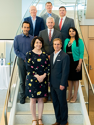
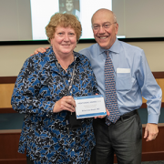
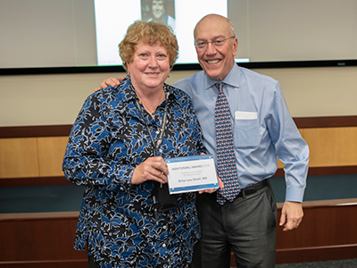
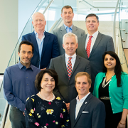
Five pediatric medical device innovators each captured $50K in funding and access to a new pediatric device accelerator program in a competition hosted April 30, 2019 by National Capital Consortium for Pediatric Device Innovation that focused on orthopedic and spine devices. Clockwise from front left: Kolaleh Eskandanian, Children’s National Health System; Cristian Atria, nView Medical; John Barrett, Auctus Surgical Inc.; Paul Mraz, ApiFix; Dan Sands, AMB Surgical II; Anuradha Dayal, BabySteps, Children’s National Health System; Paul Grand, MedTech Innovator; (center) Bill Bentley, Robert E. Fischell Institute for Biomedical Devices, University of Maryland.

The National Capital Consortium for Pediatric Device Innovation (NCC-PDI) announced five winners of its “Make Your Medical Device Pitch for Kids!” competition held on April 30 at the University of Maryland. Each winner receives $50,000 in grant funding and gains access to the consortium’s first-of-its-kind “Pediatric Device Innovator Accelerator Program” led by MedTech Innovator.

NCC-PDI, one of five FDA Pediatric Device Consortia grant programs that support the development and commercialization of pediatric medical devices, is led by the Sheikh Zayed Institute for Pediatric Surgical Innovation at Children’s National Health System and the A. James Clark School of Engineering at the University of Maryland. The consortium recently added new accelerators BioHealth Innovation and MedTech Innovator and design firm partner, Smithwise.

A panel of 32 expert judges from business, healthcare, regulatory and legal sectors selected the winners based on the clinical significance and commercial feasibility of their medical devices for children. The competition focused solely on advancing care in the pediatric orthopedics and spine sector which the FDA identified as an emerging underserved specialty lacking innovation.

The competition winners are:

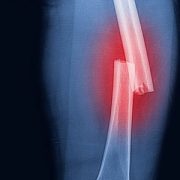
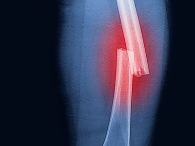
  • AMB Surgical, LLC, Dayton, Ohio – FLYTE, a device designed to reduce invasive and repetitive surgery in children and teens with orthopedic illnesses such as scoliosis and limb abnormalities
  • Auctus Surgical, Inc., San Francisco, Calif. – Auctus Surgical Dynamic Spinal Tethering System, a mechanism used to correct the scoliotic spine in pediatric patients through a tethering procedure
  • ApiFix Ltd, Boston, Mass. – ApiFix’s Minimally Invasive Deformity Correction (MID-C) System, a posterior dynamic deformity correction system for surgical treatment to provide permanent spinal curve correction while retaining flexibility
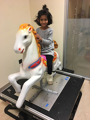
  • Children’s National Health System, Washington, D.C.– Babysteps platform to improve initial assessment of clubfoot deformity and predict the magnitude of correction
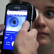
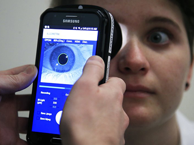
  • nView Medical, Salt Lake City, Utah – Surgical scanner using AI-based image creation to provide instant 3D imaging during surgery to improve imagery speed and accuracy

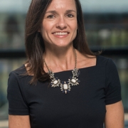
“All finalists are winners and we believe that, with NCC-PDI’s support, some of the awarded devices will be available to orthopedic and spine clinicians in the near future. That is vitally important since innovation has been stagnant in this area,” says Kolaleh Eskandanian, Ph.D., MBA, PMP, vice president and chief innovation officer at Children’s National and principal investigator of NCC-PDI. “This competition aims to increase the profile of companies by exposing them to a panel of industry leaders who may become future investors or strategic partners.”

Through the inaugural NCC-PDI “Pediatric Device Innovator Accelerator Program,” MedTech Innovator is providing winners with virtual in-depth, customized mentorship from some of the industry’s leading executives and investors. MedTech Innovator has a proven track record of identifying early-stage medical device companies with the key characteristics required for commercial success and accelerating their growth through its vast ecosystem of resources.

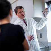
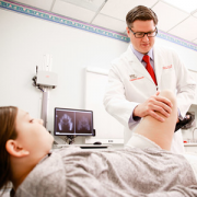
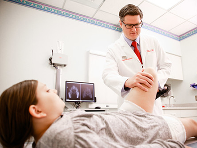
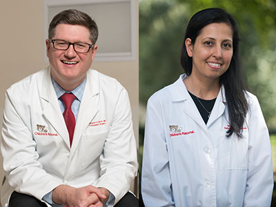
“As a pediatric orthopedic surgeon, I am encouraged by the innovations presented at this competition,” says Matthew Oetgen, M.D., division chief of Orthopaedic Surgery and Sports Medicine at Children’s National, who served on the judging panel. “We need more devices that compensate for the smaller size of children compared to adults and that can adapt as children’s bones continue to grow and develop. The finalists who competed fully embraced that challenge.”

This was NCC-PDI’s eighth competition in six years and a ninth competition is planned for fall 2019 that focuses on NICU. Including this recent round of winners, the consortium has supported 94 pediatric medical devices and helped five companies receive FDA or CE mark regulatory clearance.

To learn more about the winners and the fall 2019 pitch competition, visit the National Capital Consortium for Pediatric Device Innovation website.

Billie Lou Short and Kurt Newman at Research and Education Week

Research and Education Week honors innovative science

Billie Lou Short and Kurt Newman at Research and Education Week

Billie Lou Short, M.D., received the Ninth Annual Mentorship Award in Clinical Science.

People joke that Billie Lou Short, M.D., chief of Children’s Division of Neonatology, invented extracorporeal membrane oxygenation, known as ECMO for short. While Dr. Short did not invent ECMO, under her leadership Children’s National was the first pediatric hospital to use it. And over decades Children’s staff have perfected its use to save the lives of tiny, vulnerable newborns by temporarily taking over for their struggling hearts and lungs. For two consecutive years, Children’s neonatal intensive care unit has been named the nation’s No. 1 for newborns by U.S. News & World Report. “Despite all of these accomplishments, Dr. Short’s best legacy is what she has done as a mentor to countless trainees, nurses and faculty she’s touched during their careers. She touches every type of clinical staff member who has come through our neonatal intensive care unit,” says An Massaro, M.D., director of residency research.

For these achievements, Dr. Short received the Ninth Annual Mentorship Award in Clinical Science.

Anna Penn, M.D., Ph.D., has provided new insights into the central role that the placental hormone allopregnanolone plays in orderly fetal brain development, and her research team has created novel experimental models that mimic some of the brain injuries often seen in very preterm babies – an essential step that informs future neuroprotective strategies. Dr. Penn, a clinical neonatologist and developmental neuroscientist, “has been a primary adviser for 40 mentees throughout their careers and embodies Children’s core values of Compassion, Commitment and Connection,” says Claire-Marie Vacher, Ph.D.

For these achievements, Dr. Penn was selected to receive the Ninth Annual Mentorship Award in Basic and Translational Science.

The mentorship awards for Drs. Short and Penn were among dozens of honors given in conjunction with “Frontiers in Innovation,” the Ninth Annual Research and Education Week (REW) at Children’s National. In addition to seven keynote lectures, more than 350 posters were submitted from researchers – from high-school students to full-time faculty – about basic and translational science, clinical research, community-based research, education, training and quality improvement; five poster presenters were showcased via Facebook Live events hosted by Children’s Hospital Foundation.

Two faculty members won twice: Vicki Freedenberg, Ph.D., APRN, for research about mindfulness-based stress reduction and Adeline (Wei Li) Koay, MBBS, MSc, for research related to HIV. So many women at every stage of their research careers took to the stage to accept honors that Naomi L.C. Luban, M.D., Vice Chair of Academic Affairs, quipped that “this day is power to women.”

Here are the 2019 REW award winners:

2019 Elda Y. Arce Teaching Scholars Award
Barbara Jantausch, M.D.
Lowell Frank, M.D.

Suzanne Feetham, Ph.D., FAA, Nursing Research Support Award
Vicki Freedenberg, Ph.D., APRN, for “Psychosocial and biological effects of mindfulness-based stress reduction intervention in adolescents with CHD/CIEDs: a randomized control trial”
Renee’ Roberts Turner for “Peak and nadir experiences of mid-level nurse leaders”

2019-2020 Global Health Initiative Exploration in Global Health Awards
Nathalie Quion, M.D., for “Latino youth and families need assessment,” conducted in Washington
Sonia Voleti for “Handheld ultrasound machine task shifting,” conducted in Micronesia
Tania Ahluwalia, M.D., for “Simulation curriculum for emergency medicine,” conducted in India
Yvonne Yui for “Designated resuscitation teams in NICUs,” conducted in Ghana
Xiaoyan Song, Ph.D., MBBS, MSc, “Prevention of hospital-onset infections in PICUs,” conducted in China

Ninth Annual Research and Education Week Poster Session Awards

Basic and Translational Science
Faculty:
Adeline (Wei Li) Koay, MBBS, MSc, for “Differences in the gut microbiome of HIV-infected versus HIV-exposed, uninfected infants”
Faculty: Hayk Barseghyan, Ph.D., for “Composite de novo Armenian human genome assembly and haplotyping via optical mapping and ultra-long read sequencing”
Staff: Damon K. McCullough, BS, for “Brain slicer: 3D-printed tissue processing tool for pediatric neuroscience research”
Staff: Antonio R. Porras, Ph.D., for “Integrated deep-learning method for genetic syndrome screening using facial photographs”
Post docs/fellows/residents: Lung Lau, M.D., for “A novel, sprayable and bio-absorbable sealant for wound dressings”
Post docs/fellows/residents:
Kelsey F. Sugrue, Ph.D., for “HECTD1 is required for growth of the myocardium secondary to placental insufficiency”
Graduate students:
Erin R. Bonner, BA, for “Comprehensive mutation profiling of pediatric diffuse midline gliomas using liquid biopsy”
High school/undergraduate students: Ali Sarhan for “Parental somato-gonadal mosaic genetic variants are a source of recurrent risk for de novo disorders and parental health concerns: a systematic review of the literature and meta-analysis”

Clinical Research
Faculty:
Amy Hont, M.D., for “Ex vivo expanded multi-tumor antigen specific T-cells for the treatment of solid tumors”
Faculty: Lauren McLaughlin, M.D., for “EBV/LMP-specific T-cells maintain remissions of T- and B-cell EBV lymphomas after allogeneic bone marrow transplantation”

Staff: Iman A. Abdikarim, BA, for “Timing of allergenic food introduction among African American and Caucasian children with food allergy in the FORWARD study”
Staff: Gelina M. Sani, BS, for “Quantifying hematopoietic stem cells towards in utero gene therapy for treatment of sickle cell disease in fetal cord blood”
Post docs/fellows/residents: Amy H. Jones, M.D., for “To trach or not trach: exploration of parental conflict, regret and impacts on quality of life in tracheostomy decision-making”
Graduate students: Alyssa Dewyer, BS, for “Telemedicine support of cardiac care in Northern Uganda: leveraging hand-held echocardiography and task-shifting”
Graduate students: Natalie Pudalov, BA, “Cortical thickness asymmetries in MRI-abnormal pediatric epilepsy patients: a potential metric for surgery outcome”
High school/undergraduate students:
Kia Yoshinaga for “Time to rhythm detection during pediatric cardiac arrest in a pediatric emergency department”

Community-Based Research
Faculty:
Adeline (Wei Li) Koay, MBBS, MSc, for “Recent trends in the prevention of mother-to-child transmission (PMTCT) of HIV in the Washington, D.C., metropolitan area”
Staff: Gia M. Badolato, MPH, for “STI screening in an urban ED based on chief complaint”
Post docs/fellows/residents:
Christina P. Ho, M.D., for “Pediatric urinary tract infection resistance patterns in the Washington, D.C., metropolitan area”
Graduate students:
Noushine Sadeghi, BS, “Racial/ethnic disparities in receipt of sexual health services among adolescent females”

Education, Training and Program Development
Faculty:
Cara Lichtenstein, M.D., MPH, for “Using a community bus trip to increase knowledge of health disparities”
Staff:
Iana Y. Clarence, MPH, for “TEACHing residents to address child poverty: an innovative multimodal curriculum”
Post docs/fellows/residents:
Johanna Kaufman, M.D., for “Inpatient consultation in pediatrics: a learning tool to improve communication”
High school/undergraduate students:
Brett E. Pearson for “Analysis of unanticipated problems in CNMC human subjects research studies and implications for process improvement”

Quality and Performance Improvement
Faculty:
Vicki Freedenberg, Ph.D., APRN, for “Implementing a mindfulness-based stress reduction curriculum in a congenital heart disease program”
Staff:
Caleb Griffith, MPH, for “Assessing the sustainability of point-of-care HIV screening of adolescents in pediatric emergency departments”
Post docs/fellows/residents:
Rebecca S. Zee, M.D., Ph.D., for “Implementation of the Accelerated Care of Torsion (ACT) pathway: a quality improvement initiative for testicular torsion”
Graduate students:
Alysia Wiener, BS, for “Latency period in image-guided needle bone biopsy in children: a single center experience”

View images from the REW2019 award ceremony.

Beth Tarini

Getting to know SPR’s future President, Beth Tarini, M.D., MS

Beth Tarini

Quick. Name four pillar pediatric organizations on the vanguard of advancing pediatric research.

Most researchers and clinicians can rattle off the names of the Academic Pediatric Association, the American Academy of Pediatrics and the American Pediatric Society. But that fourth one, the Society for Pediatric Research (SPR), is a little trickier. While many know SPR, a lot of research-clinicians simply do not.

Over the next few years, Beth A. Tarini, M.D., MS, will make it her personal mission to ensure that more pediatric researchers get to know SPR and are so excited about the organization that they become active members. In May 2019 Dr. Tarini becomes Vice President of the society that aims to stitch together an international network of interdisciplinary researchers to improve kids’ health. Four-year SPR leadership terms begin with Vice President before transitioning to President-Elect, President and Past-President, each for one year.

Dr. Tarini says she looks forward to working with other SPR leaders to find ways to build more productive, collaborative professional networks among faculty, especially emerging junior faculty. “Facilitating ways to network for research and professional reasons across pediatric research is vital – albeit easier said than done. I have been told I’m a connector, so I hope to leverage that skill in this new role,” says Dr. Tarini, associate director for Children’s Center for Translational Research.

“I’m delighted that Dr. Tarini was elected to this leadership position, and I am impressed by her vision of improving SPR’s outreach efforts,” says Mark Batshaw, M.D., Executive Vice President, Chief Academic Officer and Physician-in-Chief at Children’s National. “Her goal of engaging potential members in networking through a variety of ways – face-to-face as well as leveraging digital platforms like Twitter, Facebook and LinkedIn – and her focus on engaging junior faculty will help strengthen SPR membership in the near term and long term.”

Dr. Tarini adds: “Success to me would be leaving after four years with more faculty – especially junior faculty – approaching membership in SPR with the knowledge and enthusiasm that they bring to membership in other pediatric societies.”

SPR requires that its members not simply conduct research, but move the needle in their chosen discipline. In her research, Dr. Tarini has focused on ensuring that population-based newborn screening programs function efficiently and effectively with fewer hiccups at any place along the process.

Thanks to a heel stick to draw blood, an oxygen measurement, and a hearing test, U.S. babies are screened for select inherited health conditions, expediting treatment for infants and reducing the chances they’ll experience long-term health consequences.

“The complexity of this program that is able to test nearly all 4 million babies in the U.S. each year is nothing short of astounding. You have to know the child is born – anywhere in the state – and then between 24 and 48 hours of birth you have to do testing onsite, obtain a specific type of blood sample, send the blood sample to an off-site lab quickly, test the sample, find the child if the test is out of range, get the child evaluated and tested for the condition, then send them for treatment. Given the time pressures as well as the coordination of numerous people and organizations, the fact that this happens routinely is amazing. And like any complex process, there is always room for improvement,” she says.

Dr. Tarini’s research efforts have focused on those process improvements.

As just one example, the Advisory Committee on Heritable Disorders in Newborns and Children, a federal advisory committee on which she serves, was discussing how to eliminate delays in specimen processing to provide speedier results to families. One possible solution floated was to open labs all seven days, rather than just five days a week. Dr. Tarini advocated for partnering with health care engineers who could help model ways to make the specimen transport process more efficient, just like airlines and mail delivery services. A more efficient and effective solution was to match the specimen pick-up and delivery times more closely with the lab’s operational times – which maximizes lab resources and shortens wait times for parents.

Conceptual modeling comes so easily for her that she often leaps out of her seat mid-sentence, underscoring a point by jotting thoughts on a white board, doing it so often that her pens have run dry.

“It’s like a bus schedule: You want to find a bus that not only takes you to your destination but gets you there on time,” she says.

Dr. Tarini’s current observational study looks for opportunities to improve how parents in Minnesota and Iowa are given out-of-range newborn screening test results – especially false positives – and how that experience might shake their confidence in their child’s health as well as heighten their own stress level.

“After a false positive test result, are there parents who walk away from newborn screening with lingering stress about their child’s health? Can we predict who those parents might be and help them?” she asks.

Among the challenges is the newborn screening occurs so quickly after delivery that some emotionally and physically exhausted parents may not remember it was done. Then they get a call from the state with ominous results. Another challenge is standardizing communication approaches across dozens of birthing centers and hospitals.

“We know parents are concerned after receiving a false positive result, and some worry their infant remains vulnerable,” she says. “Can we change how we communicate – not just what we say, but how we say it – to alleviate those concerns?”

Dr. Kurt Newman in front of the capitol building

Kurt Newman, M.D., shares journey as a pediatric surgeon in TEDx Talk

Kurt Newman, M.D., president and chief executive officer of Children’s National, shares his poignant journey as a pediatric surgeon, offering a new perspective for approaching the most chronic and debilitating health conditions. In this independently-organized TEDx event, Dr. Newman also shares his passion for Children’s National and the need to increase pediatric innovations in medicine.

The traction sisters

Spinal-halo-gravity traction times three

The traction sisters

Three girls received spinal-halo-gravity traction at the same time at Children’s National prior to surgery for acute idiopathic scoliosis.

Washington, D.C.’s ABC affiliate, WJLA, recently featured a story about three girls who received treatment for acute idiopathic scoliosis through the Children’s National Spinal Fusion Surgical Home, a comprehensive and effective program that has demonstrated reductions in pain medication usage and medical stays following posterior spinal fusion surgery.

All three girls had extremely severe curvatures of the spine requiring a month long inpatient stay for spinal halo-gravity traction prior to surgical intervention. Spinal curves severe enough to require traction are rare, and often impede a child’s quality of life just as severely – eating, breathing and moving are difficult. Given the long hospital stay required and the challenges of asking a child to stay in a traction frame 23 hours out of every day, the orthopaedic surgery team tries to coordinate cases so that when possible, patients can support each other throughout the process. This was the first time, however, that the team had three traction patients on similar trajectories on the unit at the same time.

Spinal halo-gravity traction can reduce the degree of surgical intervention necessary by accomplishing some pre-operative gradual straightening of the spine prior to spinal fusion procedures. For severe spinal deformities this has been shown to improve the safety and effectiveness of the final surgical procedure.

Matthew Oetgen, M.D., discusses an image of a patient’s spine.

Eliminating unnecessary radiation exposure from spinal radiography

Matthew Oetgen, M.D., discusses an image of a patient’s spine.

Chief of Orthopaedics and Sports Medicine Matthew Oetgen, M.D., discusses an image of a patient’s spine.

If a child arrives at the pediatric orthopaedic specialist for an idiopathic scoliosis evaluation without an adequate radiographic image of his or her spine, it’s often necessary to order yet another imaging study for accurate assessment.

A study published in the Journal of the American Academy of Orthopaedic Surgeons found that in a 6 month period, almost half (43 percent) of patients referred for evaluation required a repeat radiograph due to missing or poor quality existing images.

“Repeating the radiograph means these kids received another exposure to radiation, too,” says Matthew Oetgen, M.D., the study’s lead author and chief of Orthopaedic Surgery and Sports Medicine at Children’s National Health System. “It’s frustrating because in many cases, a simple change in how the initial radiograph was taken could have prevented the need for more imaging studies.”

Dr. Oetgen and the study authors note that there is currently no standardized protocol for spinal radiography of suspected idiopathic scoliosis. However, a few basic criteria could greatly reduce the number of repeat images necessary. Radiographic images that allow for proper evaluation of idiopathic scoliosis and reduce radiation exposure include:

  • A full coronal view of the spine including skull base and pelvis
  • The iliac crest as an indicator of skeletal maturity
  • A full-length lateral view of the spine

Study authors also reinforced the need to do everything possible to reduce radiation exposure for children through proper use of protective shielding for reproductive organs and digital radiograph technology.

“Orthopaedic surgeons and pediatricians share the responsibility to ensure children are exposed to as little iatrogenic radiation as possible,” Dr. Oetgen concludes. “All physicians should be sure that the radiology facilities they refer patients to for spinal radiography employ every technology and safety measure available to limit radiation exposure. Additionally, we can and should work with radiologists to define evaluation criteria and improve what’s captured by radiography on the first try.”

Dr. Benjamin Martin examines a patient

Understanding Legg-Calvé-Perthes disease

Dr. Benjamin Martin examines a patient

Legg-Calvé-Perthes disease, which affects between five and 10 of every 100,000 children each year, is so rare that it can sometimes be challenging for clinicians to know how best to care for affected patients.

That’s why in 2011 a group of pediatric orthopaedic specialists led by Texas Scottish Rite Children’s Hospital created an international study group dedicated to using research to improve the care of kids with Perthes, a hip disorder characterized by a loss of blood flow to the immature femoral head. Children’s National orthopaedic surgeon Benjamin Martin, M.D., has participated in the group since its launch.

Recently, Dr. Martin and two study group colleagues published a review study that outlines common imaging modalities used in the diagnosis and treatment of Perthes disease.

“There are many imaging options out there, including recent advances in MRI, that can add to our knowledge of the disease and how to treat it so kids have optimal outcomes,” Dr. Martin says. “Our goal was to review what’s out there, how it’s used, and identify any shortcomings of these approaches for this particular patient population.”

The authors note that imaging, in various forms, has been a crucial contributor to understanding and treatment of this disease since it was first discovered. Today, radiography remains the most common imaging technique used to diagnose and follow Perthes over time. However, some MRI applications may offer additional insight into the disorder.

Perfusion MRI allows for early understanding of extent of disease and perfusion patterns may correlate with outcomes. Diffusion weighted imaging (DWI) MRI is another promising avenue for tracking disease progression. Additionally, dynamic MRI might provide range of motion assessments that could be used in the surgical planning process.

This study was one of a handful that the international Perthes group has published so far, with several more currently under development. Exploring treatments and technology applications will enhance early diagnosis and treatment for Perthes, which is a crucial component of treatment success and improved quality of life for affected children.

young girl sitting on a bed with a cast

Creating better casts

young girl sitting on a bed with a cast

Each year, millions of children in the U.S. come to hospital emergency departments with fractures. While broken bones are commonplace, the expertise to stabilize these injuries and cast them is not, says Children’s National Health System orthopedic surgeon Shannon Kelly, M.D.

Most fractures are casted by an on-call resident without the assistance of an orthopedist, she explains. Whether that resident applies a cast successfully depends largely on how well he or she learned this skill as an intern. While most current training models have interns take calls with residents, picking up casting skills through hands-on experience from their more senior peers, they can also pick up mistakes – which get repeated once they’re caring for patients independently as residents themselves, Kelly says.

Casting mistakes aren’t trivial, she adds. They can have serious consequences for patients. For example, a cast that’s not tight enough in the right places can leave bones vulnerable to shifting, a scenario that doctors call a loss in reduction, Kelly explains. If bones aren’t in the right position to heal, doctors must reposition them either in the operating room, often exposing patients to general anesthesia, or through painful, in-office procedures.

Conversely, casts that are too tight – particularly on a fresh fracture that’s prone to swelling – can damage tissues from loss of circulation. To avoid this latter problem, doctors often create a “bivalve” cast in which the two halves are split like a clamshell, leaving room for tissues to expand. But they must use extreme care when they cut open the cast with a saw to avoid cutting patients with the rotating blade or burning them with heat generated from its friction.

“Each year, thousands of children are harmed from improper casting and must go through additional procedures to fix the damage done,” Kelly says.

That’s why she and her colleagues are developing a better way to train interns before they start their orthopedics rotation. Starting this spring, the team will be directing a series of casting workshops to train interns on the proper casting technique.

The workshops will take advantage of models that allow interns to practice without harming patients. Some of these models have simulated bones that show up on an X-ray, allowing participants to evaluate whether they achieved a good reduction once they’re finished. Other models are made of wax that melts if the heat of a cast saw becomes too intense and show nicks if the blade makes contact. Learning proper technique using this tool can help spare human patients painful burns and cuts, Kelly says.

To broaden this effort beyond Children’s National, Kelly and her colleagues received a $1,000 microgrant from the Pediatric Orthopaedic Society of North America to create videos based on material from these workshops. These videos will help trainees at medical institutions across the country learn the same pivotal casting skills.

“A broken bone is difficult enough,” Kelly says. “We’re hoping to decrease the number of times that a child has to have an unnecessary procedure on top of that from a casting mistake that could have been avoided.”

Pedbot video game

Pedbot’s next step – Home-based therapy

Pedbot video game

Pedbot’s home version adapts the same airplane-themed video game to a smaller therapeutic platform that is more affordable to build.

The novel ankle rehabilitation robot built at Children’s National to help children with cerebral palsy build ankle strength and control through video gaming is taking a big step forward. Engineers have created a smaller, more affordable version of the robotic platform using 3D printed parts, to explore the effectiveness of a home-based therapy program.

“We’re seeing preliminary success in our trial for in clinic use of the Pedbot. Now we’re hoping to see if making the technology accessible at home means that 1) Kids use it more often and 2) More frequent, regular use over time leads to better range of motion,” says Kevin Cleary, Ph.D., the Sheikh Zayed Institute for Pediatric Surgical Innovation’s bioengineering technical director and engineering lead for Pedbot.

Pedbot’s video game, designed by software engineer Hadi Fooladi, M.S., allows kids to pilot an airplane through a series of hoops at varying speeds as determined by the therapist and programmer. The game isn’t the only thing that’s unique about this therapeutic robot, however.

Just like the clinic version, the home model moves in three translational directions (x, y and z) and rotates about three axes (the x, y and z axes), similar to the movement of a flight simulator. The result is a robot that helps the patient exercise across a greater range of motion and build muscle strength in a way that more closely mimics real-life ankle function.

Pedbot Home potentially eliminates an additional major therapeutic barrier – the clinic appointment.

“The great thing about Pedbot is you’re constantly working to reach a moving target, and the therapist can vary the movement type as much or as little as needed for each patient,” says Catherine Coley, DPT, a physical therapist at Children’s National who is a member of the Pedbot development team. “We think the home version might make it easier for the child to succeed with a long term therapy program by removing the need for repeat clinic visits.”

“What if a child could come home from school and do their therapy at home after dinner? Would doing it every day for 20 minutes benefit the child more than just coming to see us once or twice a week for an hour? Can we make it easier for our patients to cooperate and follow through with therapy homework? These are some of the questions that we hope we can answer during our trial for the home version,” says Sally Evans, M.D., division chief of Pediatric Rehabilitation Medicine at Children’s National and clinical lead for the project.

The cross-functional Pedbot team includes engineers Reza Monfaredi Ph.D. and Tyler Salvador, B.S., as well as additional physical therapists, Stacey Kovelman, P.T. and Justine Belchner, P.T., and Sara Alyamani, B.A. Future expansions will include the addition of electromyography measurements in collaboration with Paola Pergami, M.D., Ph.D. and incorporation of other patient populations with Beth Wells, M.D.

Pedbot Home is currently being piloted in the home setting, with the goal of enrolling additional families to participate in a trial within the next year. The work is supported by a $500,000 federal grant from the Department of Health and Human Services’ National Institute on Disability, Independent Living, and Rehabilitation Research.

Megan Young

Orthopaedic Surgeon named 2019 LLRS Traveling Fellow

Megan Young

Megan Young, M.D.

Megan Young, M.D., has been named a 2019 Limb Lengthening and Reconstruction Society (LLRS) Traveling Fellow.

The fellowship provides leading education in limb lengthening and reconstruction, trains future leaders of LLRS and establishes mentor relationships between current society members and new surgeons.

“We are beyond proud that Dr. Young was selected for this highly competitive opportunity,” says Matthew Oetgen, M.D., chief of Orthopaedics and Sports Medicine. “This is only the latest recognition Dr. Young has received for her growing expertise in limb lengthening and complex lower extremity reconstructions.”

During her fellowship in August 2019, Dr. Young will travel to multiple limb lengthening and reconstruction surgical centers to share ideas and exchange best practices with orthopaedic surgeons at every career stage – from trainees to seasoned veterans. She will present her key takeaways from the experience at the society’s 2020 annual meeting.

Dr. Young has a special interest in caring for children with lower extremity deformity and limb length discrepancies and has developed a Limb Lengthening program at Children’s, which offers patients and their families comprehensive treatment options for these complex conditions using leading edge technology.

E coli bacteria

Urinary bacteria in spinal cord injury cases may tip balance toward UTIs

E coli bacteria

Patients with spinal cord injuries nearly universally have bacteria present in their urine regardless of whether they have a urinary tract infection.

The fallout from spinal cord injury doesn’t end with loss of mobility: Patients can have a range of other issues resulting from this complex problem, including loss of bladder control that can lead to urine retention. One of the most serious implications is urinary tract infections (UTIs), the most common cause of repeat hospitalization in people with spinal cord injuries, explains Hans G. Pohl, M.D., associate chief in the division of Urology at Children’s National Health System.

Diagnosing UTIs in people with spinal cord injuries is trickier than in people who are otherwise healthy, Dr. Pohl explains. Patients with spinal cord injuries nearly universally have bacteria present in their urine regardless of whether they have a UTI. It’s unclear whether these bacteria are innocent bystanders or precursors to UTIs in patients who don’t yet show symptoms. And although antibiotics can wipe out this bacterial population, these drugs can have undesirable side effects and frequent use can promote development of antibiotic-resistant bacteria.

Although clinical dogma has long promoted the idea that “healthy” urine is sterile, Dr. Pohl and colleagues have shown that a variety of bacteria live in urine, even in people without symptoms. These microorganisms, like the intestinal microbiome, live in harmony with their hosts and may even help promote health. However, it’s unclear what this urinary microbiome might look like for patients with spinal cord injury before, during and after UTIs.

To start investigating this question, Dr. Pohl and co-authors recently reported a case study they published online Sept. 21, 2018, in Spinal Cord Series and Cases. The case report about a 55-year-old man who had injured the thoracic segment of his spinal cord—about the level of the bottom of his shoulder blades—in a skiing accident when he was 19 was selected as “Editor’s Choice” for the journal’s October 2018 issue.  The patient had a neurogenic bladder, which doesn’t function normally due to impaired communication with the spinal cord. To compensate for this loss of function, this patient needed to have urine removed every four to six hours by catheterization.

Over eight months Dr. Pohl, the study’s senior author, and colleagues collected 12 urine samples from this patient:

  • One was collected at a time the patient didn’t show any symptoms of a UTI
  • Nine were collected when the patient had UTI symptoms, such as bladder spasticity
  • Two samples were collected when the patient had finished antibiotic treatment for the UTI.

The researchers split each sample in half. One part was put through a standard urinalysis and culture, much like what patients with a suspected UTI would receive at the doctor’s office. The other part was analyzed using a technique that searched for genetic material to identify bacteria that might be present and to estimate their abundance.

The researchers found a variety of different bacteria present in these urine samples. Regardless of the patient’s health status and symptoms, the majority of these bacterial species are known to be pathogenic or potentially pathogenic. By contrast, this patient’s urine microbiome appeared to largely lack bacterial species known to be either neutral or with potentially probiotic properties, such as Lactobacillus.

All of the bacteria that grew in culture also were identified by their genetic material in the samples. However, genetic sequencing also identified a possible novel uropathogenic species called Burkholderia fungorum that didn’t grow in the lab in five of the samples. This bacterium is ubiquitous in the environment and has been identified in soil- and plant-based samples. It also has been discovered in the respiratory secretions of patients with cystic fibrosis, in patients with a heart condition called infectious endocarditis, in the vaginal microbiota of patients with bacterial vaginosis, and in the gut of patients with HIV who have low T-cell counts. Dr. Pohl says it’s unclear whether this species played an infectious role in this patient’s UTI or whether it’s just part of his normal urine flora.

“Consistent with our previous work, this case report demonstrates that rather than healthy urine being sterile, there is a diverse urine bacterial ecosystem during various states of health and disease,” Dr. Pohl says. “Rather than UTIs resulting from the growth or overgrowth of a single organism, it’s more likely that a change in the healthy balance of the urine ecosystem might cause these infections.”

By monitoring the relative abundance of different bacteria types present in the urine of patients with spinal cord injury and combining this information with a patient’s symptoms, Dr. Pohl says doctors may be able to make more accurate UTI diagnoses in this unique population.

In addition to Dr. Pohl, study co-authors include Marcos Pérez-Losada, Ljubica Caldovic, Ph.D., Bruce Sprague and Michael H. Hsieh, M.D., Children’s National; Emma Nally, Suzanne L. Groah and Inger Ljungberg, MedStar National Rehabilitation Hospital; and Neel J. Chandel, Montefiore Medical Center.

AlgometRX

Breakthrough device objectively measures pain type, intensity and drug effects

AlgometRX

Clinical Research Assistant Kevin Jackson uses AlgometRx Platform Technology on Sarah Taylor’s eyes to measure her degree of pain. Children’s National is testing an experimental device that aims to measure pain according to how pupils react to certain stimuli. (AP Photo/Manuel Balce Ceneta)

Pediatric anesthesiologist Julia C. Finkel, M.D., of Children’s National Health System, gazed into the eyes of a newborn patient determined to find a better way to measure the effectiveness of pain treatment on one so tiny and unable to verbalize. Then she realized the answer was staring back at her.

Armed with the knowledge that pain and analgesic drugs produce an involuntary response from the pupil, Dr. Finkel developed AlgometRx, a first-of-its-kind handheld device that measures a patient’s pupillary response and, using proprietary algorithms, provides a diagnostic measurement of pain intensity, pain type and, after treatment is administered, monitors efficacy. Her initial goal was to improve the care of premature infants. She now has a device that can be used with children of any age and adults.

“Pain is very complex and it is currently the only vital sign that is not objectively measured,” says Dr. Finkel, who has more than 25 years of experience as a pain specialist. “The systematic problem we are facing today is that healthcare providers prescribe pain medicine based on subjective self-reporting, which can often be inaccurate, rather than based on an objective measure of pain type and intensity.” To illustrate her point, Dr. Finkel continues, “A clinician would never prescribe blood pressure medicine without first taking a patient’s blood pressure.”

The current standard of care for measuring pain is the 0-to-10 pain scale, which is based on subjective, observational and self-reporting techniques. Patients indicate their level of pain, with zero being no pain and ten being highest or most severe pain. This subjective system increases the likelihood of inaccuracy, with the problem being most acute with pediatric and non-verbal patients. Moreover, Dr. Finkel points out that subjective pain scores cannot be standardized, heightening the potential for misdiagnosis, over-treatment or under-treatment.

Dr. Finkel, who serves as director of Research and Development for Pain Medicine at the Sheikh Zayed Institute for Pediatric Surgical Innovation at Children’s National, says that a key step in addressing the opioid crisis is providing physicians with objective, real-time data on a patient’s pain level and type, to safely prescribe the right drug and dosage or an alternate treatment.,

She notes that opioids are prescribed for patients who report high pain scores and are sometimes prescribed in cases where they are not appropriate. Dr. Finkel points to the example of sciatica, a neuropathic pain sensation felt in the lower back, legs and buttocks. Sciatica pain is carried by touch fibers that do not have opioid receptors, which makes opioids an inappropriate choice for treating that type of pain.

A pain biomarker could rapidly advance both clinical practice and pain research, Dr. Finkel adds. For clinicians, the power to identify the type and magnitude of a patient’s nociception (detection of pain stimuli) would provide a much-needed scientific foundation for approaching pain treatment. Nociception could be monitored through the course of treatment so that dosing is targeted and personalized to ensure patients receive adequate pain relief while reducing side effects.

“A validated measure to show whether or not an opioid is indicated for a given patient could ease the health care system’s transition from overreliance on opioids to a more comprehensive and less harmful approach to pain management,” says Dr. Finkel.

She also notes that objective pain measurement can provide much needed help in validating complementary approaches to pain management, such as acupuncture, physical therapy, virtual reality and other non-pharmacological interventions.

Dr. Finkel’s technology, called AlgometRx, has been selected by the U.S. Food and Drug Administration (FDA) to participate in its “Innovation Challenge: Devices to Prevent and Treat Opioid Use Disorder.” She is also the recipient of Small Business Innovation Research (SBIR) grant from the National Institute on Drug Abuse.

Emily Niu

Osteochondritis dissecans: Deciding the best candidates for nonoperative treatment

Emily Niu

“When patients come to see me with this condition, they’re often at their lowest point. But then I get to watch them go through this transformation as they’re getting better,” says Emily Niu, M.D. “It’s really wonderful to see someone’s personality blossom over the course of treatments.”

The adage of “practice makes perfect” is true for young competitive athletes; however it also puts them at risk for overuse injuries. One of these injuries is a condition called osteochondritis dissecans (OCD), in which repeatedly overloading joints causes increased stress to certain areas of bone. This area of bone can lose its blood supply, become unhealthy and ultimately end up fragmented. In the late stages, this area of bone can break off from the surrounding healthy bone and as a result, the overlying cartilage (which relies on the bone for a foundation) can become prone to damage. This process can be likened to the formation of potholes in a road. Typically, individuals affected can have pain, limited range of motion or even arthritis down the line.

This condition can happen at different locations throughout the body, including the knees, ankles and elbows. Baseball players and gymnasts are particularly prone to getting OCD of the capitellum, or the outside of the elbow, from throwing or tumbling.

If this injury is unstable, with bone and cartilage already fragmenting, it’s typically treated with surgery, explains Emily Niu, M.D., a Children’s National orthopaedic surgeon. Stabilizing the fragment and drilling tunnels in the affected bone surface allows new bone to grow and repair the defect. But for stable OCD, the treatment path is unclear.

Sometimes nonoperative treatments, such as rest, physical therapy or bracing the joint, can allow it to fully heal over time; however, she says, some patients treated nonoperatively may not be able to heal and will still require surgery later.

What distinguishes these two groups has thus far been unclear. Dr. Niu adds that there are few studies that have looked at what characteristics might make patients better candidates for a surgical or nonoperative route. The studies that do exist are limited to very small groups of patients.

To help doctors and their patients make more informed decisions, Dr. Niu and her colleagues performed a retrospective review of 89 patients aged 18 years old and younger treated at Boston Children’s Hospital for stable OCD of the capitellum. The vast majority of these 49 male and 40 female patients were baseball players and gymnasts. Most had just a single elbow affected; four patients (all gymnasts) experienced this problem in both elbows.

Each of these patients was initially treated nonoperatively, with activity restriction, physical therapy and progressive return to activity at the discretion of the treating physician. During this time, all of the athletes had elbow radiographs, elbow MRIs or both to image the injury and follow its healing process.

The researchers report in the November 2018 that just over half of these 93 elbows healed successfully with nonoperative treatments, taking an average of about eight months for symptoms to subside and imaging to show that the bone had healed properly.

When Dr. Niu and her colleagues looked for characteristics that might have influenced whether nonoperative treatments worked or didn’t, they didn’t find any difference in the two groups with age, bone maturity, sex, hand dominance or sport. However, the healing group had symptoms for an average of four months shorter than the non-healing group before they sought treatment. Those patients with bone lesions without clear margins visible on MRI were more likely to heal than those with clear margins, as were those without cyst-like lesions on their bones – both signs of a more advanced process. In addition, those whose bone lesions were relatively small were more likely to heal than those with larger lesions compared to the size of their bones.

Dr. Niu notes that OCD can be a devastating injury for young athletes, interrupting their participation in sports on average for a minimum of six months and significantly longer if nonoperative treatments fail and surgery becomes necessary. Being able to shave some time off that schedule with better knowledge of which type of treatment is most likely to work, she says, can help her patients get back to doing what they love significantly faster.

“When patients come to see me with this condition, they’re often at their lowest point. But then I get to watch them go through this transformation as they’re getting better,” Dr. Niu says. “It’s really wonderful to see someone’s personality blossom over the course of treatments. It’s such a relief for both of us when they’re back where they want to be.”

Femoral fracture

Broken system? Pain relief for fractures differs by race/ethnicity

Femoral fracture

Data collected by a multi-institutional research team show that kids’ pain from long bone fractures may be managed differently in the emergency department depending on the child’s race and ethnicity.

Children who experience broken bones universally feel pain. However, a new multi-institutional study presented at the American Academy of Pediatrics (AAP) 2018 National Conference & Exhibition suggests that emergency treatment for this pain among U.S. children is far from equal. Data collected by the research team show that kids’ pain may be managed differently in the emergency department depending on the child’s race and ethnicity. In particular, while non-Latino black children and Latino children are more likely to receive any analgesia, non-white children with fractured bones are less likely to receive opioid pain medications, even when they arrive at the emergency department with similar pain levels.

“We know from previously published research that pain may be treated differentially based on a patient’s race or ethnicity in the emergency department setting. Our prior work has demonstrated that racial and ethnic minorities are less likely to receive opioid analgesia to treat abdominal pain, even when these patients are diagnosed with appendicitis,” says study leader Monika K. Goyal, M.D., MSCE, assistant division chief and director of Academic Affairs and Research in the Division of Emergency Medicine at Children’s National Health System. “Emergency departments delivering evidence-based care should treat all pediatric patients consistently. These findings extend our work by demonstrating that children presenting with long bone fractures also experience differential treatment of pain based on their race or ethnicity.”

The AAP calls appropriately controlling children’s pain and stress “a vital component of emergency medical care” that can affect the child’s overall emergency medical experience. Because fractures of long bones – clavicle, humerus, ulna, radius, femur, tibia, fibula – are commonly managed in the emergency department, the research team tested a hypothesis about disparities in bone fracture pain management.

They conducted a retrospective cohort study of children and adolescents 21 and younger who were diagnosed with a long bone fracture from July 1, 2014, through June 30, 2017. They analyzed deidentified electronic health records stored within the Pediatric Emergency Care Applied Research Network Registry, which collects data from all patient encounters at seven pediatric emergency departments.

During that time, 21,642 patients with long bone fractures met the study inclusion criteria and experienced moderate to severe pain, rating four or higher on a 10-point pain scale. Some 85.1 percent received analgesia of any type; 41.5 percent received opioid analgesia. Of note:

  • When compared with non-Hispanic white children, minority children were more likely to receive pain medication of any kind (i.e. non-Latino black patients were 58 percent more likely to receive any pain medication, and Latino patients were 23 percent more likely to receive any pain medication).
  • When compared with non-Latino white children, minority children were less likely to receive opioid analgesia (i.e., non-Latino black patients were 30 percent less likely to receive opioid analgesia, and Latino patients were 28 percent less likely to receive opioid analgesia).

“Even though minority children with bone fractures were more likely to receive any type of pain medication, it is striking that minority children were less likely to receive opioid analgesia, compared with white non-Latino children,” Dr. Goyal says. “While it’s reassuring that we found no racial or ethnic differences in reduction of patients’ pain scores, it is troubling to see marked differences in how that pain was managed.”

Dr. Goyal and colleagues are planning future research that will examine the factors that inform how and why emergency room physicians prescribe opioid analgesics.

American Academy of Pediatrics National Conference & Exhibition presentation

  • “Racial and ethnic differences in the management of pain among children diagnosed with long bone fractures in pediatric emergency departments.”

Monika K. Goyal, M.D., MSCE, and James M. Chamberlain, M.D., Children’s National; Tiffani J. Johnson, M.D., MSc, Scott Lorch, M.D., MSCE, and Robert Grundmeier, M.D., Children’s Hospital of Philadelphia; Lawrence Cook, Ph.D., Michael Webb, MS, and Cody Olsen, MS, University of Utah School of Medicine; Amy Drendel, DO, MS, Medical College of Wisconsin; Evaline Alessandrini, M.D., MSCE, Cincinnati Children’s Hospital; Lalit Bajaj, M.D., MPH, Denver Children’s Hospital; and Senior Author, Elizabeth Alpern, M.D., MSCE, Lurie Children’s Hospital.

Matthew Oetgen examines a patient

Surgical home program for spinal fusion achieves long-term success

Matthew Oetgen examines a patient

“Our primary goal was to improve the value of care for children with scoliosis and their families,” says Dr. Oetgen. “Even better, we’ve shown that this model can be used consistently over time to maintain the benefits it delivers to this patient population.”

“Creating an effective process that benefits patients, is sustainable long term and doesn’t increase costs is one of the most challenging parts of any new procedure, both in health care and beyond,” says Matt Oetgen, M.D., chief of Orthopaedic Surgery and Sports Medicine at Children’s National.

Dr. Oetgen’s team accomplished this feat when building the Children’s National Spinal Fusion Surgical Home. The team used LEAN process mapping at the outset to engage a broad group of care providers who established a collaborative environment that empowered and engaged everyone to take ownership over a new care pathway for every patient who undergoes posterior spinal fusion surgery at the hospital.

This unique model designed using proven business process development tools has allowed patients require fewer pain medications after surgery and have shorter stays in the hospital. Even better, the team has maintained the integrity of the pathway consistently over a longer period of time than any other pediatric spinal fusion care model to date.

“Our primary goal was to improve the value of care for children with scoliosis and their families,” says Dr. Oetgen, who was the study’s lead author. “Even better, we’ve shown that this model can be used consistently over time to maintain the benefits it delivers to this patient population.”

The team conducted a retrospective analysis of prospective data from all patients (213) undergoing posterior spinal fusion at Children’s National Health System from 2014 to 2017, a period of time that captures nearly one year  before implementation of the new pathway and 2.5 years after implementation. The outcomes were reported in the Journal of Bone and Joint Surgery.

As pressure builds to increase the value of care, many hospital systems are trying standardized care pathways for many complex conditions, in an effort to decrease care variability, improve outcomes and decrease cost. Previous research has shown the effectiveness of a variety of standardized pathways with wide ranging goals for spinal fusion procedures, however, most published studies have focused only on the initial success of these pathways. This study is the first to look at the implementation over a period of 2.5 years to gauge whether the process and its effectiveness could be maintained long term.

The authors attribute physician buy-in across disciplines and strict adherence to pathway processes as key to the success of this model. In addition, the team created standardized educational procedures for onboarding new care providers and implemented standardized electronic order sets for both orthopaedic and anesthesia services to make the pathway easy to maintain with little deviation over time. Lean process mapping at the outset included a broad group of care providers who established a collaborative environment that empowered and engaged the entire team to take ownership over the new process.

“We used proven business models for culture change that were critical to the success of this program,” Dr. Oetgen says. “We’re proud of the model we have created and think it would work well in other pediatric hospitals with similar patient populations.”

Children’s National Health System named as member of the Parent Project Muscular Dystrophy’s (PPMD) Certified Duchenne Care Centers

mitochondria

Children’s National Health System is now part of a growing Duchenne care network, becoming the newest member of the Parent Project Muscular Dystrophy’s (PPMD) Certified Duchenne Care Program.

The certification process to become a Certified Duchenne Care Center (CDCC) was grounded in the idea that comprehensive Duchenne care and services should be available and accessible to as many families as possible. By joining the network of PPMD Certified Duchenne Care Centers and standardizing care, Children’s National’s Neuromuscular Medicine Program is also improving Duchenne research and clinical trials by decreasing variability in care and increasing the quality of clinical trial outcome measures. This results in accelerating the time it takes therapies to reach the patients who need them.

By allowing neuromuscular patients of all diagnoses access to the comprehensive teams of sub-specialists serving the Duchenne population, Children’s National and other PPMD Certified Duchenne Care Centers will improve the care of all patients with neuromuscular diagnoses.

Making the grade: Children’s National is nation’s Top 5 children’s hospital

Children’s National rose in rankings to become the nation’s Top 5 children’s hospital according to the 2018-19 Best Children’s Hospitals Honor Roll released June 26, 2018, by U.S. News & World Report. Additionally, for the second straight year, Children’s Neonatology division led by Billie Lou Short, M.D., ranked No. 1 among 50 neonatal intensive care units ranked across the nation.

Children’s National also ranked in the Top 10 in six additional services:

For the eighth year running, Children’s National ranked in all 10 specialty services, which underscores its unwavering commitment to excellence, continuous quality improvement and unmatched pediatric expertise throughout the organization.

“It’s a distinct honor for Children’s physicians, nurses and employees to be recognized as the nation’s Top 5 pediatric hospital. Children’s National provides the nation’s best care for kids and our dedicated physicians, neonatologists, surgeons, neuroscientists and other specialists, nurses and other clinical support teams are the reason why,” says Kurt Newman, M.D., Children’s President and CEO. “All of the Children’s staff is committed to ensuring that our kids and families enjoy the very best health outcomes today and for the rest of their lives.”

The excellence of Children’s care is made possible by our research insights and clinical innovations. In addition to being named to the U.S. News Honor Roll, a distinction awarded to just 10 children’s centers around the nation, Children’s National is a two-time Magnet® designated hospital for excellence in nursing and is a Leapfrog Group Top Hospital. Children’s ranks seventh among pediatric hospitals in funding from the National Institutes of Health, with a combined $40 million in direct and indirect funding, and transfers the latest research insights from the bench to patients’ bedsides.

“The 10 pediatric centers on this year’s Best Children’s Hospitals Honor Roll deliver exceptional care across a range of specialties and deserve to be highlighted,” says Ben Harder, chief of health analysis at U.S. News. “Day after day, these hospitals provide state-of-the-art medical expertise to children with complex conditions. Their U.S. News’ rankings reflect their commitment to providing high-quality care.”

The 12th annual rankings recognize the top 50 pediatric facilities across the U.S. in 10 pediatric specialties: cancer, cardiology and heart surgery, diabetes and endocrinology, gastroenterology and gastrointestinal surgery, neonatology, nephrology, neurology and neurosurgery, orthopedics, pulmonology and urology. Hospitals received points for being ranked in a specialty, and higher-ranking hospitals receive more points. The Best Children’s Hospitals Honor Roll recognizes the 10 hospitals that received the most points overall.

This year’s rankings will be published in the U.S. News & World Report’s “Best Hospitals 2019” guidebook, available for purchase in late September.

Pedbot video game

New robotic therapies for cerebral palsy

Little girl on hippobot

The hippobot is a mechanical horseback riding simulator that provides hippotherapy for children.

Cerebral palsy is the most common type of movement disorder in children, affecting 1 in 500 babies born each year. For these infants, learning to sit up, stand and walk can be a big challenge which often requires years of physical therapy to stretch and strengthen their muscles. A team led by Kevin Cleary, Ph.D., technical director of the Bioengineering Initiative at Children’s National Health System, and Sally Evans, M.D., director of Pediatric Rehabilitation Medicine at Children’s National, has created two new types of robotic therapy that they hope will make physical therapy more enjoyable and accessible for children.

Hippobot equine therapy simulator

One of the most effective types of therapy for children with cerebral palsy is hippotherapy, which uses horseback riding to rehabilitate children with neurological and musculoskeletal disabilities. The movement of horses helps riders with cerebral palsy improve endurance, balance and core strength, which in turns helps them gain the ability to sit without support. If a child with cerebral palsy does not master independent sitting early in life, he or she may never gain the ability to stand or walk. Unfortunately, many children never have the chance to experience hippotherapy due to geographical constraints and cost issues.

To increase patient access to hippotherapy, the bioengineering team (Reza Monfaredi, Ph.D.; Hadi Fooladi Talari, M.S.; Pooneh Roshani Tabrizi, Ph.D.; and Tyler Salvador, B.S.) developed the hippobot — a mechanical horseback riding simulator that provides hippotherapy for children ages 4 to 10 in the office setting. To create the hippobot, the researchers mounted a carousel horse on a six-degree of freedom commercial motion platform (the platform moves in the x, y and z directions and rotates about roll, pitch and yaw axes). They then programmed the platform to simulate a horse walking, trotting and cantering.

“Several experienced horse riders have tried the motion platform and commented that it gives a realistic feel,” says Dr. Cleary.

The team then incorporated optical tracking of the hippobot rider’s spine and pelvis to monitor their posture and created a virtual reality video display that simulates a horse moving down a pier. As other animals come towards the horse, the rider must lean right or left to avoid them.  The trackers on their back show which way they are leaning and feed that information into the gaming system.

“We wanted to see how the patient’s spine reacts as the horse moves through different patterns, and if the patients get better at maintaining their posture over several sessions,” says Dr. Cleary.

To date the system has been used with several children with cerebral palsy under an IRB-approved study. All of the participants enjoyed riding the horse and came back for multiple sessions.

The hippobot system was developed in close collaboration with the Physical Medicine and Rehabilitation Division at Children’s National, including Olga Morozova, M.D., Justin Burton, M.D., and Justine Belschner, P.T.

Pedbot ankle rehabilitation system

Pedbot video game

Patients use pedbot as an input device to pilot an airplane through a series of hoops. The level of the difficulty of the game can be easily adjusted based on the patient’s capability and physical condition.

More than half of children with cerebral palsy also have gait impairment as a result of excessive plantar flexion and foot inversion/eversion, or equinovarus/equinovalgus at their ankle and foot. To help these patients, Dr. Cleary’s team developed the pedbot — a small robot platform that enables better strengthening, motor control and range of motion in the ankle joint.

“Children with cerebral palsy have difficulty walking in part because they have trouble controlling their feet,” explains Dr. Evans. “Use of pedbot as part of therapy can help to give them increased control of their feet.”

Most ankle rehabilitation robots are limited in their movements, and have only one or two degrees of freedom, focusing on ankle dorsiflexion/plantarflexion and sometimes inversion/eversion. Pedbot is unique in that it has three degrees of freedom with a remote center of motion in the ankle joint area that allows it to move in ways other devices can’t.

The pedbot platform can move in three translational directions (x, y and z) and also rotate about three axes (the x, y and z axes). As an analogy, this is similar to the movement of a flight simulator. The system also includes motors and encoders at each axis and can be used in passive and active modes.

In both modes, the patient sits on a therapy chair with their foot strapped to the robotic device. In the passive mode, the therapist assists the patient in training motions along each axis. The robot can then repeat the motion under therapist supervision while incrementally increasing the range of motion as desired by the therapist.

For the active mode or “gaming” mode, the team developed a video game based on an airplane motif. Patients use pedbot as an input device to pilot an airplane through a series of hoops. The level of the difficulty of the game can be easily adjusted based on the patient’s capability and physical condition.

To date, four patients have participated in an IRB-approved clinical trial for the pedbot. All of the patients enjoyed the game and they were willing to continue to participate as suggested by a physiotherapist.

The pedbot team, in addition to the engineers mentioned above, includes Catherine Coley, P.T.; Stacey Kovelman, P.T.; and Sara Alyamani, B.A. In future work, they plan to expand the system to include electromyography measurements with Paola Pergami, M.D.,Ph.D. They also are planning to develop a low cost, 3D printed version for the home market so children can do Pedbot therapy every day.

Benjamin Martin and Anjna Melwani

Children’s National orthopaedic surgery experts prepare for the 2018 POSNA annual meeting

The Pediatric Orthopaedic Society of North America (POSNA) will hold its 2018 annual meeting May 9-12, 2018 in Austin, TX. POSNA is dedicated to improving the care of children with musculoskeletal disorders through education, research and advocacy. Along with 1,400 othopeadic surgeons, physicians and other health care professionals, experts from Children’s National will attend and participate in the following activities:

  • Matthew Oetgen, M.D., M.B.A., Division Chief of Orthopaedic Surgery and Sports Medicine, along with hospitalists Rita Fleming, M.D., and Anjna Melwani, M.D., will give a presentation on quality, safety and value titled, “Hospitalist co-management of pediatric orthopedic patients improves outcomes and quality processes.”
  • Danielle Putur, M.D., Miguel Pelton, M.D., Niharika Patel, M.P.H., and Emily Niu, M.D., will present a poster titled, “ACL growth with age in the skeletally immature: an MRI study.”
  • Benjamin Martin, M.D., will present a poster titled, “The effectiveness of intrathecal morphine compared to oral methadone for postoperative pain control after posterior spinal fusion for adolescent idiopathic scoliosis.”
Benjamin Martin, M.D., and Anjna Melwani, M.D., are among the experts from Children’s National who will be presenting at the POSNA annual meeting.

Benjamin Martin, M.D., and Anjna Melwani, M.D., are among the experts from Children’s National who will be presenting at the POSNA annual meeting.

As a newly elected POSNA board member, Dr. Oetgen will also preside over the clinical award session, as well as chair the Spine Subspecialty Day, which is designed to update surgeons on current, cutting-edge topics and provide tips and tricks on a range of issues related to adolescent idiopathic scoliosis and moderate a discussion at this year’s meeting.

Additionally, Benjamin Martin, M.D., recently won the 2017 POSNA Clinical Trials Planning Grant – “The Treatment of Pediatric Diaphyseal Femur Fractures: A Clinical Trials Planning Grant.”

Visit the POSNA website to find out more information on this year’s conference.

Matthew Oetgen

3D printed implant used to repair knee cartilage

Matthew Oetgen

“Our preliminary study shows this novel 3D printed material is able to allow ingrowth from the bone, so the body started to grow into the material to help fix it in place,” says Matthew Oetgen, M.D., M.B.A. “These are the first step requirements for an implant like this to be acceptable for treating lesions.”

Every year, an estimated 1 million children tear the articulate cartilage that lines their knees. Unfortunately, these types of injuries are extremely hard to repair because of the cartilage’s poor healing qualities and unique physiochemical properties.

Now, a new study by Children’s National Health System researchers has found that a three dimensional (3D) printed synthetic implant can be successfully used as a scaffold to encourage the healing and repair of articulate cartilage lesions.

Three bones meet in the knee joint: the femur, the tibia and the patella. The surface of these bones is covered with articulate cartilage, which can be damaged by injury or by normal wear and tear. Because articulate cartilage has poor healing qualities, these injuries will rarely heal or regenerate on their own, especially in younger and more active patients.

“These are active 12 to 19 year olds, so it can really affect relatively normal kids,” says Matthew Oetgen, M.D., M.B.A., Division Chief of Orthopaedic Surgery and Sports Medicine at Children’s National. “While there are many ways to repair these lesions — from implanting autogenous cells to using grafts to fill the defect — none of these options are perfect, and they all have some down sides.”

To facilitate repair of these injuries, a team of researchers led by Dr. Oetgen received a grant from the Pediatric Orthopaedic Society of North America (POSNA) to design a 3D printed implant that promotes bone and cartilage growth.

To make the implant, the team used nanoporous thermoplastic polyurethane (TPU), a biodegradable material that is highly elastic and yet strong, very much like the native cartilage in the osteochondral region. TPU is also porous, which allows blood and nutrient flow through the implant.

“The implant is designed to allow native cells to repair the lesions with normal articular cartilage and not scar tissues like some repairs,” says Dr. Oetgen.

The implant itself has a stratified structure: an upper region that contains micro channels to allow for increased perfusion; a middle zone with a nanoporous structure that mimics porous cartilage and encourages stem cell recruitment, growth and differentiation; and a lower region, or articular surface, that allows for smooth transition from the articulating surface to the implant surface and minimizes adverse interactions between the articulate cartilage and the meniscus.

When tested in vitro, the implant was able to support the growth of stem cells and vascular cells, and structurally mature vascularized bone was formed around the implant after 10 days. In animal models with full thickness osteochondral lesions the implant did just as well: The scaffold was able to promote bone, soft tissue and vascular growth without eliciting an immune response.

“Our preliminary study shows this novel 3D printed material is able to allow ingrowth from the bone, so the body started to grow into the material to help fix it in place,” says Dr. Oetgen. “These are the first step requirements for an implant like this to be acceptable for treating lesions.”

Because of the ease with which 3D printing can be scaled up, Dr. Oetgen is hopeful that the implant will one day become a viable option for repairing articulate cartilage injuries. He plans on trying the implants in a larger animal model and on larger lesions, and is also looking at custom printing for the implants to match natural lesion shapes and sizes.