Nephrology

Cas9-mediated correction of metabolic liver disease

AAV.CRISPR-SaCas9

In vivo gene correction of the OTC locus in the mouse liver by AAV.CRISPR-SaCas9. Source: Nature Publishing Group copyright 2016.

What’s known

A deficiency of the enzyme ornithine transcarbamylase (OTC) in humans causes life-threatening hyperammonemic crises.  The OTC gene enables the body to make an enzyme that is a critical player in the urea cycle, a process that ensures excess nitrogen is excreted by the kidneys. Left unchecked, accumulating nitrogen becomes a toxic form of ammonia. Infants with OTC deficiency can suffer their first metabolic crisis as newborns. Up to 50 percent die or sustain severe brain injury, and survivors typically need a liver transplant by age 1. Gene therapy could cure OTC deficiency, but currently used viruses, such as adeno-associated virus (AAV), are not optimal in the neonatal setting.

What’s new

A research team led by Children’s National Health System and the University of Pennsylvania reasoned that the newborn liver may be an ideal setting for AAV-mediated gene correction using CRISPR-Cas9 gene editing. They intravenously infused two AAVs into two-day-old mice with partial OTC deficiency. One AAV expressed Cas9 and the other expressed a guide RNA and a donor OTC DNA. This resulted in correction of the mutation in 10 percent of liver cells and increased survival in mice challenged with a high-protein diet, which normally exacerbates disease. After consuming a high-protein diet for one week, the treated newborns had a 40 percent reduction in ammonia compared with the untreated group. The correction appears to last long term. The study “provides evidence for efficacy of gene editing in neonatal onset OTC deficiency,” says Mark L. Batshaw, M.D., Physician-In-Chief and Chief Academic Officer at Children’s National, and a study co-author. “This study provides convincing evidence for efficacy of in vivo genome editing in an authentic animal model of a lethal human metabolic disease,” the research team concludes.

Questions for future research

Q: More than 400 mutations can cause OTC deficiency, and each would require a separate gene-editing approach. Is it possible instead to insert the OTC genome using CRISPR-Cas9 to correct the disorder irrespective of the mutation?
Q: Will such gene editing also work in adult animal models of the OTC disorder?
Q: Do these encouraging results in animals translate to efficacy in infants?

Source: Yang, Y., L. Wang, P. Bell, D. McMenamin, Z. He, J. White, H. Yu, C. Xu, H. Morizono, K. Musunuru, M.L. Batshaw and J.M. Wilson. “A dual AAV system enables the Cas9-mediated correction of a metabolic liver disease in newborn mice.” Published Feb. 1, 2016 by Nature Biotechnology.

Fruit flies can model human genetic kidney disease

Zhe Han

Zhe Han, Ph.D., has found that a majority of human genes known to be associated with nephrotic syndrome play conserved roles in renal function, from fruit flies to humans.

Drosophila melanogaster, the common fruit fly, has played a key role in genetic research for decades. Even though D. melanogaster and humans look vastly different, researchers estimate that about 75 percent of human disease-causing genes have a functional homolog in the fly.

A Children’s National Health System research team reported in a recent issue of Human Molecular Genetics that the majority of genes associated with nephrotic syndrome (NS) in humans also play pivotal roles in Drosophila renal function, a conservation of function across species that validates transgenic flies as ideal pre-clinical models to improve understanding of human disease.

NS is a cluster of symptoms that signal kidney damage, including excess protein in urine, low protein levels in blood, elevated cholesterol and swelling. Research teams have identified mutations in more than 40 genes that cause genetic kidney disease, but knowledge gaps remain in understanding the precise roles that specific genes play in kidney cell biology and renal disease. To address those research gaps, Zhe Han, Ph.D., a principal investigator and associate professor in the Center for Cancer & Immunology Research at Children’s National, and colleagues systematically studied NS-associated genes in the Drosophila model, including seven genes whose renal function had never been analyzed in a pre-clinical model.

“Eighty-five percent of these genes are required for nephrocyte function, suggesting that a majority of human genes known to be associated with NS play conserved roles in renal function from flies to humans,” says Han, the paper’s senior author. “To hone in on functional conservation, we focused on Cindr, the fly’s version of the human NS gene, CD2AP,” Han adds. “Silencing Cindr in nephrocytes led to dramatic impairments in nephrocyte function, shortened their life span, collapsed nephrocyte lacunar channels – the fly’s nutrient circulatory system – and effaced nephrocyte slit diaphragms, which diminished filtration function.”

And, to confirm that the phenotypes they were studying truly caused human disease, they reversed the damage by expressing a wild-type human CD2AP gene. A mutant allele derived from a patient with CD2AP-associated NS did not rescue the phenotypes.

Thus, the Drosophila nephrocyte can be used to explain the clinically relevant molecular mechanisms underlying the pathogenesis of most monogenic forms of NS, the research team concludes. “This is a landmark paper for using the fly to study genetic kidney diseases,” Han adds. “For the first time, we realized that the functions of essential kidney genes could be so similar from the flies to humans.”

A logical next step will be to generate personalized in vivo models of genetic renal diseases bearing patient-specific mutations, Han says. These in vivo models can be used for drug screens to identify treatments for kidney diseases that currently lack therapeutic options, such as most of the 40 genes studies in this paper as well as the APOL1 gene that is associated with the higher risk of kidney diseases among millions of African Americans.

Lisa Guay-Woodford: minimizing kidney disease effects

Lisa M. Guay-Woodford, M.D

Lisa M. Guay-Woodford, M.D., is internationally recognized for her examination of the mechanisms that make certain inherited renal disorders particularly lethal, a research focus inspired by her patients.

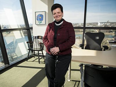
The artist chose tempera paint for her oeuvre. The flower’s petals are the color of Snow White’s buddy, the Bluebird of Happiness. Each petal is accentuated in stop light red, and the blossom’s leaves stretch up toward the sun. With its bold strokes and exuberant colors, the painting exudes life itself.

It’s the first thing Lisa M. Guay-Woodford, M.D., sees when she enters her office. It’s the last thing she sees as she leaves.

Dr. Guay-Woodford, a pediatric nephrologist, is internationally recognized for her research into the mechanisms that make certain inherited renal disorders, such as autosomal recessive polycystic kidney disease (ARPKD), particularly lethal. She also studies disparate health disorders that have a common link: Disruption to the cilia, slim hair-like structures that protrude from almost every cell in the human body and that play pivotal roles in human genetic disease.

Sarah, the artist who painted the bright blue flower more than 20 years ago when she was 8, was one of Dr. Guay-Woodford’s patients. And she’s part of the reason why Dr. Guay-Woodford has spent much of her career focused on the broader domain of disorders tied to just a single defective gene, such as ARPKD.

“It dates back to when I was a house officer and took care of kids with this disorder,” Dr. Guay-Woodford says. “Maybe 30 percent die in the newborn period. Others survive, but they have a whole range of complications.”

Two of her favorite patients died from ARPKD-related reasons in the same year. One died from uncontrolled high blood pressure. The other, Sarah, died from complications from a combined kidney and liver transplant.

“The picture she drew hangs in my office,” she says. “She was a wonderful kid who was really full of life, and what she chose really mirrored who she was as a person. We put up lots of those sorts of those things in my office. It’s a daily reminder of why we do the things we do and the end goal.”

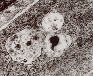
ARPKD is characterized by the growth of cysts in the liver, the kidney – which can lead to kidney failure – and complications within other structures, such as blood vessels in the heart and brain, according to the National Institutes of Health. About 1 in 20,000 live births is complicated by the genetic disorder. The age at which symptoms arise varies.

“Given the way it plays out, starting in utero, this is not a disease we are likely to cure,” she says. “But there are children who have very minimal complications. The near-term goal is to use targeted therapies to convert the children destined to have a more severe disease course to one that is less complicated so that no child suffers the full effects of the disease.”

That’s why it is essential to attain detailed knowledge about the defective gene responsible for ARPKD. To that end, Dr. Guay-Woodford participated in an international collaboration – one of three separate groups that 14 years ago identified PKHD1 as the defective gene that underlies ARPKD.

“The progress has been slow, partly because the gene and its protein products are very complex,” she says. “The good news is the gene has been identified. The daunting news is the identification did not leap us forward. It is just sort of an important step in what is going to be a fits-and-starts kind of journey.”

The field is trying to emulate the clinical successes that have occurred for patients with cystic fibrosis, which now can be treated by a drug that targets the defective gene, attacking disease at a fundamental level. Patient outcomes also have improved due to codifying care.

When she was a resident in the 1980s, children with cystic fibrosis died in their teens. “Now, they’re living well into their 40s because of careful efforts by really astute clinicians to deliver a standardized approach to care, an approach now enhanced by a terrific new drug. We measure quality care in terms of patient outcomes. That has allowed us to really understand how to effectively use antibiotics, physical therapy and how to think about nutrition – which makes a hugely important contribution that previously had been underappreciated.”

Standardizing clinical approaches dramatically improved and extended patients’ lives. “For renal cystic disease, we are beginning to do that better and better,” she adds.

There’s no targeted medicine yet for ARPKD. But thanks to an international conference that Dr. Guay-Woodford convened in Washington in 2013, such consensus expert recommendations have been published to guide diagnosis, surveillance and management of pediatric patients with ARPKD.

“There is an awful lot we can do in the way we systematically look at the clinical disease in these patients and improve our management. And, if you can overlay on top of that specific insights about why one person goes one way in disease progression versus another way, I think we can boost the baseline by developing good standards of care,” she says.

“Science does march on. There are a number of related research studies that are expanding our understanding of ARPKD. Within the next decade, we probably will be able to capitalize on not just the work in ARPKD but work in related diseases to learn the entry points for targeting therapies. That way, we can build a portfolio of markers of disease progression and test how effective these potential therapies are in slowing the course of the disease.”

Key to genetic influence of APOL1 on chronic kidney disease

Zhe Han

Drosophila melanogaster nephrocytes share structural and functional similarities with human renal cells, making the fruit fly an ideal pre-clinical model for studying how the APOL1 gene contributes to renal disease in humans.

Using the Drosophila melanogaster pre-clinical model, a Children’s National Health System research team identified a key mechanism by which the APOL1 gene contributes to chronic kidney disease in people of African descent. The model exploits the structural and functional similarities between the fruit fly’s nephrocytes and renal cells in humans to give scientists an unprecedented ability to study gene-to-cell interactions, identify other proteins that interact with APOL1 in renal disease, and target novel therapies, according to a paper published November 18 in the Journal of the American Society of Nephrology.

“This is one of the hottest research topics in the kidney field. We are the first group to generate this result in fruit flies,” says Zhe Han, Ph.D., a senior Drosophila specialist and associate professor in the Center for Cancer & Immunology Research at Children’s National. Han, senior author of the paper, presented the study results this month during Kidney Week 2016, the American Society of Nephrology’s annual gathering in Chicago that was expected to draw more than 13,000 kidney professionals from around the world.

The advantages of Drosophila for biomedical research include its rapid generation time and an unparalleled wealth of sophisticated genetic tools to probe deeply into fundamental biological processes underlying human diseases. People of African descent frequently inherit a mutant version of the APOL1 gene that affords protection from African sleeping sickness, but is associated with a 17- to 30-fold greater chance of developing certain types of kidney disease. That risk is even higher for individuals infected with the human immunodeficiency virus (HIV). Drosophila renal cells, called nephrocytes, accurately mimic pathological features of human kidney cells during APOL1-associated renal disease.

“Nephrocytes share striking structural and functional similarities with mammalian podocytes and renal proximal tubule cells, and therefore provide us a simple model system for kidney diseases,” says Han, who has studied the fruit fly for 20 years and established the fly nephrocyte as a glomerular kidney disease model in 2013 with two research papers in the Journal of the American Society of Nephrology.

In this most recent study, Han’s team cloned a mutated APOL1 gene from podocyte cells cultured from a patient with HIV-associated nephropathy. They created transgenic flies making human APOL1 in nephrocytes and observed that initially the transgene caused increased cellular functional activity. As flies aged, however, APOL1 led to reduced cellular function, increased cell size, abnormal vesicle acidification, and accelerated cell death.

“The main functions of nephrocytes are to filter proteins and remove toxins from the fly’s blood, to reabsorb protein components, and to sequester harmful toxins. It was surprising to see that these cells first became more active and temporarily functioned at higher levels,” says Han. “The cells got bigger and stronger but, ultimately, could not sustain that enhancement. After swelling to almost twice their normal size, the cells died. Hypertrophy is the way that the human heart responds to stress overload. We think kidney cells may use the same coping mechanism.”

The Children’s research team is a multidisciplinary group with members from the Center for Cancer & Immunology Research, the Center for Genetic Medicine Research, and the Division of Nephrology. The team also characterized fly phenotypes associated with APOL1 expression that will facilitate the design and execution of powerful Drosophila genetic screening approaches to identify proteins that interact with APOL1 and contribute to disease mechanisms. Such proteins represent potential therapeutic targets. Currently, transplantation is the only option for patients with kidney disease linked to APOL1.

“This is only the beginning,” Han says. “Now, we have an ideal pre-clinical model. We plan to start testing off-the-shelf therapeutic compounds, for example different kinase inhibitors, to determine whether they block any of the steps leading to renal cell disease.”

‘Trojan horse’ macrophage TNF-alpha opens door for HIV-1 to enter kidney epithelial cells, causing nephropathy

macrophage

Like a Trojan horse, the macrophage sits atop the epithelial cell with HIV hidden inside, opening a doorway into the kidney cell for high levels of HIV-1 to enter.

When nephrologist Patricio Ray, M.D., began investigating human immunodeficiency virus (HIV) as a renal fellow, children infected with the virus had a life expectancy of no more than seven years, and kids of African descent curiously were developing a type of HIV-related kidney disease.

HIV-associated nephropathy (HIVAN) is a progressive kidney disease seen in people who are both HIV-positive and of African ancestry. Kids who carry a modified protein that protects them against sleeping sickness are 80 times more likely to develop this type of kidney disease. Due to the kidney damage, they can have abnormal amounts of protein in their urine, focal segmental glomerulosclerosis, and microcystic tubular dilation, which can lead to enlarged kidneys and chronic kidney failure.

“No one understood how HIV could affect kidney cells that lack the receptors expressed in T cells and white cells,” recalls Dr. Ray, Robert Parrott Professor of Pediatrics at Children’s National Health System. Virologists said kidney epithelial cells that lacked CD4, a major receptor where HIV attaches, could not be infected with the virus. Nephrologists, meanwhile, were seeing that HIV infection was damaging these cells.

It’s taken two decades to unravel the medical mystery, aided by urine samples he coaxed kids to donate by offering them the latest music from New Kids on the Block in exchange for each urine bottle. Many of the kids died years ago, but their immortalized cells were essential in determining, through a process of elimination, which renal cell types were capable of being infected by HIV-1.

The paper represents the capstone of Dr. Ray’s career.

“This is how difficult it is to get an important contribution in science,” he says. “It’s 20 years of work involving the excellent contributions of many people, but that’s why research is called research. In the end, it’s all a learning process. But, it’s amazing how the puzzle pieces begin to fit. When the puzzle fits, it’s good.”

Dr. Ray, in collaboration with lead author Jinliang Li, Ph.D., and four additional Children’s National co-authors, published a paper November 3 in the Journal of the American Society of Nephrology that establishes a new role for transmembrane TNF-alpha, that of a facilitator that makes it easier for the HIV virus to enter certain cell types and replicate there.  Like a Trojan horse, the macrophage sits atop the epithelial cell with HIV hidden inside, opening a doorway into the kidney cell for high levels of HIV-1 to enter.

As a starting point, the research team cultured podocytes from the urine of kids with HIVAN. Through a number of steps, they isolated the unique contributions of the HIV envelope, heparan sulfate proteoglycans as attachment receptors – the glue that binds HIV to podocytes – and the essential role played by TNF-a, a 212-amino acid long type 2 transmembrane protein, in regulating at least two processes, including viral entry and fusion. They used a fluorescent marker to tag HIV-1 viruses, so it lit up bright green. Thus primed with transmembrane TNF-a, the podocytes were susceptible to HIV-1 infection when exposed to high viral loads.

Additional research is needed, such as in vitro work to help understand how HIV traffics within the cell, Dr. Ray says. Those insights could winnow the list of existing therapies that could block key steps, such as attachment to the viral envelope, which could help all people of African descent carrying the genetic mutation, including underserved kids in sub-Saharan Africa.

Another open research question is that certain cells located in the placenta and cervix express TNF-a, and may be more likely to be infected by HIV. Blocking that process could help prevent pregnant HIV-positive mothers from transmitting illness to their offspring.