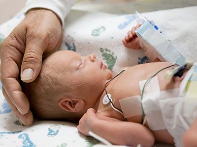
telemedicine control room

Telehealth connects pediatric heart experts about critical COVID-19 details

telemedicine control room

Telehealth is more than a doctor-to-patient tool during COVID-19. Experts in congenital heart disease meet weekly to share details about how it affects their vulnerable patients.

During the COVID-19 pandemic, telehealth has been crucial in allowing doctors to maintain safe contact with patients who require ongoing medical care without an office visit. Just as important is the role that telehealth is playing to connect care providers with each other to ensure that everyone around the world has the information they need to provide the best care possible for this swift-moving disease.

One good example of this specialist-to-specialist thought leadership connection is the ongoing weekly meeting hosted by the Children’s National Hospital cardiac critical care specialists. Since early in the spread of COVID-19, the Cardiac-ICU team, led by cardiovascular specialists including Ricardo Munoz, M.D., chief of cardiac critical care medicine and executive director of telehealth at Children’s National, have connected pediatric clinicians around the world to discuss how best to care for particularly vulnerable patients with pre-existing heart diseases, and to discuss breaking news in epidemiology of the disease and the effectiveness of various treatment approaches.

The video conference attracts hundreds of physicians and nurses who specialize in pediatric cardiac care from countries all over the world. In the last week of April, the meeting featured a late-breaking session to discuss new pediatric intensive care observations of inflammatory symptoms similar to Kawasaki disease, which were being detected in the United Kingdom, Paris and the United States. While more information is needed about this discovery, the ability of these experts to gather and compare disease phenotypes from country to country facilitates both the additional classification of pediatric-related symptoms and improves how all centers, no matter their location, can prepare to treat children who present locally with these symptoms.

In recent weeks, cardiac physicians and nurses from some of the world’s hardest hit regions, including Italy and Spain, have shared detailed information about their on-the-ground experiences to help colleagues in the U.S. and elsewhere better prepare for new developments.

“This new disease is a moving target, especially when it comes to understanding how it might impact children and adults with existing cardiac disease, particularly those with congenital heart disease,” says Dr. Munoz. “It is extremely important that we learn from each other, especially when we are able to connect with our colleagues in the epicenters of the most serious outbreaks of COVID-19. We are happy to host this important weekly meeting with the goal of helping every specialist keep as many patients with cardiac diseases as safe as possible throughout the global health emergency.”

If you would like to join these weekly telehealth meetings, please send your request to COVIDMultiCICUResponse@childrensnational.org.

CHD global outcomes set

New CHD global outcomes set released

The International Consortium for Health Outcomes Measurement (ICHOM) announced the release of a Congenital Heart Disease Standard Set (CHDSS) in late April 2020.

Gerard Martin, M.D., FAAP, FACC, FAHA, cardiologist at Children’s National Hospital, chaired the working group and contributed to the standards’ writing. In ICHOM‘s press release, he noted that, “Having a global set of outcomes that matters most to adult patients and parents of children with congenital heart disease will provide a road map for healthcare professionals and organizations engaged in setting care strategies for this population around the world. I would like to acknowledge the efforts of the Working Group and ICHOM staff for their incredible effort on this project.”

The CHDSS is a minimum core set of standards, comprised of Patient, Parent, and Clinician – Reported Outcome Measures already being collected by most practices in routine clinical care. The CHDSS measures 14 outcomes under the ICHOM framework for comprehensive outcomes measurement. These overarching domains are Overall Health, Social Health, Mental Health, and Physical Health.

Learn more about the CDHSS, the contributors and read the ICHOM press release.

CHD global outcomes set

The CHDSS measures 14 outcomes under the ICHOM overarching domains of Overall Health, Social Health, Mental Health, and Physical Health.

Patients and staff at the Uganda Heart Institute

Lifesaving heart surgeries for RHD complications in Uganda go on despite COVID-19

Patients and staff at the Uganda Heart Institute

Patients and staff at the Uganda Heart Institute for RHD-related heart surgeries in Uganda, March 2020. These patients were originally scheduled as part of the cancelled medical mission, but UHI cardiovascular surgeon successfully managed these cases without the support of the mission doctors from the U.S.

In early March as countries around the globe began to wrestle with how best to tackle the spread of COVID-19, a group of doctors, nurses, researchers and other medical staff from Children’s National Hospital were wrestling with a distinct set of challenges: What to do about the 10 Ugandan children and adults who were currently scheduled for lifesaving heart surgery (and the countless others who would benefit from the continued training of the local heart surgery team) to correct complications of rheumatic heart disease (RHD) during an impending medical mission in the country.

Rheumatic heart disease impacts over 39 million people globally and causes nearly 300,000 deaths per year. RHD is the result of frequent, untreated streptococcal throat infections in childhood that ultimately cause the body’s immune system to repeatedly damage heart valves. It is completely preventable, yet the majority of the world’s children still live in impoverished and overcrowded conditions that predispose them to RHD. Most patients present with advanced valvular heart disease. For example, in Uganda, an RHD registry includes over 600 children with clinical RHD, of which nearly 40% die within four years and the median survival time from enrollment in the registry is only nine months. For these patients, heart surgery is the only viable solution for long-term survival and normal quality of life.

Patricia: 9-year-old from Gulu

Patricia: 9-year-old from Gulu (northern Uganda), had mitral valve replacement and was doing well on a recent follow-up visit at her home.

The scheduled trip from Washington was part of a nearly 20-year partnership** between doctors, nurses, researchers and other medical staff in the United States, including Craig Sable, M.D., associate chief of cardiology, and and Pranava Sinha, M.D.,pediatric cardiovascular surgeon, at Children’s National Hospital in Washington, D.C., and the Uganda Heart Institute in Kampala, Uganda. The partnership aims to tackle RHD head-on. It provides surgical skill transfer, allows for treatment of more complex patients, and increases sustainable surgical capacity for Uganda’s RHD patients over time. As a result, over the last 15 years more than 1,000 children have received lifesaving heart surgery in Uganda, with the Uganda Heart Institute (UHI) performing one to two heart valve surgeries every two weeks over the last few years.

Jackline: 12-year-old from Gulu

Jackline: 12-year-old from Gulu, had mitral valve repair and aortic valve replacement. Jackline and Patricia were diagnosed through one of our research programs and benefit from our novel telehealth program, which helps connect patients from remote parts of Uganda to specialists at UHI.

COVID-19 was changing the current plan, however. Travel between countries was limited, and the team from the U.S. wouldn’t have been permitted to leave the U.S. and return according to schedule. The trip, and the support teams who were scheduled to arrive to help with the surgeries, were cancelled. The U.S. team members who had already arrived in Uganda were sent home after helping their UHI colleagues set up and prepare for the surgeries as much as possible. Knowing that patients and families were counting on the surgery mission to go forward after waiting for months or years to have surgery for heart valve disease, UHI decided not to cancel the majority of the surgeries. Instead, for the first time, they planned and successfully completed five valve-related cases in a single week – several of them quite complex. The cardiologists and cardiac surgeons from Children’s National who were supposed to be in-country for these procedures were forced to limit their in person assistance to the set-up activities the week prior to surgery and telehealth consult during the procedures.

“It was hard not to be able to stay  and work with the UHI team to help these families,” says Dr. Sable. “But we are so proud of the UHI team for meeting this challenge on their own. We knew they had the skills to perform at this volume and complexity. It’s a proud moment to see the team accomplish this major milestone, and to see the patients they cared for thrive.”

The patients are the most important outcome: The five who had successful open-heart surgery are all doing well, either on their way to recovery or already discharged to their communities, where they will, for the first time in memory, be able to play, exercise and go to school or work.

Longer term, this success demonstrates the UHI medical team’s ability to manage greater surgical capacity even when surgical missions from the U.S. resume. The partnership’s goal is to complete at least 1,000 annual operations (both pediatric and adult), with the majority being performed by the local team. Having this capacity available will mean the difference between life and death for many children and adults who have RHD in Uganda and the surrounding countries.

**This work is supported by the Edwards Life Sciences/Thoracic Surgery Foundation, the Emirates Airline Foundation, Samaritan’s Purse Children’s Heart Project and Gift of Life International.

muscle cells

Experimental model mimics early-stage myogenic deficit in boys with DMD

muscle cells

Muscle regeneration marked by incorporation of muscle stem cell nuclei (green) in the myofibers (red) in dystrophic muscles with low TGFβ level (upper image), but not with high TGFβ level (lower image). Inflammatory and other nuclei are labeled blue.

Boys with Duchenne muscular dystrophy (DMD) experience poor muscle regeneration, but the precise reasons for this remain under investigation. An experimental model of severe DMD that experiences a large spike in transforming growth factor-beta (TGFβ) activity after muscle injury shows that high TGFβ activity suppresses muscle regeneration and promotes fibroadipogenic progenitors (FAPs). This leads to replacement of the damaged muscle fibers by calcified and connective tissue, compromising muscle structure and function. While blocking FAP buildup provides a partial solution, a Children’s National Hospital study team identifies correcting the muscle micro-environment caused by high TGFβ as a ripe therapeutic target.

The team’s study was published online March 26, 2020, in JCI Insight.

DMD is a chronic muscle disease that affects 1 in 6,200 young men in the prime of their lives. The disorder, caused by genetic mutations leading to the inability to produce dystrophin protein, leads to ongoing muscle damage, chronic inflammation and poor regeneration of lost muscle tissue. The patients experience progressive muscle wasting, lose the ability to walk by the time they’re teenagers and die prematurely due to cardiorespiratory failure.

The Children’s National team finds for the first time that as early as preadolescence (3 to 4 weeks of age), their experimental model of severe DMD disease showed clear signs of the type of spontaneous muscle damage, regenerative failure and muscle fiber loss seen in preadolescent boys who have DMD.

“In boys, the challenge due to muscle loss exists from early in their lives, but had not been mimicked previously in experimental models,” says Jyoti K. Jaiswal, MSc, Ph.D., principal investigator in the Center for Genetic Medicine Research at Children’s National, and the study’s co-senior author. “TGFβ is widely associated with muscle fibrosis in DMD, when, in fact, our work shows its role in this disease process is far more significant.”

Research teams have searched for experimental models that replicate the sudden onset of symptoms in boys who have DMD as well as its complex progression.

“Our work not only offers insight into the delicate balance needed for regeneration of skeletal muscle, but it also provides quantitative information about muscle stem cell activity when this balanced is disturbed,” says Terence A. Partridge, Ph.D., principal investigator in the Center for Genetic Medicine Research at Children’s National, and the study’s co-senior author.

This schematic depicts the fate of injured myofibers in healthy or dystrophic muscle

This schematic depicts the fate of injured myofibers in healthy or dystrophic muscle (WT or mdx experimental models) that maintain low TGFβ level, compared with D2-mdx experimental models that experience a large increase in TGFβ level. As the legend shows, various cells are involved in this regenerative response.

“The D2-mdx experimental model is a relevant one to use to investigate the interplay between inflammation and muscle degeneration that is seen in humans with DMD,” adds Davi A.G. Mázala, co-lead study author.  “This model faithfully recapitulates many features of the complex disease process seen in humans.”

Between 3 to 4 weeks of age in the experimental models of severe DMD disease, the level of active TGFβ spiked up to 10-fold compared with models with milder disease. Intramuscular injections of an off-the-shelf drug that inhibits TGFβ signaling tamped down the number of FAPs, improving the muscle environment by lowering TGFβ activity.

“This work lays the foundation for studies that could lead to future therapeutic strategies to improve patients’ outcomes and lessen disease severity,” says James S. Novak, Ph.D., principal investigator in Children’s Center for Genetic Medicine Research, and co-lead study author. “Ultimately, our goal is to improve the ability of patients to continue to maintain muscle mass and regenerate muscle.”

In addition to Mázala, Novak, Jaiswal and Partridge, Children’s National study co-authors include Marshall W. Hogarth; Marie Nearing; Prabhat Adusumalli; Christopher B. Tully; Nayab F. Habib; Heather Gordish-Dressman, M.D.; and Yi-Wen Chen, Ph.D.

Financial support for the research described in this post was provided by the National Institutes of Health under award Nos. T32AR056993, R01AR055686 and U54HD090257; Foundation to Eradicate Duchenne; Muscular Dystrophy Association under award Nos. MDA295203, MDA480160 and MDA 477331; Parent Project Muscular Dystrophy; and Duchenne Parent Project – Netherlands.

preterm baby

Validating a better way to stratify BPD risk in vulnerable newborns

preterm baby

Factoring in the total number of days that extremely preterm infants require supplemental oxygen and tracking this metric for weeks longer than usual improves clinicians’ ability to predict respiratory outcomes according to bronchopulmonary dysplasia severity.

Factoring in the total number of days that extremely preterm infants require supplemental oxygen and tracking this metric for weeks longer than usual improves clinicians’ ability to predict respiratory outcomes according to bronchopulmonary dysplasia (BPD) severity, a research team led by Children’s National Hospital writes in Scientific Reports. What’s more, the researchers defined a brand-new category (level IV) for newborns who receive supplemental oxygen more than 120 days as a reliable way to predict which infants are at the highest risk of returning to the hospital due to respiratory distress after discharge.

About 1 in 10 U.S. infants is born preterm, before 37 weeks gestation, according to the Centers for Disease Control and Prevention. That includes extremely preterm infants who weigh about 1 lb. at birth. These very low birthweight newborns have paper thin skin, frail hearts and lungs that are not yet mature enough to deliver oxygen throughout the body as needed. Thanks to advances in neocritical care, an increasing number of them survive prematurity, and many develop BPD, a chronic lung disease characterized by abnormal development of the lungs and pulmonary vasculature.

“About half of the babies born prematurely will come back to the hospital within the first year of life with a respiratory infection. The key is identifying them and, potentially, preventing complications in this high-risk population,” says Gustavo Nino, M.D., a Children’s National pulmonologist and the study’s lead author.

For decades, the most common way to stratify BPD risk in these vulnerable newborns has been to see if they require supplemental oxygen at 36 weeks corrected gestational age.

“The problem with this classification is it doesn’t take into account the very premature babies who are on oxygen for much longer than other babies. So, we asked the question: Can we continue risk stratification beyond 36 weeks in order to identify a subset of babies who are at much higher risk of complications,” Dr. Nino says.

The longitudinal cohort study enrolled 188 infants born extremely preterm who were admitted to the neonatal intensive care unit (NICU) at Children’s National and tracked their data for at least 12 months after discharge. The team used a multidimensional approach that tracked duration of supplemental oxygen during the newborns’ NICU stay as well as scoring lung imaging as an independent marker of BPD severity. To validate the findings, these U.S.-born newborns were matched with 130 infants who were born preterm and hospitalized at two NICUs located in Bogotá, Colombia.

“Babies who are born very preterm and require oxygen beyond 120 days should have expanded ventilation of the lungs and cardiovascular pulmonary system before going home,” he notes. “We need to identify these newborns and optimize their management before they are discharged.”

And, the babies with level IV BPD risk need a different type of evaluation because the complications they experience – including pulmonary hypertension – place them at the highest risk of developing sleep apnea and severe respiratory infection, especially during the first year of life.

“The earlier we identify them, the better their outcome is likely to be,” Dr. Nino says. “We really need to change the risk stratification so we don’t call them all ‘severe’ and treat them the same when there is a subset of newborns who clearly are at a much higher risk for experiencing respiratory complications after hospital discharge.”

In addition to Dr. Nino, Children’s National study co-authors include Awais Mansoor, Ph.D., staff scientist at the Sheikh Zayed Institute for Pediatric Surgical Innovation (SZI); Geovanny F. Perez, M.D., pediatric pulmonologist; Maria Arroyo, M.D., pulmonologist; Xilei Xu Chen, M.D., postdoctoral fellow; Jered Weinstock, pediatric pulmonary fellow; Kyle Salka, MS, research technician; Mariam Said, M.D., neonatologist, and Marius George Linguraru, DPhil, MA, MSc, SZI principal investigator and senior author. Additional co-authors include Ranniery Acuña-Cordero, Universidad Militar Nueva Granada, Bogotá, Colombia; and Monica P. Sossa-Briceño and Carlos E. Rodríguez-Martínez, both of Universidad Nacional de Colombia.

Funding for research described in this post was provided by the National Institutes of Health (NIH) under award Nos. HL145669, AI130502 and HL141237. In addition, the NIH has awarded Dr. Nino an RO1 grant to continue this research.

NICU evacuation training baby on a stretcher

Innovative NICU training lauded as ‘best article’ by national journal

NICU evacuation training baby on a stretcher

“Fires, tornadoes and other natural disasters are outside of our team’s control. But it is within our team’s control to train neonatal intensive care unit (NICU) staff to master this necessary skill,” says Lisa Zell, BSN, a clinical educator at Children’s National Hospital.

Research into how to create a robust emergency evacuation preparedness plan and continually train staff that was led by Zell was lauded by editors of The Journal of Perinatal & Neonatal Nursing. The journal named the study the “best article” for the neonatal section that the prestigious journal published in 2018-19.

“We all hope for the best no matter what the situation, but we also need to extensively plan for the worse,” says Billie Lou Short, M.D., chief of the division of neonatology at Children’s National. “I’m proud that Lisa Zell and co-authors received this much-deserved national recognition on behalf of the nation’s No. 1 NICU.”

Educators worked with a diverse group within Children’s National to design and implement periodic evacuation simulations.

In addition to Zell and Lamia Soghier, M.D., FAAP, CHSE, Children’s National NICU medical unit director, study co-authors include Carmen Blake, BSN; Dawn Brittingham, MSN; and Ann-Marie Brown, MSN.

Read more
View photos showing how disaster training occurs at Children’s National

covers of books edited by Children's National faculty

We wrote the book

Children’s National Hospital is proud to have a number of faculty members who literally wrote the books on pediatric cardiology, neonatology, neurology and pulmonology. These texts, edited by experts Gil Wernovsky, M.D., Gordon Avery, M.D., Ricardo Munoz, M.D., Anastassios Koumbourlis, M.D., MPH, Robert Keating, M.D. and Roger Packer, M.D., have become the definitive references for medical students everywhere.

Through these books, generations of children worldwide will benefit from the expertise at Children’s National:

  • Anderson’s Pediatric Cardiology. Wernovsky, G., Anderson, R.H., Kumar, K., Mussatto, K.A., Redington, A.N., Tweddell, J.S., Tretter, J.T. (Eds.). (2019). Philadelphia, PA: Elsevier Publishing.
  • Avery’s Neonatology: Pathophysiology and Management of the Newborn. MacDonald, M.G., and Seshia, M.M.K. (Eds.) (2015). Philadelphia, PA: Lippincott Williams & Wilkins.
  • Critical Care of Children with Heart Disease: Basic Medical and Surgical Concepts. Munoz, R.A., More, V.O., da Cruz, E.M., Vetterly, C.G., da Silva, J.P. (Eds.). (2010) London, UK: Springer-Verlag London Ltd.
  • Diagnostic Tests in Pediatric Pulmonology. Davis, S.D., Koumbourlis, A.C., and Eber, E. (Eds.). (2015) London, UK: Springer-Verlag London Ltd.
  • Pulmonary Complications of Non-Pulmonary Pediatric Koumbourlis, A.C., and Nevin, M. (Eds.). (2018) London, UK: Springer-Verlag London Ltd.
  • Tumors of the Pediatric Central Nervous system. Keating, R.F., Goodrich, J.T., and Packer, R.J. (Eds.). (2013) New York, NY: Thieme Medical Publishers.

covers of books edited by Children's National faculty

Pediatric angiography

Congenital heart disease more deadly in low-income countries

Pediatric angiography

Even though mortality from congenital heart disease (CHD) has declined over the last three decades as diagnosis and treatments have advanced, the chances for a child to survive a CHD diagnosis significantly differs based on the country where he or she is born.

This eye-opening finding is drawn from the first comprehensive study of congenital heart disease across 195 countries, prepared using data from the Global Burden of Diseases, Injuries and Risk Factors Study 2017 (GBD), and recently published in The Lancet.

“Previous congenital heart estimates came from few data sources, were geographically narrow and did not evaluate CHD throughout the life course,” write the authors, known collectively as the 2017 GBD Congenital Heart Disease Collaborators. Co-lead author Meghan D. Zimmerman, M.D., worked on the study while completing her pediatric cardiology and American Heart Association Global Health Fellowships at Children’s National Hospital, and two pediatric cardiologists from Children’s National, Cardiology Associate Chief Craig Sable, M.D., and Gerard Martin, M.D., medical director of Global Services, provided leadership and oversight of this paper. The remaining collaborators are from more than 45 institutions around the world, spanning cardiology, public health and schools of medicine on every continent.

This is the first time the GBD study data was used along with all available data sources and previous publications – making it the most comprehensive study on congenital heart disease burden to date. Key differences between this study and prior estimates include:

  • Anatomic groupings of CHD by type, rather than simply categorized as moderate, severe or critical.
  • Inclusion of new data sources, including data from screening programs, congenital registries, administrative data and data sources in mortality and survival.
  • A control mechanism to account for cases of CHD that remit on their own to reduce the risk of overestimating prevalence.
  • Inclusion of all cases of congenital heart disease, including those with chromosomal or genetic anomalies such as Trisomy 21 that often co-occur.

This more comprehensive data set led to findings that showed lower predicted long-term survival, higher remission, and lower prevalence than previous studies that extrapolated evidence from studies of high-income countries. However, it also means these new estimates are a more accurate representation of the current global state of affairs. Overall, the study found:

  • A 34.5% decline in deaths from congenital disease between 1990 to 2017.
  • Nearly 70% of deaths caused by CHD in 2017 (180,624) were in infants less than one year old.
  • Most CHD deaths occurred in countries within the low and low-middle socio-demographic index (SDI) quintiles.
  • Mortality rates get lower as a country’s SDI rises.
  • Birth prevalence of CHD was not related to a country’s socio-demographic status, but overall prevalence was much lower in the poorest countries of the world. This is because children in these countries do not have access to life saving surgical services.
  • Nearly 12 million people are currently living with CHD globally, 18.7% more than in 1990.
  • The burden of CHD is not fully realized by just looking at prevalence and mortality. The measure “Years of Life Lost” provides deeper insight into the staggering burden of CHD, taking into account both absolute mortality and age at death.

“In high income countries like the United States, we diagnose some heart conditions prenatally during the 20-week ultrasound,” says Gerard Martin, M.D., a pediatric cardiologist at Children’s National Hospital who contributed to the study. “We catch others right after birth with a pulse oximetry screening for critical congenital heart disease. We can operate to correct a critical issue within the first week of life. And now our CHD kids are growing and thriving through adulthood and having families of their own.”

“For children born in middle- and low-income countries, these data draw stark attention to what we as cardiologists already knew from our own work in these countries – the lack of diagnostic and treatment tools leads to lower survival rates for children born with CHD,” adds Craig Sable, M.D., associate chief of cardiology at Children’s National, another primary contributor. “This is one of the most significant publications I have been a part of as it highlights the substantial loss of life to CHD in infancy around the globe.”

The authors write, “The UN has prioritized reduction of premature deaths from heart disease, but to meet the target of ‘ending preventable deaths of newborns and children under 5 years of age,’ health policy makers will need to develop specific accountability measures that address barriers and improve access to care and treatment.”

The study also includes a 400-page appendix breaking down each area by type of congenital anomaly, world region and country.

Pediatric device competition

Premier annual pediatric medical device competition now accepting submissions

Pediatric device competition

Pediatric innovators pitch for grant awards and participation in a special accelerator program.

The official call for submissions is underway for the premiere annual pediatric medical device competition, sponsored by National Capital Consortium for Pediatric Device Innovation (NCC-PDI). The competition is led by Sheikh Zayed Institute for Pediatric Surgical Innovation at Children’s National Hospital, the A. James Clark School of Engineering at the University of Maryland and non-profit accelerator MedTech Innovator. The three organizations are all an integral part of the FDA-funded NCC-PDI, which aims to facilitate the development, production and distribution of pediatric medical devices. Additional NCC-PDI members include accelerator BioHealth Innovation and design firm Archimedic.

The competition focuses on pediatric devices in three areas of critical need: cardiovascular, orthopedic and spine, and neonatal intensive care (NICU) and is now accepting applications. Contestants will pitch for a share of up to $250K in grant awards and the opportunity to participate in the MedTech Innovator 2020 Accelerator – Pediatric Track.

The first stage of competition will be held on March 23 at the University of Maryland and will include up to 30 companies selected from all submissions received. Up to 10 finalists selected from that event will move on to the “Make Your Medical Device Pitch for Kids!” finals on October 4, 2020 in Toronto, Canada. Finalists from the March qualifying round will be notified in May, 2020.

“While there is a great need for pediatric devices in many specialty areas, the development and commercialization process is very challenging because of the small market size and dynamic characteristics of the patient population,” says Kolaleh Eskandanian, Ph.D., MBA, PMP, vice president and chief innovation officer at Children’s National Hospital and principal investigator of NCC-PDI. “To provide pediatric innovators with greater support in meeting these unique challenges, we must go beyond grant funding, which is why we are collaborating with MedTech Innovator to offer an accelerator program with a pediatric track.”

To date, NCC-PDI has mentored over 100 medical device sponsors to help advance their pediatric innovations, notes Eskandanian, with six devices having received either their FDA market clearance or CE marking. She says the success of NCC-PDI’s portfolio companies is attributed to funding, mentorship, support from partners, facilitated interactions between device innovators and potential investors, and being discovered during their presentations at the signature “Make Your Medical Device Pitch for Kids!” competitions.

While advancements have been made in some pediatric specialties, there is still a critical need for novel devices in cardiovascular, orthopedic and spine, and NICU areas. On average over the past decade, only 24 percent of life-saving medical devices approved by FDA – those that go through PMA and HDE regulatory pathways – have an indication for pediatric use. Of those, most are designated for children age 12 or older. “Devices designed specifically for the younger pediatric population are vitally needed and, at this early stage of the intervention, can significantly improve developmental outcomes for a child,” Eskandanian said.

Enhancing access to resources for pediatric innovators is also one of the aims of the Children’s National Research and Innovation Campus, a first-of-its-kind focused on pediatric healthcare innovation, currently under development on the former Walter Reed Army Medical Center campus in Washington, D.C. and opening in December, 2020. With its proximity to federal research institutions and agencies, universities, academic research centers, as well as on site accelerator Johnson and Johnson Innovation – JLABS, the campus will create a rich ecosystem of public and private partners which, like the NCC-PDI network, will help bolster pediatric innovation and commercialization.

NOTE: The deadline for submissions has been extended to February 22 at midnight EST.

Catherine Limperopoulos

Stressful pregnancies can leave fingerprint on fetal brain

Catherine Limperopoulos

“We were alarmed by the high percentage of pregnant women with a diagnosis of a major fetal heart problem who tested positive for stress, anxiety and depression,” says Catherine Limperopoulos, Ph.D., director of the Center for the Developing Brain at Children’s National and the study’s corresponding author.

When a diagnosis of fetal congenital heart disease causes pregnant moms to test positive for stress, anxiety and depression, powerful imaging can detect impaired development in key fetal brain regions, according to Children’s National Hospital research published online Jan. 13, 2020, in JAMA Pediatrics.

While additional research is needed, the Children’s National study authors say their unprecedented findings underscore the need for universal screening for psychological distress as a routine part of prenatal care and taking other steps to support stressed-out pregnant women and safeguard their newborns’ developing brains.

“We were alarmed by the high percentage of pregnant women with a diagnosis of a major fetal heart problem who tested positive for stress, anxiety and depression,” says Catherine Limperopoulos, Ph.D., director of the Center for the Developing Brain at Children’s National and the study’s corresponding author. “Equally concerning is how prevalent psychological distress is among pregnant women generally. We report for the first time that this challenging prenatal environment impairs regions of the fetal brain that play a major role in learning, memory, coordination, and social and behavioral development, making it all the more important for us to identify these women early during pregnancy to intervene,” Limperopoulos adds.

Congenital heart disease (CHD), structural problems with the heart, is the most common birth defect. Still, it remains unclear how exposure to maternal stress impacts brain development in fetuses with CHD.

The multidisciplinary study team enrolled 48 women whose unborn fetuses had been diagnosed with CHD and 92 healthy women with uncomplicated pregnancies. Using validated screening tools, they found:

  • 65% of pregnant women expecting a baby with CHD tested positive for stress
  • 27% of women with uncomplicated pregnancies tested positive for stress
  • 44% of pregnant women expecting a baby with CHD tested positive for anxiety
  • 26% of women with uncomplicated pregnancies tested positive for anxiety
  • 29% of pregnant women expecting a baby with CHD tested positive for depression and
  • 9% women with uncomplicated pregnancies tested positive for depression

All told, they performed 223 fetal magnetic resonance imaging sessions for these 140 fetuses between 21 and 40 weeks of gestation. They measured brain volume in cubic centimeters for the total brain as well as volumetric measurements for key regions such as the cerebrum, cerebellum, brainstem, and left and right hippocampus.

Maternal stress and anxiety in the second trimester were associated with smaller left hippocampi and smaller cerebellums only in pregnancies affected by fetal CHD. What’s more, specific regions — the hippocampus head and body and the left cerebellar lobe – were more susceptible to stunted growth. The hippocampus is key to memory and learning, while the cerebellum controls motor coordination and plays a role in social and behavioral development.

The hippocampus is a brain structure that is known to be very sensitive to stress. The timing of the CHD diagnosis may have occurred at a particularly vulnerable time for the developing fetal cerebellum, which grows faster than any other brain structure in the second half of gestation, particularly in the third trimester.

“None of these women had been screened for prenatal depression or anxiety. None of them were taking medications. And none of them had received mental health interventions. In the group of women contending with fetal CHD, 81% had attended college and 75% had professional educations, so this does not appear to be an issue of insufficient resources,” Limperopoulos adds. “It’s critical that we routinely to do these screenings and provide pregnant women with access to interventions to lower their stress levels. Working with our community partners, Children’s National is doing just that to help reduce toxic prenatal stress for both the health of the mother and for the future newborns. We hope this becomes standard practice elsewhere.”

Adds Yao Wu, Ph.D., a research associate working with Limperopoulos at Children’s National and the study’s lead author: “Our next goal is exploring effective prenatal cognitive behavioral interventions to reduce psychological distress felt by pregnant women and improve neurodevelopment in babies with CHD.”

In addition to Limperopoulos and Wu , Children’s National study co-authors include Kushal Kapse, MS, staff engineer; Marni Jacobs, Ph.D., biostatistician; Nickie Niforatos-Andescavage, M.D., neonatologist; Mary T. Donofrio, M.D., director, Fetal Heart Program; Anita Krishnan, M.D., associate director, echocardiography; Gilbert Vezina, M.D., director, Neuroradiology Program; David Wessel, M.D., Executive Vice President and Chief Medical Officer; and Adré  J. du Plessis, M.B.Ch.B., director, Fetal Medicine Institute. Jessica Lynn Quistorff, MPH, Catherine Lopez, MS, and Kathryn Lee Bannantine, BSN, assisted with subject recruitment and study coordination.

Financial support for the research described in this post was provided by the National Institutes of Health under grant No. R01 HL116585-01 and the Thrasher Research Fund under Early Career award No. 14764.

newborn baby

Directly measuring function in tiny hearts

newborn baby

The amount of blood the heart pumps in one minute can be directly measured safely in newborns by monitoring changes in blood velocity after injecting saline, indicates the first clinical study of direct cardiac output measurement in newborns.

The amount of blood that the heart pumps in one minute (cardiac output) can be directly measured safely in newborns by monitoring changes in blood velocity after injecting saline, indicates a paper published online Dec. 17, 2019 in the Journal of Pediatrics and Neonatal Medicine. The research, conducted by Children’s National Hospital faculty, is believed to be the first clinical study of direct cardiac output measurement in newborns.

Right now, cardiac output is measured indirectly in the nation’s neonatal intensive care units (NICU) using newborns’ blood pressure, heart rate, urine output and other indirect measures. However, these techniques can produce imprecise readings in children. And the field lacks a feasible “gold standard” to measure cardiac output in newborns.

The COstatus monitor already uses ultrasound dilution – the expected decrease in the velocity of blood when saline is injected, producing a dilution curve. A Children’s National research team used ultrasound dilution in their small pilot study to gauge the feasibility of directly measuring cardiac output in newborns.

“Infants who stand to benefit most from directly monitoring cardiac hemodynamics are often so sick they already have central venous access,” says Khodayar Rais-Bahrami, M.D., an attending neonatologist at Children’s National and the study’s senior author. “Using the COstatus monitor in these children would enable the clinical team to personalize care based on the newborn’s current hemodynamic status, while introducing minimal fluid during measurements,” Dr. Rais-Bahrami adds.

COstatus monitor

The COstatus Monitor uses an extracorporeal loop attached to arterial and venous lines to measure cardiac output using ultrasound dilution. The research team injected 1mL/kg of body temperature saline into the loop and performed up to two measurement sessions daily.

The research team recruited 12 newborns younger than 2 weeks old who already had central venous and arterial access. The venous line of the arteriovenous AV loop is connected to the umbilical venous catheter while the COstatus monitor’s arterial line is connected to the umbilical arterial catheter. During measurement sessions, two injections of solution are injected into the venous loop, allowing for two measures of cardiac output, cardiac index, active circulating volume index, central blood volume index and systemic vascular resistance index.

Infants enrolled in the pilot study underwent up to two measurement sessions per day for up to four days, for a total of 54 cardiac hemodynamic measurements. The newborns ranged from 720 to 3,740 grams in weight and 24 to 41.3 weeks in gestational age.

The infants’ mean cardiac output was 0.43L/min and increased with gestational age. By contrast, the mean cardiac index was 197mL/kg/min and changed little with infants’ increasing maturity – either by gestational age or postnatal age. Two of the study participants were undergoing therapeutic cooling for hypoxic-ischemic encephalopathy and had their measurements taken during cooling and after rewarming.

“Although this study size is small, it demonstrates that this minimally invasive technique can safely be used in newborns to directly measure cardiac hemodynamics,” says Simranjeet S. Sran, M.D., a Children’s National neonatalogist and the study’s lead author. “This technology may allow for more precise and personalized care of critically ill newborns in a range of disease states – real-world utility in NICUs that serve some of the youngest and sickest newborns,” Dr. Sran adds.

The research team notes that direct measurement by ultrasound dilution revealed a stark increase in cardiac index as infants undergoing therapeutic hypothermia were rewarmed, raising questions about whether indirect measures using other technology, such as echocardiography, underestimate hypothermia’s effect on hemodynamics.

In addition to Drs. Rais-Bahrami and Sran, Mariam Said, M.D., also a Children’s National neonatalogist, was a study co-author.

Michael Tsifansky

Lung transplant expert Michael Tsifansky, M.D., F.A.A.P., joins Children’s

Michael Tsifansky

Earlier this year Michael Tsifansky, M.D., F.A.A.P., joined Children’s National Hospital as an attending physician in the Cardiac Intensive Care Unit and in the Division of Pulmonology and Sleep Medicine. He brings to Children’s National a unique mix of expertise in critical care and pulmonary medicine. That passion for these two subspecialties has also made him one of the country’s leading experts in lung transplant procedures and the recovery from them.

Dr. Tsifansky shared more information about caring for patients with complex lung diseases, especially those with end-stage lung disease. He outlines the patient population for pediatric lung transplants and the arduous process patients endure while waiting for a transplant, undergoing this major procedure, and then recovering from it.

What types of patients undergo lung transplant surgeries?

Lung transplantation in children is indicated when the following criteria are met:

  • End-stage lung disease
  • No reasonable alternative to the established diagnosis
  • No medical or surgical alternative to the current course of treatment
  • No other organ failure
  • Stable social environment

Could you describe the surgery process?

Pediatric lung transplantation may be performed on cardiopulmonary bypass, on extracorporeal membrane oxygenation (ECMO) or off extracorporeal cardiopulmonary support (ECS). The donor’s lungs are kept chilled prior to transplantation and should be transplanted within six to eight hours after removal from the donor. The donor’s main-stem bronchi and pulmonary arteries are connected to those of the recipient, and the donor’s pulmonary venous drainage is connected to the recipient’s left atrium using the donor’s left atrial roof tissue. This procedure typically takes six to eight hours.

Could you describe the recovery process?

Typically, pediatric lung transplant recipients are extubated and encouraged to sit up four to six hours after the transplant procedure and walk soon afterward. It is important that they be out of bed and moving as soon as possible, and our colleague from Rehabilitation Services (physical and occupational therapists and rehabilitation physicians) will be working with the children toward these goals. After transplantation, pediatric patients will be given discharge instructions with individualized guidelines for a healthy lifestyle. Patients should return to near-normal life approximately three to six months after transplantation.

How long does the recovery process take?

The patient will remain hospitalized for 11-14 days following surgery for acute rehab, titration of antirejection meds and initial healing.

You’ve mentioned that it’s important for transplant patients to get moving as part of recovery. When can a patient begin walking again?

Lung recipients will be assisted into a chair soon after the transplant. Within the first 24-36 hours, the patient is encouraged to take short walks, increasing the distance each day. A physical therapist will work with the patient during their hospitalization to meet their goals. We also encourage patients to exercise on the treadmill regularly while hospitalized. By the time the patient is ready to go home, he or she will be able to easily move around by themselves and do most of their care without assistance. They feel so much better than before transplant and have so much energy that we almost always have to gently limit their activity for a short while to allow their chest incision to heal properly.

What do you see as the next step in pulmonary care for end stage lung disease at Children’s National Hospital?

The development of a pediatric-specific lung transplant and respiratory failure program is the natural extension of the hospital’s cystic fibrosis program, heart transplant program and programs in pulmonary hypertension, bronchopulmonary dysplasia and extracorporeal membrane oxygenation for respiratory failure.

At present, there is no local option for a pediatric-specific program that can perform the transplant and provide the necessary comprehensive wrap-around services for patients in infancy up to age 18. As a top children’s hospital, Children’s National is uniquely positioned to provide the highest level of pediatric-specific care to this patient population and allow patients and their families to spend more time at home while undergoing this and other lifesaving treatments.

Dr. Tsifansky hopes to launch a comprehensive pediatric lung transplant and respiratory failure program at Children’s National in the very near future. Stay tuned for future developments from this area.

brain network illustration

$2.5M to protect the brain from metabolic insult

brain network illustration

The brain comprises only 2% of the body’s volume, but it uses more than 20% of its energy, which makes this organ particularly vulnerable to changes in metabolism.

More than 30 million Americans have diabetes, with the vast majority having Type 2 disease. Characterized by insulin resistance and persistently high blood sugar levels, poorly controlled Type 2 diabetes has a host of well-recognized complications: compared with the general population, a greatly increased risk of kidney disease, vision loss, heart attacks and strokes and lower limb amputations.

But more recently, says Nathan A. Smith, MS, Ph.D., a principal investigator in Children’s National Research Institute’s Center for Neuroscience Research, another consequence has become increasingly apparent. With increasing insulin resistance comes cognitive damage, a factor that contributes significantly to dementia diagnoses as patients age.

The brain comprises only 2% of the body’s volume, but it uses more than 20% of its energy, Smith explains – which makes this organ particularly vulnerable to changes in metabolism. Type 2 diabetes and even prediabetic changes in glucose metabolism inflict damage upon this organ in mechanisms with dangerous synergy, he adds. Insulin resistance itself stresses brain cells, slowly depriving them of fuel. As blood sugar rises, it also increases inflammation and blocks nitric oxide, which together narrow the brain’s blood vessels while also increasing blood viscosity.

When the brain’s neurons slowly starve, they become increasingly inefficient at doing their job, eventually succumbing to this deprivation. These hits don’t just affect individual cells, Smith adds. They also affect connectivity that spans across the brain, neural networks that are a major focus of his research.

While it’s well established that Type 2 diabetes significantly boosts the risk of cognitive decline, Smith says, it’s been unclear whether this process might be halted or even reversed. It’s this question that forms the basis of a collaborative Frontiers grant, $2.5 million from the National Science Foundation split between his laboratory; the lead institution, Stony Brook University; and Massachusetts General Hospital/Harvard Medical School.

Smith and colleagues at the three institutions are testing whether changing the brain’s fuel source from glucose to ketones – byproducts from fat metabolism – could potentially save neurons and neural networks over time. Ketones already have shown promise for decades in treating some types of epilepsy, a disease that sometimes stems from an imbalance in neuronal excitation and inhibition. When some patients start on a ketogenic diet – an extreme version of a popular fat-based diet – many can significantly decrease or even stop their seizures, bringing their misfiring brain cells back to health.

Principal Investigator Smith and his laboratory at the Children’s National Research Institute are using experimental models to test whether ketones could protect the brain against the ravages of insulin resistance. They’re looking specifically at interneurons, the inhibitory cells of the brain and the most energy demanding. The team is using a technique known as patch clamping to determine how either insulin resistance or insulin resistance in the presence of ketones affect these cells’ ability to fire.

They’re also looking at how calcium ions migrate in and out of the cells’ membranes, a necessary prerequisite for neurons’ electrical activity. Finally, they’re evaluating whether these potential changes to the cells’ electrophysiological properties in turn change how different parts of the brain communicate with each other, potentially restructuring the networks that are vital to every action this organ performs.

Colleagues at Athinoula A. Martinos Center for Biomedical Imaging at Massachusetts General Hospital and Harvard Medical School, led by Principal Investigator Eva-Maria Ratai, Ph.D.,  will perform parallel work in human subjects. They will use imaging to determine how these two fuel types, glucose or ketones, affect how the brain uses energy and produces the communication molecules known as neurotransmitters. They’re also investigating how these factors might affect the stability of neural networks using techniques that investigate the performance of these networks both while study subjects are at rest and performing a task.

Finally, colleagues at the Laufer Center for Physical and Quantitative Biology at Stony Brook University, led by Principal Investigator Lilianne R. Mujica-Parodi, Ph.D., will use results generated at the other two institutions to construct computational models that can accurately predict how the brain will behave under metabolic stress: how it copes when deprived of fuel and whether it might be able to retain healthy function when its cells receive ketones instead of glucose.

Collectively, Smith says, these results could help retain brain function even under glucose restraints. (For this, the research team owes a special thanks to Mujica-Parodi, who assembled the group to answer this important question, thus underscoring the importance of team science, he adds.)

“By supplying an alternate fuel source, we may eventually be able to preserve the brain even in the face of insulin resistance,” Smith says.

Dr. Kurt Newman in front of the capitol building

Making healthcare innovation for children a priority

Dr. Kurt Newman in front of the capitol building

Recently, Kurt Newman, M.D., president and CEO of Children’s National Hospital, authored an opinion piece for the popular political website, The Hill. In the article, he called upon stakeholders from across the landscape to address the significant innovation gap in children’s healthcare versus adults.

As Chair of the Board of Trustees of the Children’s Hospital Association,  Dr. Newman knows the importance of raising awareness among policy makers at the federal and state level about the healthcare needs of children. Dr. Newman believes that children’s health should be a national priority that is addressed comprehensively. With years of experience as a pediatric surgeon, he is concerned by the major inequities in the advancements of children’s medical devices and technologies versus those for adults. That’s why Children’s National is working to create collaborations, influence policies and facilitate changes that will accelerate the pace of pediatric healthcare innovation for the benefit of children everywhere. One way that the hospital is tackling this challenge is by developing the Children’s National Research & Innovation Campus, which will be the nation’s first innovation campus focused on pediatric research.

Research & Innovation Campus

Children’s National welcomes Virginia Tech to its new campus

Children’s National Hospital and Virginia Tech create formal partnership that includes the launch of a Virginia Tech biomedical research facility within the new Children’s National Research & Innovation Campus.

Children’s National Hospital and Virginia Tech recently announced a formal partnership that will include the launch of a 12,000-square-foot Virginia Tech biomedical research facility within the new Children’s National Research & Innovation Campus. The campus is an expansion of Children’s National that is located on a nearly 12-acre portion of the former Walter Reed Army Medical Center in Washington, D.C. and is set to open its first phase in December 2020. This new collaboration brings together Virginia Tech, a top tier academic research institution, with Children’s National, a U.S. News and World Report top 10 children’s hospital, on what will be the nation’s first innovation campus focused on pediatric research.

Research & Innovation Campus

“Virginia Tech is an ideal partner to help us deliver on what we promised for the Children’s National Research & Innovation Campus – an ecosystem that enables us to accelerate the translation of potential breakthrough discoveries into new treatments and technologies,” says Kurt Newman, M.D., president and CEO, Children’s National. “Our clinical expertise combined with Virginia Tech’s leadership in engineering and technology, and its growing emphasis on biomedical research, will be a significant advance in developing much needed treatment and cures to save children’s lives.”

Earlier this year, Children’s National announced a collaboration with Johnson & Johnson Innovation LLC to launch JLABS @ Washington, DC at the Research & Innovation Campus. The JLABS @ Washington, DC site will be open to pharmaceutical, medical device, consumer and health technology companies that are aiming to advance the development of new drugs, medical devices, precision diagnostics and health technologies, including applications in pediatrics.

“We are proud to welcome Virginia Tech to our historic Walter Reed campus – a campus that is shaping up to host some of the top minds, talent and innovation incubators in the world,” says Washington, D.C. Mayor Muriel Bowser. “The new Children’s National Research & Innovation Campus will exemplify why D.C. is the capital of inclusive innovation – because we are a city committed to building the public and private partnerships necessary to drive discoveries, create jobs, promote economic growth and keep D.C. at the forefront of innovation and change.”

Faculty from the Children’s National Research Institute and the Fralin Biomedical Research Institute at Virginia Tech Carilion (VTC) have worked together for more than a decade, already resulting in shared research grants, collaborative publications and shared intellectual property. Together, the two institutions will now expand their collaborations to develop new drugs, medical devices, software applications and other novel treatments for cancer, rare diseases and other disorders.

“Joining with Children’s National in the nation’s capital positions Virginia Tech to improve the health and well-being of infants and children around the world,” says Virginia Tech President Tim Sands, Ph.D. “This partnership resonates with our land-grant mission to solve big problems and create new opportunities in Virginia and D.C. through education, technology and research.”

The partnership with Children’s National adds to Virginia Tech’s growing footprint in the Washington D.C. region, which includes plans for a new graduate campus in Alexandria, Va. with a human-centered approach to technological innovation. Sands said the proximity of the two locations – just across the Potomac – will enable researchers to leverage resources, and will also create opportunities with the Virginia Tech campus in Blacksburg, Va. and the Virginia Tech Carilion Health Science and Technology campus in Roanoke, Va.

Carilion Clinic and Children’s National have an existing collaboration for provision of certain specialized pediatric clinical services. The more formalized partnership between Virginia Tech and Children’s National will drive the already strong Virginia Tech-Carilion Clinic partnership, particularly for children’s health initiatives and facilitate collaborations between all three institutions in the pediatric research and clinical service domains.

Children’s National and Virginia Tech will engage in joint faculty recruiting, joint intellectual property, joint training of students and fellows, and collaborative research projects and programs according to Michael Friedlander, Ph.D., Virginia Tech’s vice president for health sciences and technology, and executive director of the Fralin Biomedical Research Institute at VTC.

“The expansion and formalization of our partnership with Children’s National is extremely timely and vital for pediatric research innovation and for translating these innovations into practice to prevent, treat and ultimately cure nervous system cancer in children,” says Friedlander, who has collaborated with Children’s National leaders and researchers for more than 20 years. “Both Virginia Tech and Children’s National have similar values and cultures with a firm commitment to discovery and innovation in the service of society.”

“Brain and other nervous system cancers are among the most common cancers in children (alongside leukemia),” says Friedlander. “With our strength in neurobiology including adult brain cancer research in both humans and companion animals at Virginia Tech and the strength of Children’s National research in pediatric cancer, developmental neuroscience and intellectual disabilities, this is a perfect match.”

The design of the Children’s National Research & Innovation Campus not only makes it conducive for the hospital to strengthen its prestigious partnerships with Virginia Tech and Johnson & Johnson, it also fosters synergies with federal agencies like the Biomedical Advanced Research and Development Authority, which will collaborate with JLABS @ Washington, DC to establish a specialized innovation zone to develop responses to health security threats. As more partners sign on, this convergence of key public and private institutions will accelerate discoveries and bring them to market faster for the benefit of children and adults.

“The Children’s National Research & Innovation Campus pairs an inspirational mission to find new treatments for childhood illness and disease with the ideal environment for early stage companies. I am confident the campus will be a magnet for big ideas and will be an economic boost for Washington DC and the region,” says Jeff Zients, who was appointed chair of the Children’s National Board of Directors effective October 1, 2019. As a CEO and the former director of President Obama’s National Economic Council, Zients says that “When you bring together business, academia, health care and government in the right setting, you create a hotbed for innovation.”

Ranked 7th in National Institutes of Health research funding among pediatric hospitals, Children’s National continues to foster collaborations as it prepares to open its first 158,000-square-foot phase of its Research & Innovation Campus. These key partnerships will enable the hospital to fulfill its mission of keeping children top of mind for healthcare innovation and research while also contributing to Washington D.C.’s thriving innovation economy.

Nikki Gillum Posnack

Research team develops new and improved method for studying cardiac function

Nikki Gillum Posnack

While researching how plastic affects heart function in sensitive populations, such as children born with congenital heart defects, Children’s National researcher Nikki Posnack, Ph.D., led a team that developed a new and improved, replicable method of performing simultaneous dual optical mapping to examine electrical activity and calcium for the study of cardiac function.

Since arriving at the Sheikh Zayed Institute for Pediatric Surgical Innovation, researcher Nikki Gillum Posnack, Ph.D., a principal investigator with the institute and assistant professor of pediatrics at the George Washington University School of Medicine and Health Sciences, has been focused on examining how exposure to plastic affects heart function in sensitive populations, such as children born with congenital heart defects. She performs optical mapping to conduct this research, but the industry standard approaches of either using dual cameras or sequential single cameras were cost prohibitive and technically challenging while also diminishing the quality of the imaging results.

Fast forward to July 2019 when Dr. Posnack and her team published “Plasticizer Interaction With the Heart” in the journal Arrhythmia and Electrophysiology, which used imaging techniques to reveal the impact of plastic chemicals on the electrical activity of the heart. Dr. Posnack’s laboratory has since expanded this technique and revealed a new replicable method of performing simultaneous dual optical mapping to examine electrical activity and calcium handling in the heart.

Sharing a new method for studying cardiac function

This groundbreaking method is itself the focus of a new BMC Biomedical Engineering journal article titled “Lights, camera, path splitter: a new approach for truly simultaneous dual optical mapping of the heart with a single camera.”

The article compares and contrasts the current standard for dual camera simultaneous configurations and single camera sequential configurations to Dr. Posnack’s new single camera simultaneous configuration.

Simultaneous dual mapping systems use two probes and dual dyes – one for electrical voltage and the other for calcium. While dual-dye combinations like Di-4-ANEPPS with Indo-1, Di-2-ANEPEQ and calcium green have been developed to separate fluorescence signals by emission, these dye combinations can have spectral overlap, creating the need for non-ideal emission bandpass to negate spectral overlap and/or the inclusion of a calcium probe with an inferior dissociation constant. Additionally, dual-sensor systems require proper alignment to ensure that fluorescence signals are being analyzed from the same tissue region on each individual detector, which could lead to erroneous results. The dual-camera optical setup is expensive, technically challenging and requires a large physical footprint that is often not feasible for basic science and teaching laboratories conducting critical research.

As an alternative, some researchers use a single camera configuration to sequentially image the voltage and calcium probes using excitation light patterning. This approach also has limitations. These single-sensor designs use dual-dye combinations that require two or more excitation light sources, but share a single emission band. Like the dual camera system, this platform design is also technically challenging since the different excitation light wavelengths require light source triggering, camera synchronization and frame interleaving. Due to timing coordination, decreased frame rates, excitation light ramp up/down times and shutter open/close times, single system setups require shorter exposure times compared to dual sensor setups, diminishing the signal-to-noise quality without offering the same temporal fidelity. There is a cost advantage to the single camera system, however, because the additional camera is often one of the most expensive components.

This new single camera, simultaneous dual optical mapping approach is the first multiparametric mapping system that simultaneously acquires calcium and voltage signals from cardiac preparations, using a commercially available optical path splitter, single camera and single excitation light. Using a large field of view sCMOS sensor that is faster and more sensitive, this configuration separates the two emission bands for voltage and calcium probes and simultaneously directs them to either sides of the single, large camera sensor. This protocol employs a commonly used dual-dye combination (RH237 and Rhod2-AM). In contrast, other protocols may require genetically-encoded indicators or fluorescent probes that are not yet commercially available.

The team validated the utility of the approach by performing high-speed simultaneous dual imaging with sufficient signal-to-noise ratio for calcium and voltage signals and specificity of emission signals with negligible cross-talk. Demonstrating the need for simultaneous electrical and calcium sensors, they found that when ventricular tachycardia is induced, there is spatially discordant calcium alternans present in different regions of the heart even when the electrical alternans remain concordant.

Having eliminated the second camera as well as the need for multiple excitation light sources, light pattering and frame interleaving, this system is more cost effective, simpler, and can be easily setup by various types of researchers, not just those with engineering backgrounds.

With a limited research budget and a background in physiology, Dr. Posnack worked collaboratively with her post-doctoral fellow Rafael Jaimes III, an engineer in the Sheikh Zayed Institute for Pediatric Surgical Innovation, to develop a cost-effective system that would enable her to truly study the effects of plastics on the heart.

Multidisciplinary approach

“We’re fortunate to have a multidisciplinary team in the Sheikh Zayed Institute so that I could work with an engineer to develop the technology and system we needed to propel our research,” said Dr. Posnack. “There are so many researchers who have the science background, but not necessarily the technical aptitude, and they get stymied in their research, so we’re proud that this paper will help other researchers replicate the system to study cardiac function.”

The research paper was funded by a grant from the National Institutes of Health as well as support from the Children’s Research Institute, Children’s National Heart Institute and the Sheikh Zayed Institute for Pediatric Surgical Innovation.

The applications for this optical mapping system are significant and Dr. Posnack has been consulted by other research teams looking to implement it in their labs. Additionally, Dr. Posnack has collaborated with several neuroscience teams at Children’s National Hospital, including one that is investigating the effects of hypoxia on brain and heart development, and another that is interested in using image modalities and data processing to analyze calcium as an indicator of neuron firing.

Dr. Posnack continues to use this new dual optical mapping system to further her research as she anticipates the publication of a new article about age-dependent changes in cardiac electrophysiology and calcium handling.

telemedicine

Children’s National partners with Sabará Hospital Infantil to provide pediatric telehealth services in Brazil

telemedicine

Through a new partnership with Sabará Hospital Infantil in São Paulo, Brazil, Children’s National Hospital will provide access to pediatric cardiac intensive care specialists and consultations via telehealth. This is the first international telehealth offering from Children’s National for pediatric cardiac critical care.

The partnership includes sharing care proposals, second opinion for complex cardiology cases, alignment with international benchmarks, adoption of diagnostic and therapeutic protocols, development of critical mass for continuous process improvement and continued training. Joint multidisciplinary visits will also be carried out to help Sabará validate and improve existing protocols and learn about innovations and service improvement opportunities.

Children’s National will also provide teleconference-based training for Sabará nursing staff and second opinions through medical teleconsultation with specialists in all areas of pediatric cardiology, based on each patient’s individual needs.

“It is an honor to partner with Sabará Hospital lnfantil,” says Ricardo Munoz, M.D., executive director of Telemedicine and chief of Cardiac Critical Care at Children’s National. “We look forward to working together toward our shared goal of providing the best health care possible for the children in Brazil.”

doctor's stethescope coming out of a computer

Virtual cardiology follow-ups may save families time and money

doctor's stethescope coming out of a computer

Virtual cardiology follow-ups via computer or smartphone are a feasible alternative to in-person patient follow-ups for some pediatric cardiac conditions.

A poster presentation at the AHA Scientific Sessions shows successful implementation of virtual care delivered directly to patients and families via technology.

Health provider follow-ups delivered via computer or smartphone is a feasible alternative to in-person patient follow-ups for some pediatric cardiac conditions, according to the findings of a pilot study presented at the AHA Scientific Sessions this week.

“We’ve used telemedicine in pediatric cardiology for physician-to-physician communications for years at Children’s National, thanks to cardiologists like Dr. Craig Sable,” says Ashraf Harahsheh, M.D., cardiologist at Children’s National Hospital and senior author of the study. “But this is the first time we’ve really had the appropriate technology to speak directly to patients and their families in their homes instead of requiring an in-person visit.”

“We developed it [telemedicine] into a primary every day component of reading echocardiograms around the region and the globe,” says Craig Sable, M.D., associate chief of cardiology at Children’s National. “Telemedicine has enabled doctors at Children’s National to extend our reach to improve the care of children and avoid unnecessary transport, family travel and lost time from work.”

Participants in the virtual visit pilot study were previously established patients with hyperlipidemia, hypercholesterolemia, syncope, or who needed to discuss cardiac testing results. The retrospective sample included 18 families who met the criteria and were open to the virtual visit/telehealth follow up option between 2016 and 2019. Six months after their virtual visit, none of the participants had presented urgently with a cardiology issue. While many (39%) had additional visits with cardiology scheduled as in person, none of those subsequent in-person visits were a result of a deficiency related to the virtual visit.

“There are many more questions to be answered about how best to appropriately use technology advances that allow us to see and hear our patients without requiring them to travel a great distance,” adds Dr. Harahsheh. “But my team and I were encouraged by the results of our small study, and by the anecdotal positive reviews from families who participated. We’re looking forward to determining how we can successfully and cost-effectively implement these approaches as additional options for our families to get the care they need.”

The project was supported by the Research, Education, Advocacy, and Child Health Care (REACH) program within the Children’s National Hospital Pediatric Residency Program.

###

Direct-to-Consumer Cardiology Telemedicine: A Single Large Academic Pediatric Center Experience
Aaron A. Phillips, M.D., Craig A. Sable, M.D., FAAP; Christina Waggaman, M.S.; and Ashraf S. Harahsheh, M.D., F.A.C.C., F.A.A.P.
Poster Presentation by first author Aaron Phillips, M.D., a third-year resident at Children’s National
CH.APS.12 – Man vs. Machine: Tech in Kids
AHA Scientific Sessions 2019
November 17, 2019
12:30 -1:00 p.m.

baby with tubes

BPA analogues may be less likely to disrupt heart rhythm

Some chemical alternatives to plastic bisphenol-a (BPA), which is still commonly used in medical settings such as operating rooms and intensive care units, may be less disruptive to heart electrical function than BPA.

A poster at the AHA Scientific Sessions suggests bisphenol-s (BPS) and bisphenol-f (BPF) may have less impact on heart function than bisphenol-a (BPA).

Some chemical alternatives to plastic bisphenol-a (BPA), which is still commonly used in medical settings such as operating rooms and intensive care units, may be less disruptive to heart electrical function than BPA, according to a pre-clinical study that explored how the structural analogues bisphenol-s (BPS) and bisphenol-f (BPF) interact with the chemical and electrical functions of heart cells.

The findings suggest that in terms of toxicity for heart function, these chemicals that are similar in structure to BPA may actually be safer for medically fragile heart cells, such as those in children with congenital heart disease. Previous research has found a high likelihood that BPA exposure may impact the heart’s electrical conductivity and disrupt heart rhythm, and patients are often exposed to the plastic via clinical equipment found in intensive care and in the operating room.

“There are still many questions that need to be answered about the safety and efficacy of using chemicals that look and act like BPA in medical settings, especially in terms of their potential contribution to endocrine disruption,” says Nikki Gillum Posnack, Ph.D., the poster’s senior author and a principal investigator in the Sheikh Zayed Institute for Pediatric Surgical Innovation at Children’s National Hospital. “What we can say is that, in this initial pre-clinical investigation, it appears that these structural analogues have less of an impact on the electrical activity within the heart and therefore, may be less likely to contribute to dysrhythmias.”

Future studies will seek to quantify the risk that these alternative chemicals pose in vulnerable populations, including pediatric cardiology and cardiac surgery patients. Since pediatric patients’ hearts are still growing and developing, the interactions may be different than what was seen in this pilot study.

Learn more the impacts of exposure to plastics such as bisphenol-A and plasticizers such as DEHP and MEHP that are commonly used in medical devices:

###

Bisphenol-a Analogues May Be Safer Alternatives For Plastic Medical Products
Rafael Jaimes, Damon McCullough, Luther M Swift, Marissa Reilly, Morgan Burke, Jiansong Sheng, Javier Saiz, Nikki G Posnack
Poster Presentation by senior author Nikki G Posnack
CH.APS.01 – Translational Research in Congenital Heart Disease
AHA Scientific Sessions
November 16, 2019
1:30 p.m. – 2:00 p.m.

Newborn baby laying in crib

Can cells collected from bone marrow stimulate generation of new neurons in babies with CHD?

Newborn baby laying in crib

The goal of the study will be to optimize brain development in babies with congenital heart disease (CHD) who sometimes demonstrate delay in the development of cognitive and motor skills.

An upcoming clinical trial at Children’s National Hospital will harness cardiopulmonary bypass as a delivery mechanism for a novel intervention designed to stimulate brain growth and repair in children who undergo cardiac surgery for congenital heart disease (CHD).

The NIH has awarded Children’s National $2.5 million to test the hypothesis that mesenchymal stromal cells (MSCs), which have been shown to possess regenerative properties and the ability to modulate immune responses in a variety of diseases, collected from allogeneic bone marrow, may promote regeneration of damaged neuronal and glial cells in the early postnatal brain. If successful, the trial will determine the safety of the proposed treatment in humans and set the stage for a Phase 2 efficacy trial of what could potentially be the first treatment for delays in brain development that happen before birth as a consequence of congenital heart disease. The study is a single-center collaboration between three Children’s National physician-researchers: Richard Jonas, M.D.Catherine Bollard, M.B.Ch.B., M.D. and Nobuyuki Ishibashi, M.D.

Dr. Jonas, chief of cardiac surgery at Children’s National, will outline the trial and its aims on Monday, November 18, 2019, at the American Heart Association’s Scientific Sessions 2019. Dr. Jonas was recently recognized by the Cardiac Neurodevelopmental Outcome Collaborative for his lifelong research of how cardiac surgery impacts brain growth and development in children with CHD.

Read more about the study: Researchers receive $2.5M grant to optimize brain development in babies with CHD.

###

Regenerative Cell Therapy in Congenital Heart Disease – Protecting the Immature Brain
Presented by Richard Jonas, M.D.
AHA Scientific Sessions
Session CH.CVS.608 Congenital Heart Disease and Pediatric Cardiology Seminar: A Personalized Approach to Heart Disease in Children
9:50 a.m. to 10:05 a.m.
November 18, 2019