Interventional cardiac magnetic resonance team welcomes new specialist

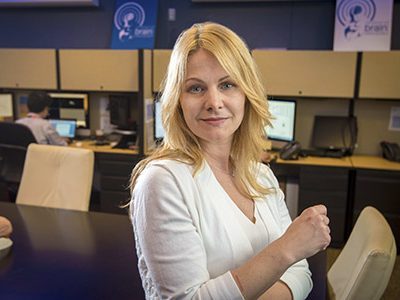
The Interventional Cardiac Magnetic Resonance (ICMR) Program at Children’s National is actively developing newer and safer ways to perform cardiac procedures on young patients, with some of the world’s leading experts in cardiac catheterization and imaging. Elena Grant, M.D., a former pediatric cardiology fellow at Children’s National, is the newest member to join the team that pioneered real-time MRI-guided radiation-free cardiac catheterization for children.
In addition to clinical work as a Children’s National Interventional Cardiologist, Dr. Grant will perform preclinical research at the National Institutes of Health to develop new procedures, techniques, and devices that can be translated to clinical practice to treat children and adults with congenital heart disease.
Dr. Grant specializes in interventional cardiology. She received her medical degree from the University of Dundee Medical School in Dundee, Scotland, followed by Foundation Training in Edinburgh, Scotland. She completed her pediatric residency at Massachusetts General Hospital, her Pediatric Cardiology fellowship at Children’s National, and she recently finished an advanced fellowship in interventional pediatric cardiology at Children’s Healthcare of Atlanta and Emory University.
Advances in interventional cardiovascular MRI
Children’s National is at the forefront of this exciting new field and is currently the only institution in the United States to perform radiation-free MRI-guided cardiac catheterization procedures in children.
ICMR is a partnership with the National Institutes of Health that brings together researchers, clinicians, engineers, and physicists to provide radiation-free, less invasive, and more precise diagnostics and treatment options for pediatric patients and adults with congenital heart disease.
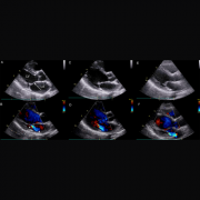
The ICMR approach to heart catheterization uses real-time MRI, instead of X-ray, in pediatric research subjects undergoing medically necessary heart catheterization. This research study is intended as a step toward routine MRI-guided catheterization in children, which attempts to avoid the hazards of ionizing radiation (X-ray).
In 2015, after working with NIH to explore how interventional cardiovascular MRI could be integrated into pediatric practices, the ICMR team, including Dr. Grant, Russell Cross, M.D., Joshua Kanter, M.D., and Laura Olivieri, M.D., performed the first radiation-free MRI-guided right heart catheterization on a 14-year-old girl at Children’s National. Since then, nearly 50 such procedures have been successfully completed, and the team is working to broaden the age range and cardiac disease complexity of patients who can undergo the procedure.
About 1 percent of newborns are born with a heart condition, and the team at Children’s performs more than 450 X-ray guided cardiac catheterizations and over 500 cardiac MRI scans per year.