Lee Beers, M.D., F.A.A.P, begins term as AAP president

Lee Beers

“The past year has been a stark reminder about the importance of partnership and working together toward common goals,” says Dr. Beers. “I am humbled and honored to be taking on this role at such a pivotal moment for the future health and safety of not only children, but the community at large.”

Lee Savio Beers, M.D., F.A.A.P., medical director of Community Health and Advocacy at the Child Health Advocacy Institute (CHAI) at Children’s National Hospital, has begun her term as president of the American Academy of Pediatrics (AAP). The AAP is an organization of 67,000 pediatricians committed to the optimal physical, mental and social health and well-being for all children – from infancy to adulthood.

“The past year has been a stark reminder about the importance of partnership and working together toward common goals,” says Dr. Beers. “I am humbled and honored to be taking on this role at such a pivotal moment for the future health and safety of not only children, but the community at large.”

Dr. Beers has pledged to continue AAP’s advocacy and public policy efforts and to further enhance membership diversity and inclusion. Among her signature issues:

  • Partnering with patients, families, communities, mental health providers and pediatricians to co-design systems to bolster children’s resiliency and to alleviate growing pediatric mental health concerns.
  • Continuing to support pediatricians during the COVID-19 pandemic with a focus on education, pediatric practice support, vaccine delivery systems and physician wellness.
  • Implementation of the AAP’s Equity Agenda and Year 1 Equity Workplan.

Dr. Beers is looking forward to continuing her work bringing together the diverse voices of pediatricians, children and families as well as other organizations to support improving the health of all children.

“Dr. Beers has devoted her career to helping children,” says Kurt Newman, M.D., president and chief executive officer of Children’s National. “She has developed a national advocacy platform for children and will be of tremendous service to children within AAP national leadership.”

Read more about Dr. Beer’s career and appointment as president of the AAP.

Family love and the right care for neurofibromatosis type 1 give Maddox a fresh start

Maddox and family

Maddox and his family in early 2020.

13-year-old Maddox Gibson is learning to cook. He says he wants to be a chef and wants to make meals for people who need it most — the homeless and the hungry.

It makes sense that he’s eager to help people who need it. As a young child growing up in a group home in his native country of China, he knows firsthand how important that support can be. In 2017 at age 10, he found his own endless supply of love and support when he met and was adopted by the Gibson family.

Zhen Chao, now called Maddox, was born in China with a genetic condition called neurofibromatosis type 1 that can cause painful or disfiguring tumors called plexiform neurofibromas. Zhen Chao had two on his head when he arrived — on his scalp and on his left optic nerve — which had been largely untreated for most of his life in China. On top of that, his right leg had been fractured and not fixed properly years before, causing him pain and weakness that left him wheelchair bound.

Adoptive mom Lindsey, a registered nurse, knew he would need special care to meet all the unique challenges he faced, and she’d done her homework — he needed the expertise of Miriam Bornhorst, M.D.,  and the Gilbert Family Neurofibromatosis Institute at Children’s National Hospital to help him thrive in his new life in the U.S. Since shortly after he came to the U.S., Lindsey has been driving Maddox the 6-plus hours from their home in North Carolina to Washington, D.C., regularly, to get care for all of his health challenges.

Maddox’s optic neurofibroma was too large when he arrived at Children’s National for a simple surgical removal. Due to her role as the lead investigator on a cutting edge clinical trial for the orphan drug selumetinib — a so-called MEK inhibitor that has shown early promise at reducing the cell growth of tumors like plexiform neurofibromas, Dr. Bornhorst enrolled Maddox in a compassionate use program for the drug, an opportunity that is not widely available. The drug was initially developed for something completely different — treatment of melanoma and non-small cell lung cancer in adults–but has been adapted through its FDA orphan drug designation for pediatric clinical trials in NF1. In the time since Maddox started taking it, it was approved for use in NF1 patients by the FDA.

The trial drug did its job — in late 2019, Maddox’s tumor had shrunk enough that chief neurosurgeon Robert Keating, M.D., and plastic surgeon Michael Boyajian, M.D., were able to successfully remove it. Follow-up procedures led by that team have also worked to repair the tissue that was impacted by the optic neurofibroma.

In addition to treatment of his neurofibromas, Maddox and his mom are able to see every service they need during one stay in D.C. The Neurofibromatosis Institute works closely across specialties, so his corrective surgery for his leg from Children’s chief of orthopaedics, Matthew Oetgen, M.D., MBA, in September 2019. He was assessed and prescribed physical therapy early in the process and even before surgery, so now he’s stronger than ever and walking. Learning difficulties, including autism and ADHD are common in NF1 patients, and so the NF Institute’s neuropsychology team has evaluated him and worked with the family to find resources and strategies near home that will support him. It should be noted, those learning difficulties only became apparent after Maddox taught himself English from scratch in only two years’ time with the help of his school’s ESOL program.

This kind of full spectrum care, from clinical assessment to surgical treatment and psychological supports, is crucial to the lives of patients with neurofibromatosis type 1 and is only available at a pediatric specialty care institution like Children’s National. The hospital has gathered some of the preeminent researchers, surgeons, and physicians within the NF Institute to make sure that the care families will travel hundreds of miles to receive is the best possible, using the latest evidence-based treatments for every challenge they face.

Though his care and follow-ups will continue at Children’s National Hospital and his condition may pose  new challenges in the future, for now, Maddox is able to focus on exploring new things and doing what he loves — playing outdoors with his family, learning to cook and building with Legos.

New grant to help establish maternal mental health telehealth program

depressed mom holding baby

Children’s National has received a $76,000 grant from the Health Resources & Services Administration (HRSA) which will allow a cross-functional team of neonatologists and psychologists to establish a parental mental telehealth program.

Worldwide about 10% of pregnant women and 13% of women who have just given birth experience a mental health disorder, primarily depression, according to the World Health Organization.

“This is a topic that is quickly garnering attention but remains extremely underfunded,” says Lamia Soghier, M.D., F.A.A.P., C.H.S.E., medical director of the Neonatal Intensive Care Unit (NICU) at Children’s National Hospital. “We tend to focus on the babies but don’t pay enough attention to the parents.”

Dr. Soghier’s focus has been on NICU parents who experience postpartum mood and anxiety disorders (PMADs), often due to their uniquely stressful experiences.

“We have been screening on a small scale for many years and have noticed a 33-45% rate of postpartum depression symptoms in our NICU families,” she says.

Maternal mental disorders are treatable with effective screening and interventions. Children’s National has received a $76,000 grant from the Health Resources & Services Administration (HRSA) which will allow a cross-functional team of neonatologists and psychologists to establish a parental mental telehealth program to expand screening and provide diagnosis, therapy and counseling to NICU parents who experience postpartum mood and anxiety disorders.

Dr. Soghier, along with Ololade ‘Lola’ Okito, M.D., neonatologist at Children’s National, and Erin Sadler, Psy.D., psychologist in the Division of Psychology and Behavioral Health at Children’s National, discuss the importance of this work.

Q: Tell us more about the program you’re establishing.

A: Dr. Soghier: This program will allow us to hire a licensed psychologist who will see families both in the NICU and through follow-up telehealth visits. It provides a one-stop shop for our families, which is particularly important during the COVID-19 pandemic. The grant will also allow us to develop an iPad loaner program to give loaner iPads to low income families who do not have access to a device or to reliable internet services so that they can receive therapy at home.

Dr. Sadler: We’ll be examining how the implementation of these services can increase accessibility and reduce barriers that prevent assessment and initiation of crucial mental health services for at-risk mothers. Our partnerships will be key. Mothers experiencing barriers to participating in care services in the NICU will also have access to an in-house, licensed psychologist through telehealth services within the comfort of their homes. Families experiencing problems accessing telehealth technology due to economic limits would get the loaner iPad. We’re meeting our families where they are in order to provide these critical services.

Q: Why is grant funding to important in this space?

A: Dr. Okito: Access to perinatal mental health services is limited at the local and national levels, particularly for vulnerable parents of infants admitted to the NICU. Little is known about the effect of interventions to address depression and anxiety among NICU parents, and this grant will allow us to contribute to this very important area of research.

Dr. Sadler: It is not enough to recognize the health disparities that exist amongst communities in our nation. It is imperative that we’re able to explore and examine solutions that can aid in enhancing the equity of care for children and adults alike. As Dr. Okito mentions, there is little to no research available that looks at the feasibility of the support programs we intended to put in place. We hope to create a viable model that could be used to help NICU families across the country.

Q: How is Children’s National uniquely positioned to do this work?

A: Dr. Soghier: Healthy moms and healthy dads equal happy babies. That’s why we will be taking care of the family as a whole. This is truly family-centered care and at the heart of what Children’s National is all about.

Dr. Sadler: The Children’s National NICU team has an established postpartum depression screening program. Through the piloted work, staff have identified notable barriers to universal screening, access to perinatal mental health support and the impact of PMADs on parent engagement in newborn care.  As a result, Children’s National is uniquely positioned to directly address such barriers and provide specialized care.

Q: What excites you about this work?

A: Dr. Sadler: As a specialist in perinatal and infant mental health, I look forward to being able to demonstrate the lasting impact maternal mental health services can provide for not only newborns and their families, but for care providers as well. I am excited to have additional opportunities to advocate for the integration of perinatal and infant mental health in non-traditional spaces.

Dr. Okito: I am most excited about the potential to expand universal depression screening among NICU parents. Having done this work for the past three years, I know there are limitations in screening because we’ve only been able to screen parents that are at the patient’s bedside. More screening will lead to more parents getting the referrals and services that they need.

Boeing gives $5 million to support Research & Innovation Campus

Research & Innovation Campus

Children’s National Hospital announced a $5 million gift from The Boeing Company that will help drive lifesaving pediatric discoveries at the new Children’s National Research & Innovation Campus.

Children’s National Hospital announced a $5 million gift from The Boeing Company that will help drive lifesaving pediatric discoveries at the new Children’s National Research & Innovation Campus. The campus, now under construction, is being developed on nearly 12 acres of the former Walter Reed Army Medical Center. Children’s National will name the main auditorium in recognition of Boeing’s generosity.

“We are deeply grateful to Boeing for their support and commitment to improving the health and well-being of children in our community and around the globe,” said Kurt Newman, M.D., president and CEO of Children’s National “The Boeing Auditorium will help the Children’s National Research & Innovation campus become the destination for discussion about how to best address the next big healthcare challenges facing children and families.”

The one-of-a-kind pediatric hub will bring together public and private partners for unprecedented collaborations. It will accelerate the translation of breakthroughs into new treatments and technologies to benefit kids everywhere.

“Children’s National Hospital’s enduring mission of positively impacting the lives of our youngest community members is especially important today,” said Boeing President and CEO David Calhoun. “We’re honored to join other national and community partners to advance this work through the establishment of their Research & Innovation Campus.”

Children’s National Research & Innovation Campus partners currently include Johnson & Johnson Innovation – JLABS, Virginia Tech, the National Institutes of Health (NIH), Food & Drug Administration (FDA), U.S. Biomedical Advanced Research and Development Authority (BARDA), Cerner, Amazon Web Services, Microsoft, National Organization of Rare Diseases (NORD) and local government.

The 3,200 square-foot Boeing Auditorium will be the focal point of the state-of-the-art conference center on campus. Nationally renowned experts will convene with scientists, medical leaders and diplomats from around the world to foster collaborations that spur progress and disseminate findings.

Boeing’s $5 million commitment deepens its longstanding partnership with Children’s National. The company has donated nearly $2 million to support pediatric care and research at Children’s National through Chance for Life and the hospital’s annual Children’s Ball. During the coronavirus pandemic, Boeing fabricated and donated 2,000 face shields to help keep patients and frontline care providers at Children’s National safe.

Children use both brain hemispheres to understand language

Illustration of brain hemispheres

New research finds young children process language in both hemispheres of the brain, which could help compensation after a neural injury. This is unlike adults who process most language tasks in one side (usually the left) of their brain’s two hemispheres. It suggests a possible reason why children often seem to recover from brain injury more easily than adults.

New research finds young children process language in both hemispheres of the brain, which could help compensate after a neural injury. The study, published Sept. 8, 2020, in PNAS, says this is unlike adults who process most language tasks in one side (usually the left) of their brain’s two hemispheres. It suggests a possible reason why children often seem to recover from brain injury more easily than adults.

We talked with researcher William D. Gaillard, M.D., chief of the Divisions of Child Neurology, Epilepsy and Neurophysiology at Children’s National Hospital, to discuss the importance of this work.

Q: Tell us a little bit about this study.

A: This is a study we did with our colleagues at Georgetown University Medical Center, using fMRI to map brain regions that are used to process language across development. What we found was that younger children have more bilateral “activation” in language processing regions, the traditional left and homotopic regions in the right. With aging there is consolidation that becomes more left lateralized. This process is most clearly seen in the frontal brain regions, called Broca’s area, where the right activation diminishes over age

Q: Why are these findings important?

A: It’s important because this work provides evidence for how cognitive systems, and the neural networks that underlie them, become consolidated and lateralized over time during development. It provides insights into principles of the development of cognitive systems.

The timeline for lateralization of language systems means that the cognitive systems that sustain language are “plastic” – that is the right hemisphere can sustain language functions in the setting of injury to the left hemisphere until around 10 years of age.

Q: What excites you about this work?

A: This is part of a larger collaborative effort that is mapping out the consolidation of cognitive systems across development (language, visual spatial, memory and working memory). This work will help us to understand the limits of brain plasticity in the setting of injury caused by stroke or epilepsy, which could have benefits down the road to helping patients recover from these types of events.

Q: How is Children’s National leading the ongoing discovery in this space?

A: It is a true team effort. We are working with colleagues at Georgetown University Medical Center, MedStar National Rehabilitation Network and Johns Hopkins Medicine. Team members come from diverse backgrounds and scientific skills. We are one of the leading groups using advanced functional imaging to investigate brain development of critical cognitive systems and their response to brain injury.

You can find the full study published in PNAS. Learn more about the Children’s National Research Institute Center for Neuroscience Research.


Children’s National ranked a top 10 children’s hospital and No. 1 in newborn care nationally by U.S. News

US News Badges

Children’s National Hospital in Washington, D.C., was ranked No. 7 nationally in the U.S. News & World Report 2020-21 Best Children’s Hospitals annual rankings. This marks the fourth straight year Children’s National has made the list, which ranks the top 10 children’s hospitals nationwide.

In addition, its neonatology program, which provides newborn intensive care, ranked No.1 among all children’s hospitals for the fourth year in a row.

For the tenth straight year, Children’s National also ranked in all 10 specialty services, with seven specialties ranked in the top 10.

“Our number one goal is to provide the best care possible to children. Being recognized by U.S. News as one of the best hospitals reflects the strength that comes from putting children and their families first, and we are truly honored,” says Kurt Newman, M.D., president and CEO of Children’s National Hospital.

“This year, the news is especially meaningful, because our teams — like those at hospitals across the country — faced enormous challenges and worked heroically through a global pandemic to deliver excellent care.”

“Even in the midst of a pandemic, children have healthcare needs ranging from routine vaccinations to life-saving surgery and chemotherapy,” said Ben Harder, managing editor and chief of Health Analysis at U.S. News. “The Best Children’s Hospitals rankings are designed to help parents find quality medical care for a sick child and inform families’ conversations with pediatricians.”

The annual rankings are the most comprehensive source of quality-related information on U.S. pediatric hospitals. The rankings recognize the nation’s top 50 pediatric hospitals based on a scoring system developed by U.S. News. The top 10 scorers are awarded a distinction called the Honor Roll.

The bulk of the score for each specialty service is based on quality and outcomes data. The process includes a survey of relevant specialists across the country, who are asked to list hospitals they believe provide the best care for patients with the most complex conditions.

Below are links to the seven Children’s National specialty services that U.S. News ranked in the top 10 nationally:

The other three specialties ranked among the top 50 were cardiology and heart surgery, gastroenterology and gastro-intestinal surgery, and urology.

Children’s National Research Institute releases annual report

Vittorio Gallo and Marc Batshaw

Children’s National Research Institute directors Vittorio Gallo, Ph.D., and Mark Batshaw, M.D.

The Children’s National Research Institute recently released its 2019-2020 academic annual report, titled 150 Years Stronger Through Discovery and Care to mark the hospital’s 150th birthday. Not only does the annual report give an overview of the institute’s research and education efforts, but it also gives a peek in to how the institute has mobilized to address the coronavirus pandemic.

“Our inaugural research program in 1947 began with a budget of less than $10,000 for the study of polio — a pressing health problem for Washington’s children at the time and a pandemic that many of us remember from our own childhoods,” says Vittorio Gallo, Ph.D., chief research officer at Children’s National Hospital and scientific director at Children’s National Research Institute. “Today, our research portfolio has grown to more than $75 million, and our 314 research faculty and their staff are dedicated to finding answers to many of the health challenges in childhood.”

Highlights from the Children’s National Research Institute annual report

  • In 2018, Children’s National began construction of its new Research & Innovation Campus (CNRIC) on 12 acres of land transferred by the U.S. Army as part of the decommissioning of the former Walter Reed Army Medical Center campus. In 2020, construction on the CNRIC will be complete, and in 2012, the Children’s National Research Institute will begin to transition to the campus.
  • In late 2019, a team of scientists led by Eric Vilain, M.D., Ph.D., director of the Center for Genetic Medicine Research, traveled to the Democratic Republic of Congo to collect samples from 60 individuals that will form the basis of a new reference genome data set. The researchers hope their project will generate better reference genome data for diverse populations, starting with those of Central African descent.
  • A gift of $5.7 million received by the Center for Translational Research’s director, Lisa Guay-Woodford, M.D., will reinforce close collaboration between research and clinical care to improve the care and treatment of children with polycystic kidney disease and other inherited renal disorders.
  • The Center for Neuroscience Research’s integration into the infrastructure of Children’s National Hospital has created a unique set of opportunities for scientists and clinicians to work together on pressing problems in children’s health.
  • Children’s National and the National Institute of Allergy and Infectious Diseases are tackling pediatric research across three main areas of mutual interest: primary immune deficiencies, food allergies and post-Lyme disease syndrome. Their shared goal is to conduct clinical and translational research that improves what we know about those conditions and how we care for children who have them.
  • An immunotherapy trial has allowed a little boy to be a kid again. In the two years since he received cellular immunotherapy, Matthew has shown no signs of a returning tumor — the longest span of time he’s been tumor-free since age 3.
  • In the past 6 years, the 104 device projects that came through the National Capital Consortium for Pediatric Device Innovation accelerator program raised $148,680,256 in follow-on funding.
  • Even though he’s watched more than 500 aspiring physicians pass through the Children’s National pediatric residency program, program director Dewesh Agrawal, M.D., still gets teary at every graduation.

Understanding and treating the novel coronavirus (COVID-19)

In a short period of time, Children’s National Research Institute has mobilized its scientists to address COVID-19, focusing on understanding the virus and advancing solutions to ameliorate the impact today and for future generations. Children’s National Research Institute Director Mark Batshaw, M.D., highlighted some of these efforts in the annual report:

  • Eric Vilain, M.D., Ph.D., director of the Center for Genetic Medicine Research, is looking at whether or not the microbiome of bacteria in the human nasal tract acts as a defensive shield against COVID-19.
  • Catherine Bollard, M.D., MBChB, director of the Center for Cancer and Immunology Research, and her team are seeing if they can “train” T cells to attack the invading coronavirus.
  • Sarah Mulkey, M.D., Ph.D., an investigator in the Center for Neuroscience Research and the Fetal Medicine Institute, is studying the effects of, and possible interventions for, coronavirus on the developing brain.

You can view the entire Children’s National Research Institute academic annual report online.

Clinicians and transgender autistic youth create support model

girl talking to doctor

Young people with the co-occurrence of autism and gender diversity and their families partner with clinical researchers to understand care needs and how care providers can meet those needs.

The first ever set of specific recommendations to support transgender autistic young people was co-created by these youth and their families working hand-in-hand with clinical experts. The resulting model offers clinicians a set of concrete ways to provide this unique population the support they need.

The recommendations, A Clinical Program for Transgender and Gender-Diverse Neurodiverse/Autistic Adolescents Developed through Community-Based Participatory Design, were published by the Journal of Clinical Child and Adolescent Psychology on May 4, 2020.

“The idea of patients helping to co-design their own care isn’t new, but including the perspectives of autistic youth in their own care is quite new,” says John Strang, Psy.D., who directs the Gender and Autism Program within the Center for Autism Spectrum Disorders at Children’s National Hospital.

He continues, “And for the many youth who are both transgender and autistic, including their voices and perspectives in their clinical care is critical. Without their input, there is a great risk for misunderstanding their needs – and for marginalization.”

“This was an important process in which to participate, and will hopefully help those — autistic people, trans people, and autistic trans people alike — who often only see themselves represented by cisgender, neurotypical researchers and providers. It is a relief to be a part of creating something like this,” adds Marisa Alexa McCool, a co-researcher who is an autistic transgender woman.

The new publication builds on previously published broad clinical guidelines for providers, now integrating the perspectives of autistic transgender individuals themselves. The program includes specific approaches for supporting young people in their diverse needs, and identifies three key components central to this care:

  1. Helping autistic, gender-diverse young people build community together, which they need and often want, in contrast to clichés about lack of desire for social contact in autism. Many autistic transgender young people prior to entering clinical care have never met another person who is transgender and autistic. The connections that they built with one another through this new clinical care model were critical in helping them develop a positive sense of identity and to know that they are not alone in this world.
  2. Introducing the youth to a broad spectrum of gender diverse and/or neurodiverse role models helps make possibilities for their future more concrete, and builds a sense of hopefulness and pride. Abstract concepts such as gender or future gender can be particularly challenging for autistic youth. The new care model addresses this by providing these youth opportunities to meet and interact with a range of living role models who represent various gender identities as well as neurodiversity experiences. “Being able to see and hear about the diverse journeys of adults who have already navigated gender and/or autism-related diversity has been helpful in making the various options more tangible for gender diverse autistic youth,” says Dr. Strang. “The chance to meet role models with different gender-related experiences – transgender, cisgender, exploring – has helped autistic gender diverse youth to better figure out what is most true for them and what they need from us.”
  3. Supporting the gender expression needs of autistic transgender youth through gender style coaching. Because of autism-related sensory sensitivities and problems with planning and social understanding, autistic transgender young people often have difficulty achieving their desired gender transition. Gender-style coaching can help autistic youth reach their gender-related goals in ways that accommodate and support the young person’s autism-related challenges.

The perspectives included in the new clinical program were from a range of ages and backgrounds, as well as across multiple points in time to make sure that as youths’ own views evolved, their evolving needs were captured as well.

The authors created a specific clinical guide to complement the publication, which is available on the Children’s National website.

“We’re so happy to have been able to partner with self-advocates from the autistic transgender and gender diverse communities, youth who are living this experience, and their families, to co-create a community-driven model that can be used for kids seeking guidance and support,” says Dr. Strang.

“We hear over and over again that what parents and care providers really need are concrete tools to support young people with co-occurring autism and gender diversity, so that’s what we sought to do here,” he concludes. “It’s exciting because, for the first time, we have some simple tools to support these kids. And this is critical, because although the co-occurrence of autism and gender diversity has been of great interest to researchers, nearly all studies to date have focused on how many transgender youth are autistic, instead of how to help and support this poorly understood group.”

Special issue of “Neurochemical Research” honors Vittorio Gallo, Ph.D.

Vittorio Gallo

Investigators from around the world penned manuscripts that were assembled in a special issue of “Neurochemical Research” that honors Vittorio Gallo, Ph.D., for his leadership in the field of neural development and regeneration.

At a pivotal moment early in his career, Vittorio Gallo, Ph.D., was accepted to work with Professor Giulio Levi at the Institute for Cell Biology in Rome, a position that leveraged courses Gallo had taken in neurobiology and neurochemistry, and allowed him to work in the top research institute in Italy directed by the Nobel laureate, Professor Rita Levi-Montalcini.

For four years as a student and later as Levi’s collaborator, Gallo focused on amino acid neurotransmitters in the brain and mechanisms of glutamate and GABA release from nerve terminals. Those early years cemented a research focus on glutamate neurotransmission that would lead to a number of pivotal publications and research collaborations that have spanned decades.

Now, investigators from around the world who have worked most closely with Gallo penned tributes in the form of manuscripts that were assembled in a special issue of “Neurochemical Research” that honors Gallo “for his contributions to our understanding of glutamatergic and GABAergic transmission during brain development and to his leadership in the field of neural development and regeneration,” writes guest editor Arne Schousboe, of the University of Copenhagen in Denmark.

Dr. Gallo as a grad student

Vittorio Gallo, Ph.D. as a 21-year-old mustachioed graduate student.

“In spite of news headlines about competition in research and many of the negative things we hear about the research world, this shows that research is also able to create a community around us,” says Gallo, chief research officer at Children’s National Hospital and scientific director for the Children’s National Research Institute.

As just one example, he first met Schousboe 44 years ago when Gallo was a 21-year-old mustachioed graduate student.

“Research can really create a sense of community that we carry on from the time we are in training, nurture as we meet our colleagues at periodic conferences, and continue up to the present. Creating community is bi-directional: influencing people and being influenced by people. People were willing to contribute these 17 articles because they value me,” Gallo says. “This is a lot of work for the editor and the people who prepared papers for this special issue.”

In addition to Gallo publishing more than 140 peer-reviewed papers, 30 review articles and book chapters, Schousboe notes a number of Gallo’s accomplishments, including:

  • He helped to develop the cerebellar granule cell cultures as a model system to study how electrical activity and voltage-dependent calcium channels modulate granule neuron development and glutamate release.
  • He developed a biochemical/neuropharmacological assay to monitor the effects of GABA receptor modulators on the activity of GABA chloride channels in living neurons.
  • He and Maria Usowicz used patch-clamp recording and single channel analysis to demonstrate for the first time that astrocytes express glutamate-activated channels that display functional properties similar to neuronal counterparts.
  • He characterized one of the spliced isoforms of the AMPA receptor subunit gene Gria4 and demonstrated that this isoform was highly expressed in the cerebellum.
  • He and his Children’s National colleagues demonstrated that glutamate and GABA regulate oligodendrocyte progenitor cell proliferation and differentiation.
Purkinje cells

Purkinje cells are large neurons located in the cerebellum that are elaborately branched like interlocking tree limbs and represent the only source of output for the entire cerebellar cortex.

Even the image selected to grace the special issue’s cover continues the theme of continuity and leaving behind a legacy. That image of Purkinje cells was created by a young scientist who works in Gallo’s lab, Aaron Sathyanesan, Ph.D. Gallo began his career working on the cerebellum – a region of the brain important for motor control – and now studies with a team of scientists and clinician-scientists Purkinje cells’ role in locomotor adaptive behavior and how that is disrupted after neonatal brain injury.

“These cells are the main players in cerebellar circuitry,” Gallo says. “It’s a meaningful image because goes back to my roots as a graduate student and is also an image that someone produced in my lab early in his career. It’s very meaningful to me that Aaron agreed to provide this image for the cover of the special issue.”

Celiac disease linked to psychosocial distress

gluten free cupcakes

A recent study found elevated rates of psychosocial distress among children with celiac disease compared to the general population.

Shayna Coburn, Ph.D., assistant professor and psychologist at Children’s National Hospital, is the lead author of a recent article on the first study to report mental health disorders (MHD) in North American children with celiac disease (CeD). The study found elevated rates of psychosocial distress among the children compared to the general population.

The study is based on electronic surveys of patients’ MHD history, psychological symptoms and experiences with the gluten-free diet (GFD) as well as follow-up visits to the Multidisciplinary Celiac Disease Clinic at Children’s National between spring 2017 and spring 2018. The survey participants included 73 parents of children ages 3 to 18 attending the clinic. The researchers calculated rates of MHD in the children and compared them to National Institute of Mental Health population-level data.

Thirty-four percent of the children had at least one MHD. Their rates of anxiety disorders (16%) and attention-deficit/hyperactivity disorder (ADHD, 16%,) were more common than general population rates. More than one-quarter of parents reported current psychosocial distress in their child (28-39%), and approximately half reported their own stress (51%) and worry about the financial burden (46%) associated with the GFD – the only treatment for the disease.

The findings are detailed in an article titled “Mental Health Disorders and Psychosocial Distress in Pediatric Celiac Disease,” which appears on the website of the Journal of Pediatric Gastroenterology and Nutrition. The article is scheduled to appear in the May 2020 print edition of the journal, which will be available April 24.

Coburn and her co-authors also compared the experiences of children diagnosed with CeD less than three months prior to the study with those diagnosed more than three months prior. They were surprised to find that patients’ rates of comorbid CeD and MHD didn’t differ depending on the time of diagnosis, says Coburn.

Parents of children with new CeD diagnoses were less confident in the GFD, but the timing of a CeD diagnosis did not affect the rates of MHD, stress and financial burden. Children with MHD had more anxiety, anger and overall distress as well as parents who were suffering with distress than those without MHD.

The researchers’ findings about the timing of diagnosis “seemed to indicate that perhaps there’s a chronic stress burden on families that doesn’t necessarily improve with time and might be exacerbated in children who have mental health disorders,” says Coburn, who directs psychosocial services for the hospital’s Celiac Disease Program.

Overall, the findings emphasize the importance of ongoing routine screening and treatment for psychosocial distress associated with CeD and the GFD.

The start of the study coincided with the establishment of the clinic, where Coburn and her colleagues were seeing patients with comorbid CeD and MHD. At the clinic, patients and their families are treated by a gastroenterologist as well as the clinic’s nutritionist, education team, psychologist, neurologist and neuropsychologist during an integrative multidisciplinary appointment.

Coburn notes that generally the psychosocial impact on patients with CeD has been overlooked or viewed as a minor condition. “Our work is showing that there are a lot of psychosocial vulnerabilities in children and adults with celiac disease.”

As she continues her research, Coburn sees a need “to advocate for incorporating psychological screening into routine medical treatment of patients with celiac disease. We’d like this to be part of best practices and want to develop behavioral treatments for patients so they’re succeeding with the gluten-free diet.”

“With ADHD there are problems with impulse control, which can make it extra hard to maintain a gluten-free diet,” says Coburn. The co-principal investigators want to study in-depth some of the families who participated in the earlier study to gauge how effectively they’re able to manage ADHD symptoms in order to maintain a gluten-free diet.

Coburn and Maegan Sady, a neuropsychologist at Children’s National, have received a $25,000 grant from the Lambert Family Foundation to study comorbid ADHD and CeD and how they affect a patient’s ability to adhere to the GFD.

Religiousness linked to improved quality of life for people with HIV

people sitting in a circle holding hands

Adults living with HIV in Washington, D.C., were more likely to feel higher levels of emotional and physical well-being if they attended religious services regularly, prayed daily, felt “God’s presence,” and self-identified as religious or spiritual.

Adults living with HIV in Washington, D.C., were more likely to feel higher levels of emotional and physical well-being if they attended religious services regularly, prayed daily, felt “God’s presence,” and self-identified as religious or spiritual, according to research published online Jan. 29, 2020, in Psychology of Religion and Spirituality. By contrast, patients living with HIV who had the lowest levels of quality of life and more mental health challenges were privately religious, potentially eschewing organized religion due to fears about being stigmatized or ostracized.

“These findings are significant because they point to the untapped potential of encouraging patients living with HIV who are already religious to attend religious services regularly.  Scientific evidence suggests that religions that present God as all-powerful, personal, responsive, loving, just and forgiving make a difference in health-related quality of life. By contrast, belief systems and religions that see God as punishing, angry, vengeful and distant and isolate members from their families and the larger community do not have health benefits or contribute to health-related quality of life. People who identify as spiritual also benefit from improved overall health-related quality of life,” says Maureen E. Lyon, Ph.D., FABPP, a clinical health psychologist at Children’s National Hospital, and senior study author.

“In general, patients living with HIV have reported that they wished their health care providers acknowledged their religious beliefs and spiritual struggles. Additional research is needed to gauge whether developing faith-based interventions or routine referrals to faith-based programs that welcome racial and sexual minorities improve satisfaction with treatment and health outcomes,” Lyon adds.

More than 1 million people in the U.S. live with HIV, and in 2018, 37,832 people received an HIV diagnosis in the U.S., according to the Centers for Disease Control and Prevention (CDC). In 2017, the Washington, D.C., region recorded one of nation’s highest rates of new cases of HIV: 46.3 diagnoses per 100,000 people, according to the CDC.

A research team that includes current and former Children’s National faculty wanted to learn more about the degree of religiousness and spirituality reported by people living with HIV and the interplay between religion and health-related quality of life. They recruited patients to participate in a clinical trial about family-centered advance care planning and enrolled 223 patient/family dyads in this study.

Fifty-six percent of patients were male. Eighty-six percent were African American, and their mean age was 50.8. Seventy-five percent were Christian.

The researchers identified three distinct classes of religious beliefs:

  • Class 1, the highest level of religiousness/spirituality, applied to people more likely to attend religious services in person each week, to pray daily, to “feel God’s presence” and to self-identify as religious and spiritual. Thirty-five percent of study participants were Class 1 and tended to be older than 40.
  • Class 2 applied to privately religious people who engaged in religious activities at home, like praying, and did not attend services regularly. Forty-seven percent of study participants were Class 2.
  • Class 3 participants self-identified as spiritual but were not involved in organized religion. Nearly 18 percent of study participants were Class 3, the lowest overall level of religiousness/spirituality.

Class 1 religiousness/spirituality was associated with increased quality of life, mental health and improved health status.

“Being committed to a welcoming religious group provides social support, a sense of identity and a way to cope with stress experienced by people living with HIV,” Lyon says. “We encourage clinicians to capitalize on patients’ spiritual beliefs that improve health – such as prayer, meditation, reading spiritual texts and attending community events – by including them in holistic treatment programs in a non-judgmental way.”

What’s more, the research team encourages clinicians to appoint a member of the team who is responsible for handling religiousness/spirituality screening and providing referrals to welcoming hospital-based chaplaincy programs or community-based religious groups.

“This is particularly challenging for HIV-positive African American men who have sex with men, as this group faces discrimination related to race and sexual orientation. Because HIV infection rates are increasing for this group, this additional outreach is all the more important,” she adds.

In addition to Lyon, study co-authors include Biostatistician Jichuan Wang, Ph.D., and Yao I. Cheng, MS., both of Children’s National; and Lead Author Katherine B. Grill, Ph.D., the former clinical coordinator for this randomized clinical trial who is currently an adjunct professor at the California Institute of Integral Studies.

Financial support for research described in this post was provided by the National Institutes of Health under award Nos. R01NR014-052-05 and UL1RR031988.


Maternal mental health alters structure and biochemistry of developing fetal brain

Even when pregnant women have uncomplicated pregnancies and high socioeconomic status, when they experience elevated anxiety, stress or depression these prenatal stressors can alter the structure of the developing fetal brain and disrupt its biochemistry, according to Children’s National Hospital research published online Jan. 29, 2020, in JAMA Network Open.

The Children’s National research findings “have enormous scientific, clinical and public health implications,” Charles A. Nelson III, Ph.D.,  Boston Children’s Hospital, writes in a companion editorial.

“Previously we found that 65% of pregnant women who received a diagnosis of fetal congenital heart disease had elevated levels of stress. It’s concerning but not surprising that pregnant women who wonder if their baby will need open heart surgery would feel stress,” says Catherine Limperopoulos, Ph.D., director of the Center for the Developing Brain at Children’s National and the study’s senior author. “In this latest study, we ran the same panel of questionnaires and were surprised to find a high proportion of otherwise healthy pregnant women whose unborn babies are doing well also report high levels of stress.”

Anxiety and depression are the most common mental health problems during pregnancy. To learn more about the implications for the developing fetal brain, the Children’s National research team recruited 119 healthy volunteers with low-risk pregnancies from obstetric clinics in Washington, D.C., from Jan. 1, 2016, to April 17, 2019. The women’s mean age was 34.4 years old. All were high school graduates, 83% were college graduates, and 84% reported professional employment.

T2-Weighted Magnetic Resonance (MR) Imaging Brain Segmentation.

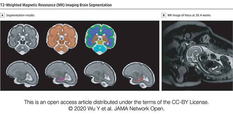
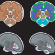
T2-Weighted Magnetic Resonance (MR) Imaging Brain Segmentation. Segmentation results of total brain (orange), cortical gray matter (green), white matter (blue), deep gray matter (brown), brainstem (yellow), cerebellum (light blue), left hippocampus (purple) and right hippocampus (red) on a 3-Dimensional reconstructed T2-weighted MR image of a fetus at 26.4 gestational weeks. The hippocampus plays a central role in memory and behavioral inhibition and contains high concentrations of corticosteroid receptors and, thus, this brain region is sensitive to stress. Credit: JAMA Network Open.

The team performed 193 fetal brain magnetic resonance imaging (MRI) sessions between 24-40 weeks gestation and measured the volume of the total fetal brain as well as the cortical gray matter, white matter, deep gray matter, cerebellum, brainstem and hippocampus volumes. On the same day as their MRI visit, the pregnant women completed validated questionnaires to measure maternal stress, anxiety and depression, answering questions such as “how do you feel right now,” “how do you generally feel” as well as the degree of stressful feelings they experienced the month prior.

Of the pregnant women in the study:

  • 27% tested positive for stress
  • 26% tested positive for anxiety
  • 11% tested positive for depression
  • Maternal anxiety and stress were associated with increased fetal cortical gyrification
  • Elevated maternal depression was associated with decreased creatine and choline levels in the fetal brain
  • Maternal stress scores decreased with increasing gestational age, while anxiety and depression did not

“We report for the first time that maternal psychological distress may be associated with increased fetal local gyrification index in the frontal and temporal lobes,” says Yao Wu, Ph.D., a research associate working with Limperopoulos at Children’s National and the study’s lead author. “We also found an association with left fetal hippocampal volume, with maternal psychological distress selectively stunting the left hippocampal volumetric growth more than the right. And elevated maternal depression was associated with decreased creatine and choline levels in the fetal brain,” Wu adds.

Late in pregnancy – at the time these women were recruited into the cohort study – the fetal brain grows exponentially and key metabolite levels also rise. Creatine facilitates recycling of adenosine triphosphate, the cell’s energy currency. Typically, levels of this metabolite rise, denoting rapid changes and higher cellular maturation; creatine also is known to support cognitive function. Choline levels also typically rise, marking cell membrane turnover as new cells are generated and support memory, mental focus and concentration.

“These women were healthy, and of high socioeconomic status and educational level, leading us to conclude that the prevalence of prenatal maternal psychological distress may be underestimated,” Limperopoulos adds. “While stress is an everyday reality for most of us, this is different because elevated stress during pregnancy can alter fetal brain programming. Our findings underscore the critical need to universally screen all pregnant women for prenatal psychological distress, even young mothers whose pregnancies wouldn’t otherwise raise red flags.”

In addition to Limperopoulos and Wu, Children’s National study co-authors include Yuan-Chiao Lu, Ph.D., research associate; Marni Jacobs, Ph.D., biostatistician; Subechhya Pradhan, Ph.D., research faculty; Kushal Kapse, MS, staff engineer; Li Zhao, Ph.D., research faculty; Nickie Niforatos-Andescavage, M.D., neonatologist; Gilbert Vezina, M.D., director of the neuroradiology program; and Adré  J. du Plessis, M.B.Ch.B., director, Fetal Medicine Institute. Research coordinators Catherine Lopez, MS, Kathryn Lee Bannantine, BSN, and Jessica Lynn Quistorff, MPH, assisted with subject recruitment.

Financial support for the research described in this post was provided by the National Institutes of Health under grant No. RO1 HL116585-01 and the Thrasher Research Fund under Early Career award No. 14764.

Journal Reference:
Yao Wu, Yuan-Chiao Lu, Marni Jacobs, Subechhya Pradhan, Kushal Kapse, Li Zhao, Nickie Niforatos-Andescavage, Gilbert Vezina, Adré J. du Plessis, Catherine Limperopoulos. “Association of prenatal maternal psychological distress with fetal brain growth, metabolism and cortical maturation,” JAMA Network Open, 3(1): e1919940, 2020

Stressful pregnancies can leave fingerprint on fetal brain

Catherine Limperopoulos

“We were alarmed by the high percentage of pregnant women with a diagnosis of a major fetal heart problem who tested positive for stress, anxiety and depression,” says Catherine Limperopoulos, Ph.D., director of the Center for the Developing Brain at Children’s National and the study’s corresponding author.

When a diagnosis of fetal congenital heart disease causes pregnant moms to test positive for stress, anxiety and depression, powerful imaging can detect impaired development in key fetal brain regions, according to Children’s National Hospital research published online Jan. 13, 2020, in JAMA Pediatrics.

While additional research is needed, the Children’s National study authors say their unprecedented findings underscore the need for universal screening for psychological distress as a routine part of prenatal care and taking other steps to support stressed-out pregnant women and safeguard their newborns’ developing brains.

“We were alarmed by the high percentage of pregnant women with a diagnosis of a major fetal heart problem who tested positive for stress, anxiety and depression,” says Catherine Limperopoulos, Ph.D., director of the Center for the Developing Brain at Children’s National and the study’s corresponding author. “Equally concerning is how prevalent psychological distress is among pregnant women generally. We report for the first time that this challenging prenatal environment impairs regions of the fetal brain that play a major role in learning, memory, coordination, and social and behavioral development, making it all the more important for us to identify these women early during pregnancy to intervene,” Limperopoulos adds.

Congenital heart disease (CHD), structural problems with the heart, is the most common birth defect. Still, it remains unclear how exposure to maternal stress impacts brain development in fetuses with CHD.

The multidisciplinary study team enrolled 48 women whose unborn fetuses had been diagnosed with CHD and 92 healthy women with uncomplicated pregnancies. Using validated screening tools, they found:

  • 65% of pregnant women expecting a baby with CHD tested positive for stress
  • 27% of women with uncomplicated pregnancies tested positive for stress
  • 44% of pregnant women expecting a baby with CHD tested positive for anxiety
  • 26% of women with uncomplicated pregnancies tested positive for anxiety
  • 29% of pregnant women expecting a baby with CHD tested positive for depression and
  • 9% women with uncomplicated pregnancies tested positive for depression

All told, they performed 223 fetal magnetic resonance imaging sessions for these 140 fetuses between 21 and 40 weeks of gestation. They measured brain volume in cubic centimeters for the total brain as well as volumetric measurements for key regions such as the cerebrum, cerebellum, brainstem, and left and right hippocampus.

Maternal stress and anxiety in the second trimester were associated with smaller left hippocampi and smaller cerebellums only in pregnancies affected by fetal CHD. What’s more, specific regions — the hippocampus head and body and the left cerebellar lobe – were more susceptible to stunted growth. The hippocampus is key to memory and learning, while the cerebellum controls motor coordination and plays a role in social and behavioral development.

The hippocampus is a brain structure that is known to be very sensitive to stress. The timing of the CHD diagnosis may have occurred at a particularly vulnerable time for the developing fetal cerebellum, which grows faster than any other brain structure in the second half of gestation, particularly in the third trimester.

“None of these women had been screened for prenatal depression or anxiety. None of them were taking medications. And none of them had received mental health interventions. In the group of women contending with fetal CHD, 81% had attended college and 75% had professional educations, so this does not appear to be an issue of insufficient resources,” Limperopoulos adds. “It’s critical that we routinely to do these screenings and provide pregnant women with access to interventions to lower their stress levels. Working with our community partners, Children’s National is doing just that to help reduce toxic prenatal stress for both the health of the mother and for the future newborns. We hope this becomes standard practice elsewhere.”

Adds Yao Wu, Ph.D., a research associate working with Limperopoulos at Children’s National and the study’s lead author: “Our next goal is exploring effective prenatal cognitive behavioral interventions to reduce psychological distress felt by pregnant women and improve neurodevelopment in babies with CHD.”

In addition to Limperopoulos and Wu , Children’s National study co-authors include Kushal Kapse, MS, staff engineer; Marni Jacobs, Ph.D., biostatistician; Nickie Niforatos-Andescavage, M.D., neonatologist; Mary T. Donofrio, M.D., director, Fetal Heart Program; Anita Krishnan, M.D., associate director, echocardiography; Gilbert Vezina, M.D., director, Neuroradiology Program; David Wessel, M.D., Executive Vice President and Chief Medical Officer; and Adré  J. du Plessis, M.B.Ch.B., director, Fetal Medicine Institute. Jessica Lynn Quistorff, MPH, Catherine Lopez, MS, and Kathryn Lee Bannantine, BSN, assisted with subject recruitment and study coordination.

Financial support for the research described in this post was provided by the National Institutes of Health under grant No. R01 HL116585-01 and the Thrasher Research Fund under Early Career award No. 14764.

From the mouths of babes: Lessons in humility

Alexandra M. Sims

A poem written by Alexandra M. Sims, M.D., FAAP, was published Jan. 7, 2020, in JAMA, as part of its series of works by artists and physicians that explore the meaning of healing and illness.

Each encounter is like a single shard in a mosaic that, taken as a whole, presents a picture of amazing optimism despite myriad challenges.

Alexandra M. Sims, M.D., FAAP, a General Academics Pediatric Fellow at Children’s National Hospital, captured the anonymized vignettes in her journal, using writing as a way to help process both the unbounded joy and sobering trauma experienced by her young patients.

Dr. Sims distilled the snippets into a 27-line poem published Jan. 7, 2020, in JAMA, as part of “Poetry and Medicine,” poems penned by artists and physicians to explore the meaning of healing and illness.

One of the vignettes collapses eye-opening comments she heard during a number of clinical encounters, including a childhood immunization session for a 4-year-old: Doesn’t flinch with the vaccines, but tells me not to call them ‘shots’ / His classmate was shot last year / And she died

“When I’m talking about a ‘shot,’ the first thing that comes to my mind – because of what I do for a living and how my life has unfolded – is a vaccination,” Dr. Sims explains. “That caught me off guard. Even though I have been doing this job for a while, I can always learn from patients and families. It really made me shift the language I use, avoiding words that I might think are innocuous that can be translated in ways that can be scary for a child.”

And the poem’s title, “Keep That Same Energy,” was inspired by a young man who, like many patients, calls her Dr. Seuss, and ended his visit by doing 10 pushups: Keep that same energy, sweet Black boy, I silently pray / That agency, that confidence / When the world tries to tell you who you are

During each clinical encounter, Dr. Sims says she tries to instill a sense of pride and competence in the hopes it helps her patients continue to persevere in the face of adversity.

“The patients we see here experience trauma in a lot of big and small ways,” she says. “I’m blown away by their positivity and resilience and ability to deal with a lot of things life is throwing at them. My worry is when – and if – the resiliency will wear down and what things we should be doing as providers to build up that self-efficacy and resiliency so it will last a lifetime.”

LISTEN: Dr. Sims reads “Keep That Same Energy”

False negatives: Delayed Zika effects in babies who appeared normal at birth

sleeping baby

Colombian infants exposed to Zika virus in the womb showed neurodevelopmental delays as toddlers, despite having “normal” brain imaging and head circumference at birth, a finding that underscores the importance of long-term neurodevelopmental follow-up for Zika-exposed infants.

Colombian infants exposed to Zika virus in the womb showed neurodevelopmental delays as toddlers, despite having “normal” brain imaging and head circumference at birth, a finding that underscores the importance of long-term neurodevelopmental follow-up for Zika-exposed infants, according to a cohort study published online Jan. 6, 2020, in JAMA Pediatrics.

“These infants had no evidence of Zika deficits or microcephaly at birth. Neurodevelopmental deficits, including declines in mobility and social cognition, emerged in their first year of life even as their head circumference remained normal,” says Sarah B. Mulkey, M.D. Ph.D., a fetal/neonatal neurologist at Children’s National Hospital and the study’s first author. “About one-third of these newborns who underwent postnatal head ultrasound had nonspecific imaging results, which we believe are the first published results finding a link between subtle brain injuries and impaired neuromotor development in Zika-exposed children.”

The multi-institutional research group led by Children’s National enrolled pregnant women in Atlántico Department, which hugs the Caribbean coast of Colombia, who had been exposed to Zika, and performed a series of fetal magnetic resonance images (MRI) and ultrasounds as their pregnancies progressed.

Even though their mothers had laboratory-confirmed Zika infections, 77 out of 82 of their offspring were born with no sign of congenital Zika syndrome, a constellation of birth defects that includes severe brain abnormalities, eye problems and congenital contractures, and 70 underwent additional testing of neurodevelopment during infancy. These apparently normal newborns were born between Aug. 1, 2016, and Nov. 30, 2017, at the height of the Zika epidemic, and had normal head circumference.

When they were 4 to 8 months or 9 to 18 months of age, the infants’ neurodevelopment was evaluated using two validated tools, the Warner Initial Developmental Evaluation of Adaptive and Functional Skills (a 50-item test of such skills as self-care, mobility, communication and social cognition) and the Alberta Infant Motor Scale (a motor examination of infants in prone, supine, sitting and standing positions). Some infants were assessed during each time point.

Women participating in the study were highly motivated, with 91% following up with appointments, even if it meant traveling hours by bus. In addition to Children’s National faculty traveling to Colombia to train staff how to administer the screening instruments, videotaped assessments, MRIs and ultrasounds were read, analyzed and scored at Children’s National. According to the study team, the U.S. scoring of Alberta Infant Motor Scale tests administered in Colombia is also unprecedented for a research study and offers the potential of remote scoring of infants’ motor skill maturity in regions of the world where pediatric specialists, like child neurologists, are lacking.

“Normally, neurodevelopment in infants and toddlers continues for years, building a sturdy neural network that they later use to carry out complex neurologic and cognitive functions as children enter school,” Dr. Mulkey adds. “Our findings underscore the recommendations by the Centers for Disease Control and Prevention (CDC) that all infants exposed to Zika in the womb undergo long-term follow-up, providing an opportunity to intervene earlier.”

An accompanying editorial by CDC staffers concurs, saying the study reported “intriguing data” that add “to the growing evidence of the need for long-term follow-up for all children with Zika virus exposure in utero to ensure they receive the recommended clinical evaluations even when no structural defects are identified at birth.”

In addition to Dr. Mulkey, study co-authors include Margarita Arroyave-Wessel, MPH, Dorothy I. Bulas, M.D., chief of Diagnostic Imaging and Radiology, JiJi Jiang, MS, Stephanie Russo, BS, Robert McCarter, ScD, research section head, design and biostatistics,  Adré J. du Plessis, M.B.Ch.B., MPH, chief of the Division of Fetal and Transitional Medicine, and co-Senior Author, Roberta L. DeBiasi, MD, MS, chief of the Division of Pediatric Infectious Diseases, all of Children’s National; Colleen Peyton, PT, DPT, of Northwestern University; Yamil Fourzali, M.D., of Sabbag Radiologos, Barranquilla, Colombia; Michael E. Msall, M.D., of University of Chicago Comer Children’s Hospital; and co-Senior Author, Carlos Cure, M.D., BIOMELab, Barranquilla, Colombia.

Funding for the research described in this post was provided by the Thrasher Research Fund, the National Institutes of Health under award Nos. UL1TR001876 and KL2TR001877, and the Leadership Education in Neurodevelopmental and Related Disorders Training Program under grant HRSA/MCHB T73 MC11047.

Making healthcare innovation for children a priority

Dr. Kurt Newman in front of the capitol building

Recently, Kurt Newman, M.D., president and CEO of Children’s National Hospital, authored an opinion piece for the popular political website, The Hill. In the article, he called upon stakeholders from across the landscape to address the significant innovation gap in children’s healthcare versus adults.

As Chair of the Board of Trustees of the Children’s Hospital Association,  Dr. Newman knows the importance of raising awareness among policy makers at the federal and state level about the healthcare needs of children. Dr. Newman believes that children’s health should be a national priority that is addressed comprehensively. With years of experience as a pediatric surgeon, he is concerned by the major inequities in the advancements of children’s medical devices and technologies versus those for adults. That’s why Children’s National is working to create collaborations, influence policies and facilitate changes that will accelerate the pace of pediatric healthcare innovation for the benefit of children everywhere. One way that the hospital is tackling this challenge is by developing the Children’s National Research & Innovation Campus, which will be the nation’s first innovation campus focused on pediatric research.

Children’s National welcomes Virginia Tech to its new campus

Children’s National Hospital and Virginia Tech create formal partnership that includes the launch of a Virginia Tech biomedical research facility within the new Children’s National Research & Innovation Campus.

Children’s National Hospital and Virginia Tech recently announced a formal partnership that will include the launch of a 12,000-square-foot Virginia Tech biomedical research facility within the new Children’s National Research & Innovation Campus. The campus is an expansion of Children’s National that is located on a nearly 12-acre portion of the former Walter Reed Army Medical Center in Washington, D.C. and is set to open its first phase in December 2020. This new collaboration brings together Virginia Tech, a top tier academic research institution, with Children’s National, a U.S. News and World Report top 10 children’s hospital, on what will be the nation’s first innovation campus focused on pediatric research.

Research & Innovation Campus

“Virginia Tech is an ideal partner to help us deliver on what we promised for the Children’s National Research & Innovation Campus – an ecosystem that enables us to accelerate the translation of potential breakthrough discoveries into new treatments and technologies,” says Kurt Newman, M.D., president and CEO, Children’s National. “Our clinical expertise combined with Virginia Tech’s leadership in engineering and technology, and its growing emphasis on biomedical research, will be a significant advance in developing much needed treatment and cures to save children’s lives.”

Earlier this year, Children’s National announced a collaboration with Johnson & Johnson Innovation LLC to launch JLABS @ Washington, DC at the Research & Innovation Campus. The JLABS @ Washington, DC site will be open to pharmaceutical, medical device, consumer and health technology companies that are aiming to advance the development of new drugs, medical devices, precision diagnostics and health technologies, including applications in pediatrics.

“We are proud to welcome Virginia Tech to our historic Walter Reed campus – a campus that is shaping up to host some of the top minds, talent and innovation incubators in the world,” says Washington, D.C. Mayor Muriel Bowser. “The new Children’s National Research & Innovation Campus will exemplify why D.C. is the capital of inclusive innovation – because we are a city committed to building the public and private partnerships necessary to drive discoveries, create jobs, promote economic growth and keep D.C. at the forefront of innovation and change.”

Faculty from the Children’s National Research Institute and the Fralin Biomedical Research Institute at Virginia Tech Carilion (VTC) have worked together for more than a decade, already resulting in shared research grants, collaborative publications and shared intellectual property. Together, the two institutions will now expand their collaborations to develop new drugs, medical devices, software applications and other novel treatments for cancer, rare diseases and other disorders.

“Joining with Children’s National in the nation’s capital positions Virginia Tech to improve the health and well-being of infants and children around the world,” says Virginia Tech President Tim Sands, Ph.D. “This partnership resonates with our land-grant mission to solve big problems and create new opportunities in Virginia and D.C. through education, technology and research.”

The partnership with Children’s National adds to Virginia Tech’s growing footprint in the Washington D.C. region, which includes plans for a new graduate campus in Alexandria, Va. with a human-centered approach to technological innovation. Sands said the proximity of the two locations – just across the Potomac – will enable researchers to leverage resources, and will also create opportunities with the Virginia Tech campus in Blacksburg, Va. and the Virginia Tech Carilion Health Science and Technology campus in Roanoke, Va.

Carilion Clinic and Children’s National have an existing collaboration for provision of certain specialized pediatric clinical services. The more formalized partnership between Virginia Tech and Children’s National will drive the already strong Virginia Tech-Carilion Clinic partnership, particularly for children’s health initiatives and facilitate collaborations between all three institutions in the pediatric research and clinical service domains.

Children’s National and Virginia Tech will engage in joint faculty recruiting, joint intellectual property, joint training of students and fellows, and collaborative research projects and programs according to Michael Friedlander, Ph.D., Virginia Tech’s vice president for health sciences and technology, and executive director of the Fralin Biomedical Research Institute at VTC.

“The expansion and formalization of our partnership with Children’s National is extremely timely and vital for pediatric research innovation and for translating these innovations into practice to prevent, treat and ultimately cure nervous system cancer in children,” says Friedlander, who has collaborated with Children’s National leaders and researchers for more than 20 years. “Both Virginia Tech and Children’s National have similar values and cultures with a firm commitment to discovery and innovation in the service of society.”

“Brain and other nervous system cancers are among the most common cancers in children (alongside leukemia),” says Friedlander. “With our strength in neurobiology including adult brain cancer research in both humans and companion animals at Virginia Tech and the strength of Children’s National research in pediatric cancer, developmental neuroscience and intellectual disabilities, this is a perfect match.”

The design of the Children’s National Research & Innovation Campus not only makes it conducive for the hospital to strengthen its prestigious partnerships with Virginia Tech and Johnson & Johnson, it also fosters synergies with federal agencies like the Biomedical Advanced Research and Development Authority, which will collaborate with JLABS @ Washington, DC to establish a specialized innovation zone to develop responses to health security threats. As more partners sign on, this convergence of key public and private institutions will accelerate discoveries and bring them to market faster for the benefit of children and adults.

“The Children’s National Research & Innovation Campus pairs an inspirational mission to find new treatments for childhood illness and disease with the ideal environment for early stage companies. I am confident the campus will be a magnet for big ideas and will be an economic boost for Washington DC and the region,” says Jeff Zients, who was appointed chair of the Children’s National Board of Directors effective October 1, 2019. As a CEO and the former director of President Obama’s National Economic Council, Zients says that “When you bring together business, academia, health care and government in the right setting, you create a hotbed for innovation.”

Ranked 7th in National Institutes of Health research funding among pediatric hospitals, Children’s National continues to foster collaborations as it prepares to open its first 158,000-square-foot phase of its Research & Innovation Campus. These key partnerships will enable the hospital to fulfill its mission of keeping children top of mind for healthcare innovation and research while also contributing to Washington D.C.’s thriving innovation economy.

Novel approach to detect fetal growth restriction

doctor checking pregnant woman's belly

Morphometric and textural analyses of magnetic resonance imaging can point out subtle architectural deviations associated with fetal growth restriction during the second half of pregnancy, a first-time finding that has the promise to lead to earlier intervention.

Morphometric and textural analyses of magnetic resonance imaging (MRI) can point out subtle architectural deviations that are associated with fetal growth restriction (FGR) during the second half of pregnancy. The first-time finding hints at the potential to spot otherwise hidden placental woes earlier and intervene in a more timely fashion, a research team led by Children’s National Hospital faculty reports in Pediatric Research.

“We found reduced placental size, as expected, but also determined that the textural metrics are accelerated in FGR when factoring in gestational age, suggesting premature placental aging in FGR,” says Nickie Andescavage, M.D., a neonatologist at Children’s National and the study’s lead author. “While morphometric and textural features can discriminate placental differences between FGR cases with and without Doppler abnormalities, the pattern of affected features differs between these sub-groups. Of note, placental insufficiency with abnormal Doppler findings have significant differences in the signal-intensity metrics, perhaps related to differences of water content within the placenta.”

The placenta, an organ shared by the pregnant woman and the developing fetus, delivers oxygen and nutrients to the developing fetus and ferries away waste products. Placental insufficiency is characterized by a placenta that develops poorly or is damaged, impairing blood flow, and can result in still birth or death shortly after birth. Surviving infants may be born preterm or suffer early brain injury; later in life, they may experience cardiovascular, metabolic or neuropsychiatric problems.

Because there are no available tools to help clinicians identify small but critical changes in placental architecture during pregnancy, placental insufficiency often is found after some damage is already done. Typically, it is discovered when FGR is diagnosed, when a fetus weighs less than 9 of 10 fetuses of the same gestational age.

“There is a growing appreciation for the prenatal origin of some neuropsychiatric disorders that manifest years to decades later. Those nine months of gestation very much define the breath of who we later become as adults,” says Catherine Limperopoulos, Ph.D., director of MRI Research of the Developing Brain at Children’s National and the study’s senior author. “By identifying better biomarkers of fetal distress at an earlier stage in pregnancy and refining our imaging toolkit to detect them, we set the stage to be able to intervene earlier and improve children’s overall outcomes.”

The research team studied 32 healthy pregnancies and compared them with 34 pregnancies complicated by FGR. These women underwent up to two MRIs between 20 weeks to 40 weeks gestation. They also had abdominal circumference, fetal head circumference and fetal femur length measured as well as fetal weight estimated.

In pregnancies complicated by FGR, placentas were smaller, thinner and shorter than uncomplicated pregnancies and had decreased placental volume. Ten of 13 textural and morphometric features that differed between the two groups were associated with absolute birth weight.

“Interestingly, when FGR is diagnosed in the second trimester, placental volume, elongation and thickness are significantly reduced compared with healthy pregnancies, whereas the late-onset of FGR only affects placental volume,” Limperopoulos adds. “We believe with early-onset FGR there is a more significant reduction in the developing placental units that is detected by gross measures of size and shape. By the third trimester, the overall shape of the placenta seems to have been well defined so that primarily volume is affected in late-onset FGR.”

In addition to Dr. Andescavage and Limperopoulos, study co-authors include Sonia Dahdouh, Sayali Yewale, Dorothy Bulas, M.D., chief of the Division of Diagnostic Imaging and Radiology, and Biostatistician, Marni Jacobs, Ph.D., MPH, all of Children’s National; Sara Iqbal, of MedStar Washington Hospital Center; and Ahmet Baschat, of Johns Hopkins Center for Fetal Therapy.

Financial support for research described in this post was provided by the National Institutes of Health under award number 1U54HD090257, R01-HL116585, UL1TR000075 and KL2TR000076, and the Clinical-Translational Science Institute-Children’s National.

$5M in federal funding to help patients with urea cycle disorders

Andrea Gropman

Andrea L. Gropman, M.D.: We have collected many years of longitudinal clinical data, but with this new funding now we can answer questions about these diseases that are meaningful on a day-to-day basis for patients with urea cycle disorders.

An international research consortium co-led by Andrea L. Gropman, M.D., at Children’s National Hospital has received $5 million in federal funding as part of an overall effort to better understand rare diseases and accelerate potential treatments to patients.

Urea cycle disorder, one such rare disease, is a hiccup in a series of biochemical reactions that transform nitrogen into a non-toxic compound, urea. The six enzymes and two carrier/transport molecules that accomplish this essential task reside primarily in the liver and, to a lesser degree, in other organs.

The majority of patients have the recessive form of the disorder, meaning it has skipped a generation. These kids inherit one copy of an abnormal gene from each parent, while the parents themselves were not affected, says Dr. Gropman, chief of the Division of Neurodevelopmental Pediatrics and Neurogenetics at Children’s National. Another more common version of the disease is carried on the X chromosome and affects boys more seriously that girls, given that boys have only one X chromosome.

Regardless of the type of urea cycle disorder, when the urea cycle breaks down, nitrogen converts into toxic ammonia that builds up in the body (hyperammonemia), particularly in the brain. As a result, the person may feel lethargic; if the ammonia in the bloodstream reaches the brain in high concentrations, the person can experience seizures, behavior changes and lapse into a coma.

Improvements in clinical care and the advent of effective medicines have transformed this once deadly disease into a more manageable chronic ailment.

“It’s gratifying that patients diagnosed with urea cycle disorder now are surviving, growing up, becoming young adults and starting families themselves. Twenty to 30 years ago, this never would have seemed conceivable,” Dr. Gropman says. “We have collected many years of longitudinal clinical data, but with this new funding now we can answer questions about these diseases that are meaningful on a day-to-day basis for patients with urea cycle disorders.”

In early October 2019, the National Institutes of Health (NIH) awarded the Urea Cycle Disorders Consortium for which Dr. Gropman is co-principal investigator a five-year grant. This is the fourth time that the international Consortium of physicians, scientists, neuropsychologists, nurses, genetic counselors and researchers has received NIH funding to study this group of conditions.

Dr. Gropman says the current urea cycle research program builds on a sturdy foundation built by previous principal investigators Mendel Tuchman, M.D., and Mark Batshaw, M.D., also funded by the NIH. While previous rounds of NIH funding powered research about patients’ long-term survival prospects and cognitive dysfunction, this next phase of research will explore patients’ long-term health.

Among the topics they will study:

Long-term organ damage. Magnetic resonance elastrography (MRE) is a state-of-the-art imaging technique that combines the sharp images from MRI with a visual map that shows body tissue stiffness. The research team will use MRE to look for early changes in the liver – before patients show any symptoms – that could be associated with long-term health impacts. Their aim is spot the earliest signs of potential liver dysfunction in order to intervene before the patient develops liver fibrosis.

Academic achievement. The research team will examine gaps in academic achievement for patients who appear to be underperforming to determine what is triggering the discrepancy between their potential and actual scholastics. If they uncover issues such as learning difficulties or mental health concerns like anxiety, there are opportunities to intervene to boost academic achievement.

“And if we find many of the patients meet the criteria for depression or anxiety disorders, there are potential opportunities to intervene.  It’s tricky: We need to balance their existing medications with any new ones to ensure that we don’t increase their hyperammonemia risk,” Dr. Gropman explains.

Neurologic complications. The researchers will tap continuous, bedside electroencephalogram, which measures the brain’s electrical activity, to detect silent seizures and otherwise undetectable changes in the brain in an effort to stave off epilepsy, a brain disorder that causes seizures.

“This is really the first time we will examine babies’ brains,” she adds. “Our previous imaging studies looked at kids and adults who were 6 years and older. Now, we’re lowering that age range down to infants. By tracking such images over time, the field has described the trajectory of what normal brain development should look like. We can use that as a background and comparison point.”

In the future, newborns may be screened for urea cycle disorder shortly after birth. Because it is not possible to diagnose it in the womb in cases where there is no family history, the team aims to better counsel families contemplating pregnancy about their possible risks.

Research described in this post was underwritten by the NIH through its Rare Diseases Clinical Research Network.

Autism spectrum disorder risk linked to insufficient placental steroid

allopregnanolone molecule

A study led by Children’s National Hospital and presented during Neuroscience 2019 finds that loss of allopregnanolone, a key hormone supplied by the placenta, leads to long-term structural alterations of the cerebellum – a brain region essential for smooth motor coordination, balance and social cognition – and increases the risk of developing autism.

An experimental model study suggests that allopregnanolone, one of many hormones produced by the placenta during pregnancy, is so essential to normal fetal brain development that when provision of that hormone decreases – as occurs with premature birth – offspring are more likely to develop autism-like behaviors, a Children’s National Hospital research team reports at the Neuroscience 2019 annual meeting.

“To our knowledge, no other research team has studied how placental allopregnanolone (ALLO) contributes to brain development and long-term behaviors,” says Claire-Marie Vacher, Ph.D., lead author. “Our study finds that targeted loss of ALLO in the womb leads to long-term structural alterations of the cerebellum – a brain region that is essential for motor coordination, balance and social cognition ­– and increases the risk of developing autism,” Vacher says.

According to the Centers for Disease Control and Prevention, about 1 in 10 infants is born preterm, before 37 weeks gestation; and 1 in 59 children has autism spectrum disorder.

In addition to presenting the abstract, on Monday, Oct. 21, Anna Penn, M.D., Ph.D., the abstract’s senior author, will discuss the research with reporters during a Neuroscience 2019 news conference. This Children’s National abstract is among 14,000 abstracts submitted for the meeting, the world’s largest source of emerging news about brain science and health.

ALLO production by the placenta rises in the second trimester of pregnancy, and levels of the neurosteroid peak as fetuses approach full term.

To investigate what happens when ALLO supplies are disrupted, a research team led by Children’s National created a novel transgenic preclinical model in which they deleted a gene essential in ALLO synthesis. When production of ALLO in the placentas of these experimental models declines, offspring had permanent neurodevelopmental changes in a sex- and region-specific manner.

“From a structural perspective, the most pronounced cerebellar abnormalities appeared in the cerebellum’s white matter,” Vacher adds. “We found increased thickness of the myelin, a lipid-rich insulating layer that protects nerve fibers. From a behavioral perspective, male offspring whose ALLO supply was abruptly reduced exhibited increased repetitive behavior and sociability deficits – two hallmarks in humans of autism spectrum disorder.”

On a positive note, providing a single ALLO injection during pregnancy was enough to avert both the cerebellar abnormalities and the aberrant social behaviors.

The research team is now launching a new area of research focus they call “neuroplacentology” to better understand the role of placenta function on fetal and newborn brain development.

“Our team’s data provide exciting new evidence that underscores the importance of placental hormones on shaping and programming the developing fetal brain,” Vacher notes.

  • Neuroscience 2019 presentation
    Sunday, Oct. 20, 9:30 a.m. (CDT)
    “Preterm ASD risk linked to cerebellar white matter changes”
    Claire-Marie Vacher, lead author; Sonia Sebaoui, co-author; Helene Lacaille, co-author; Jackie Salzbank, co-author; Jiaqi O’Reilly, co-author; Diana Bakalar, co-author; Panagiotis Kratimenos, M.D., neonatologist and co-author; and Anna Penn, M.D., clinical neonatologist and developmental neuroscientist and senior author.