Fighting lymphoma with targeted T-cells
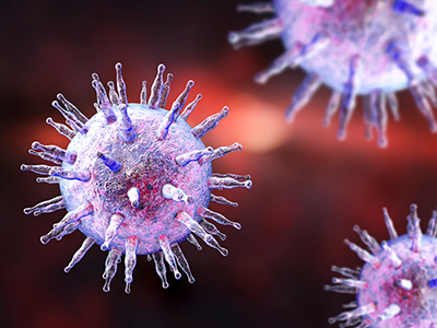
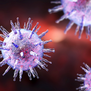
The Epstein-Barr virus (EBV) is best known as the cause of mononucleosis, the ubiquitous “kissing disease” that most people contract at some point in their life. But in rare instances, this virus plays a more sinister role as the impetus of lymphomas, cancers that affect the white blood cells known as lymphocytes.
The Epstein-Barr virus (EBV) is best known as the cause of mononucleosis, the ubiquitous “kissing disease” that most people contract at some point in their life. But in rare instances, this virus plays a more sinister role as the impetus of lymphomas, cancers that affect the white blood cells known as lymphocytes. EBV-associated lymphomas account for about 40% of Hodgkin lymphomas, 20% of diffuse large B-cell lymphomas, and more than 90% of natural killer/T-cell lymphomas. This latter type of lymphoma typically has a very poor prognosis even with the “standard of care” lymphoma treatments such as chemotherapy and/or radiation.
When these interventions fail, the only curative approach is an allogeneic hematopoietic stem cell transplant from a healthy donor, a treatment that’s tough on patients’ bodies and carries significant risks, says Lauren P. McLaughlin, M.D., a pediatrician specializing in hematology and oncology at Children’s National in Washington, D.C. Patients who receive these allogenic transplants are immune-compromised until the donor cells engraft; the grafts can attack patients’ healthy cells in a phenomenon called graft versus host disease; and if patients relapse or don’t respond to this treatment, few options remain.
To help improve outcomes, Dr. McLaughlin and colleagues tested an addition to the allogeneic hematopoietic stem cell transplant procedure for patients with EBV-associated lymphomas: infusion of a type of immune cell called T cells specifically trained to fight cells infected with EBV.
Dr. McLaughlin, along with Senior Author Catherine M. Bollard, M.D., M.B.Ch.B., director of the Center for Cancer and Immunology Research and the Program for Cell Enhancement and Technologies for Immunotherapy at Children’s National, and colleagues tested this therapy in 26 patients treated at Children’s National or Baylor College of Medicine. They published these results online on Sept. 27, 2018, in the journal Blood. The study was a Phase I clinical trial, meaning that the therapy was tested primarily for safety, with efficacy as a secondary aim.
Seven patients who received the therapy had active disease that had not responded to conventional therapies. The other 19 were patients deemed to be at high risk for relapse.
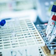
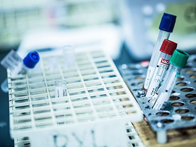
Before each patient received their stem cell transplant, their donors gave an additional blood sample to generate the cancer-fighting T cells. Over the next 8 to 10 weeks, the researchers painstakingly manufactured the immune cells known as T-cells that specifically targeted EBV, growing these cells into numbers large enough for clinical use. Then, as early as 30 days after transplant, the researchers infused these T-cells into patients administering at least two doses, spaced two weeks apart.
Over the next several weeks, the researchers at CNMC and Baylor College of Medicine monitored patients with comprehensive exams to see how they fared after these transplants. The results showed that adverse effects from the treatment were exceedingly rare. There were no immediate infusion-related toxicities to the T-cell therapy and only one incident of dose-limiting toxicity.
This therapy may be efficacious, depending on the individual patients’ circumstances, Dr McLaughlin adds. For those in complete remission but at high risk of relapsing, the two-year survival rate was 78%, suggesting that the administration of this novel T-cell therapy may give the immune system a boost to prevent the lymphoma from returning after transplant. For patients with active T-cell lymphomas, two-year survival rates were 60%. However, even these lower rates are better than the historical norm of 30-50%, suggesting that the targeted T-cell therapies could help fight disease in patients with this poor prognosis lymphoma.
Dr. McLaughlin, the study’s lead author and a Lymphoma Research Foundation grantee, notes that researchers have more work to do before this treatment becomes mainstream. For example, this treatment will need to be tested in larger populations of patients with EBV-related lymphoma to determine who would derive the most benefit, the ideal dose and dose timing. It also may be possible to extend targeted T-cell treatments like this to other types of cancers. In the future, Dr. McLaughlin adds, it may be possible to develop T-cells that could be used “off the shelf”—in other words, they wouldn’t need to come from a matched donor and would be ready to use whenever a recipient needs them. Another future goal is using this therapy as one of the first lines of treatment rather than as a last resort.
“Our ultimate goal is to find a way to avoid chemotherapy and/or radiation therapy while still effectively treating a patient’s cancer,” she says. “Can you use the immune system to do that job? We’re working to answer that question.”
In addition to Drs. McLaughlin and Bollard, study co-authors include Rayne Rouce, Stephen Gottschalk, Vicky Torrano, George Carrum, Andrea M. Marcogliese, Bambi Grilley, Adrian P. Gee, Malcolm K. Brenner, Cliona M. Rooney and Helen E. Heslop, all of Baylor College of Medicine; Meng-Fen Wu from the Dan L. Duncan Comprehensive Cancer Center; and Fahmida Hoq and Patrick J. Hanley, Ph.D. from Children’s National in Washington, D.C.