newborn in incubator

Tracking oxygen saturation with vital signs to identify vulnerable preemies


Khodayar-Rais-Bahrami

What’s known

Critically ill infants in neonatal intensive care units (NICU) require constant monitoring of their vital signs. Invasive methods, such as using umbilical arterial catheters to check blood pressure, are the gold standard but pose significant health risks. Low-risk noninvasive monitoring, such as continuous cardiorespiratory monitors, can measure heart rate, respiratory rate and blood oxygenation. A noninvasive technique called near-infrared spectroscopy (NIRS) can gauge how well tissues, including the brain, are oxygenated. While NIRS long has been used to monitor oxygenation in conditions in which blood flow is altered, such as bleeding in the brain, how NIRS values relate to other vital sign measures in NICU babies was unknown.

What’s new

A research team led by Khodayar Rais-Bahrami, M.D., a neonatologist at Children’s National Health System, investigated this question in 27 babies admitted to Children’s NICU. The researchers separated these subjects into two groups: Low birth weight (LBW, less than 1.5 kg or 3.3 pounds) and moderate birth weight (MBW, more than 1.5 kg). Then, they looked for correlations between information extracted from NIRS, such as tissue oxygenation (specific tissue oxygen saturation, StO2) and the balance between oxygen supply and consumption (fractional tissue oxygen extraction, FTOE), and various vital signs. They found that StO2 increased with blood pressure for LBW babies but decreased with blood pressure for MBW babies. Brain and body FTOE in LBW babies decreased with blood pressure. In babies with abnormal brain scans, brain StO2 increased with blood pressure and brain FTOE decreased with blood pressure. Together, the researchers suggest, these measures could give a more complete picture of critically ill babies’ health.

Questions for future research

Q: Can NIRS data be used as a surrogate for other forms of monitoring?

Q: How could NIRS data help health care professionals intervene to improve the health of critically ill infants in the NICU?

Source: Significant correlation between regional tissue oxygen saturation and vital signs of critically ill infants.” B. Massa-Buck, V. Amendola, R. McCloskey and K. Rais-Bahrami. Published by Frontiers in Pediatrics Dec. 21, 2017.

Carlos Ferreira Lopez

Researchers discover new gene variant for inherited amino acid-elevating disease

Carlos Ferreira Lopez

PDF Version

What’s known

Hypermethioninemia is a rare condition that causes elevated levels of methionine, an essential amino acid in humans. This condition stems from genetic variations inherited from one or both parents. Some forms of hypermethioninemia are recessive, meaning that two copies of defective genes are necessary to cause this disease. Other forms are dominant, meaning that only one copy can cause hypermethioninemia. Recessive forms of the disease tend to have more serious consequences, causing elevated methionine levels throughout life and leading to changes in the brain’s white matter visible on magnetic resonance imaging that can cause neurological problems. The dominant forms are generally thought to be largely benign and require minimal follow-up.

What’s new

A research team led by Carlos Ferreira Lopez, M.D., a medical geneticist at Children’s National Health System, discovered a new gene variant that had not been associated with hypermethioinemia previously when an infant who had tested positive for elevated methionine on newborn blood-spot screening came in for a follow-up evaluation. While the majority of dominant hypermethioninemia are caused by a genetic mutation known as MAT1A p.Arg264His, the child didn’t have this or any of the common recessive hypermethioninemia mutations. Genetic testing showed that she carried a different mutation to the MAT1A gene known as p.Ala259Val, of which she carried only a single copy. The child fit the typical profile of having the dominant form of the disease, with methionine levels gradually declining over time. Testing of her mother showed that she carried the same gene variant, with few consequences other than a hepatitis-like illness as a child. Because liver disease can accompany dominant hypermethioninemia, the infant’s doctors will continue periodic follow-up to ensure she remains healthy.

Questions for future research

Q: Besides the potential for harmful liver effects, does dominant hypermethioninemia have other negative consequences?

Q: How common is this gene variant, and are certain people at more risk for carrying it?

Source: Confirmation that MAT1A p.Ala259Val mutation causes autosomal dominant hypermethioninemia. Muriello, M.J., S. Viall, T. Bottiglieri, K. Cusmano-Ozog and C. R. Ferreira. Published by Molecular Genetics and Metabolism Reports December 2017.

Irf6

IRF6 gene plays roles in tissue development beyond epithelium

Irf6

Kousa, Y. A., Moussa, D. and Schutte, B. C. (2017), IRF6 expression in basal epithelium partially rescues Irf6 knockout mice. Developmental Dynamics, 2017. © 2017 Wiley Periodicals, Inc.

PDF Version

What’s known

IRF6 is a gene that plays a key role in the development of epithelium, the tissue that lines the cavities and surfaces of blood vessels and organs throughout the body. Mutations in this gene are known to contribute to human diseases, including van der Woude syndrome and popliteal pterygium syndrome, both of which are characterized by cleft lip and palate, skin abnormalities and limb defects. Experimental models that are genetically modified to lack this gene typically have systemic defects so severe that they die at birth. However, it’s been unclear whether these defects are all due to problems with the epithelium and their related consequences, or if IRF6 also plays a role in other tissues during fetal development.

What’s new

A research team led by Youssef A. Kousa, M.S., D.O., Ph.D., a pediatric resident in the child neurology track at Children’s National Health System, investigated where IRF6’s activity is important by partially “rescuing” experimental models altered to lack this gene – or selectively restoring its activity – in just the epithelium. When the resulting animals were born, they survived for hours, unlike animals that lack IRF6 completely. However, the partially rescued experimental models had physical characteristics that were intermediate between animals that were not genetically modified and those that totally lacked IRF6. These partially rescued animals still had cleft palates, skin abnormalities and limb defects, but these defects were not as severe as the modified animals that weren’t rescued at all. The findings suggest that IRF6 plays a role in development of tissue types beyond epithelium. Gaining a better understanding of how mutations in this gene exert their effects on this array of tissues eventually may help researchers develop ways to prevent related disorders or to treat them early in development.

Questions for future research

Q: What function is IRF6 playing in tissues beyond epithelium?
Q: Which defects lead to premature death in experimental models whose IRF6 function is restored in the epithelium?
Q: Is there a way to prevent defects caused by IRF6 mutations without the need to restore IRF6 function directly?

Source:IRF6 expression in basal epithelium partially rescues Irf6 knockout mice.” Kousa, Y.A., D. Moussa and B.C. Schutte. Published online by Developmental Dynamics June 23, 2017.
Angioletta Rheumatic Heart Disease

Rheumatic Heart Disease Center Launches with $3.7 Million AHA Grant

Angioletta Rheumatic Heart Disease

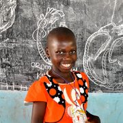
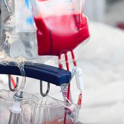
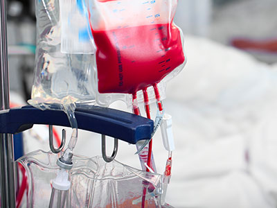
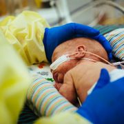
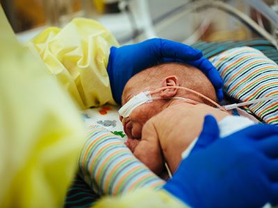
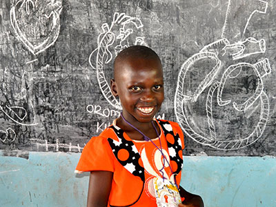
Ten-year-old Angioletta was clinically diagnosed with rheumatic heart disease in 2014 (severe leakage of her mitral valve). She’s been medically managed at the clinic Children’s helps support and conducts research at in Gulu, and she is a very active participant in the support group led by Children’s National research assistant, Amy Scheel. Angioletta hasn’t had any major complications, but her only hope for long-term survival is to undergo open heart surgery to replace her abnormal valve. Experts are looking towards the research from the new Center to help prevent future generations of children like Angioletta from developing RHD.

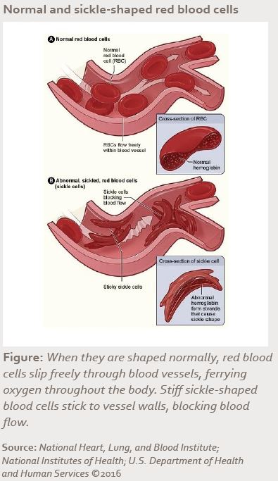
Known as the ‘world’s forgotten disease,’ Rheumatic Heart Disease (RHD) is caused by untreated streptococcal throat infections that progress into acute rheumatic fever (ARF) and eventually weaken the valves of the heart. It is the most common cardiovascular disease in children and young adults globally – affecting nearly 33 million people and causing 345,000 deaths annually – yet, it is preventable with early detection and access to penicillin.

To help end the epidemic, Children’s National Health System has been awarded a $3.7 million grant from the American Heart Association (AHA) to launch a Rheumatic Heart Disease Center, with the goal of developing innovative strategies and economic incentives to improve the prevention and diagnosis of RHD in high-risk, financially disadvantaged countries and low-income communities across the United States.

Children’s National is one of four centers in the AHA’s Strategically Focused Children’s Research Network, which is dedicated to improving children’s heart health and reducing the global burden of cardiovascular disease and stroke. AHA selected Children’s for the grant based on its proven record of global collaboration to solve complex health issues and the potential impact of this research. The program will use Children’s robust telemedicine infrastructure to connect co-collaborators around the world, as well as train the next generation of globally minded cardiovascular researchers.

“While it’s often thought that we’ve already beaten rheumatic heart disease, data shows there’s nearly no decrease in mortality rates in low-income countries. The disease is endemic in Sub-Saharan Africa, and some poverty-stricken communities in the U.S. are hit nearly as hard,” said Craig Sable, M.D., associate division chief of cardiology. “We are thrilled to receive this funding from the AHA, which will help us close the research gap for this neglected disease and change the plight of millions of children around the world.”

About the center and research focus areas

Over the next four years, the Rheumatic Heart Disease Center, led by Children’s National Heart Institute experts Dr. Sable and Andrea Beaton, M.D., cardiologist, along with RHD leaders around the globe, will develop evidence-based strategies to strengthen the health system’s response to RHD through synergistic basic, clinical and population science research along the entire spectrum of the disease.

Andrea Beaton and Craig Sable

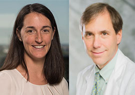
The Rheumatic Heart Disease Center, led by Children’s National Heart Institute experts Andrea Beaton, M.D., and Craig Sable, M.D., along with RHD leaders around the globe, will develop evidence-based strategies to strengthen the health system’s response to RHD.

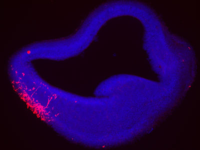
The basic research project, led by James Dale, M.D., chief of the division of infectious disease at the University of Tennessee in Memphis, will work to better define the immune system response to Group A Streptococcal (GAS) infection, or strep throat, paving the way for vaccine development. In collaboration with a partner site in Cape Town, South Africa, experts will recruit 300 children ages 5-15 to participate for 24 months in a study capturing and classifying various strains of the GAS bacteria. Similar to the common flu, the strains of GAS bacteria vary from region to region and year to year. By identifying immune system targets, or how our bodies fight GAS, the research can inform the creation of effective and long-lasting vaccines.

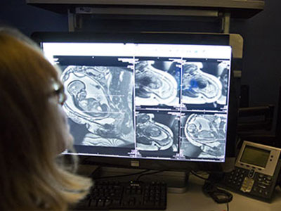
Dr. Beaton will lead the clinical project that will work to improve understanding and detection of ARF, the precursor to RHD. According to Dr. Beaton, the current, outdated paradigm is that patients with RHD at one point experienced a full-blown episode of ARF – including fever, severe joint pains and rash. These symptoms should be unmistakable and prompt treatment, but in truth the disease remains vastly underdiagnosed in high-risk regions. Through an on-the-ground partnership with experts at Mulago National Referral Hospital in Uganda, the clinical project will work to enroll over 1,000 children ages 3-18 with more subtle symptoms, potentially suggestive of ARF, in order to paint a more accurate picture of the disease in Africa today.

“The gap between the low number of children diagnosed with ARF and the high number of young adults with advanced RHD remains one of the most challenging mysteries and barriers to improved RHD prevention,” said Dr. Beaton. “For the first time, we will systematically characterize the clinical, laboratory and echocardiographic features of ARF in low-resource settings, with the goal of developing a biological signature for ARF that can be translated into a diagnostic test and improve detection.”

Dr. Beaton expects that this research could benefit other related diseases too, such as kidney disease or serious skin infections.

The population research project, led by David Watkins, M.D., M.P.H., an expert in epidemiological and economic modeling at the University of Washington in Seattle, will work to build an economic case for prevention around the world, using the data from the basic and clinical work. The goal is to identify local gaps in delivery of health services for disease prevention and treatment and to measure the cost-effectiveness of RHD interventions, as well as the cost of inaction – especially as patients suffering from advanced RHD are often in the prime of their productive, adult lives. Researchers anticipate the findings will provide effective tools for addressing RHD in other endemic countries too.

Chima Oluigbo

A novel way to treat intractable epilepsy caused by hemimegalencephaly

Chima Oluigbo

A multidisciplinary team led by Chima Oluigbo, M.D., F.R.C.S.C., pioneered a novel technique to preserve newborns’ healthy brain tissue, buying time until the infants became old enough to undergo a hemispherectomy.

PDF Version

What’s known

Hemimegalencephaly is an extremely rare birth defect in which one side of the brain grows larger than the other. This anomaly typically leads to severe, recurrent seizures that can be difficult to control solely with medications. While the seizures themselves are detrimental to the developing brain, the amount of medications used to reduce seizure frequency often come with significant side effects and have the potential to hamper brain growth. Hemispherectomy, a radical surgery in which one half of the brain is removed, is often the most successful way to treat severe and intractable epilepsy. However, this surgery can be challenging to perform successfully in very young babies.

What’s new

In this case report, the Children’s National Health System Epilepsy Team led by Chima Oluigbo, M.D., F.R.C.S.C., a pediatric neurosurgeon; Tammy N. Tsuchida, M.D., PhD., a pediatric surgical epileptologist; Monica Pearl, M.D., a pediatric interventional neuroradiologist; Taeun Chang, M.D., a neonatal neurointensivist; and the neonatal intensive care team explored the possibility of using minimally invasive surgery to cut off the blood supply to the brain hemisphere responsible for generating seizures in newborns with hemimegalencephaly. This procedure, they reasoned, could buy time for babies to mature and become more resilient to withstand the future hemispherectomy while also lessening the damage caused by uncontrolled, recurrent seizures. The case report focused on the first two patients with hemimegalencephaly who had sequential procedures to gradually restrict blood flow to the affected brain hemisphere within their first few weeks of life, followed by hemispherectomies at a few months of age. This novel approach significantly lessened their seizures until hemispherectomy, allowing these children to continue to grow and develop seizure-free.

Questions for future research

Q: Which patients are best suited for this surgical procedure?
Q: How can surgeons reduce the risk of excessive blood loss during hemispherectomy caused by the growth of additional blood vessels after flow through the brain’s major vessels has been blocked?
Q: What are the long-term outcomes for infants who undergo these procedures?

Source: “ ‘Endovascular embolic hemispherectomy’: A strategy for the initial management of catastrophic holohemispheric epilepsy in the neonate.” Oluigbo, C., M.S. Pearl, T.N. Tsuchida, T. Chang, C.-Y. Ho and W. D. Gaillard. Published by Child’s Nervous System October 29, 2016.
Nobuyuki Ishibashi

Congenital heart disease and the brain

Nobuyuki Ishibashi

In a recent review article published in Circulation Research, Nobuyuki Ishibashi, M.D., and his colleagues at Children’s National Health System summarized what is currently known about how congenital heart disease affects brain maturation.

What’s known

Among all known birth defects, congenital heart disease (CHD) is the leading cause of death in infants. Fortunately, advances in surgical techniques and treatments are improving the outlook for these children, and more and more are reaching adulthood. However, because of this increased longevity, it has become increasingly clear that children born with CHD are at risk of developing life-long neurological deficits. Several risk factors for these neurodevelopmental abnormalities have been identified, but direct links between specific factors and neurological defects have yet to be established.

What’s new

In a recent review article published in Circulation Research, a team from Children’s National Health System summarized what is currently known about how CHD affects brain maturation. Drawing from studies conducted at Children’s National as well as other research institutions, Paul D. Morton, Ph.D., Nobuyuki Ishibashi, M.D., and Richard A. Jonas, M.D., write that clinical findings in patients, improvements in imaging analysis, advances in neuromonitoring techniques and the development of animal models have greatly contributed to our understanding of the neurodevelopmental changes that occur with CHD.

Findings from Children’s National include:

  • An assessment of the intraoperative effects of cardiopulmonary bypass surgery on white matter using neonatal piglets.
  • An arterial spin labeling MRI study that showed newborns with complex CHD have a significant reduction in global cerebral blood flow.
  • A rodent study that modeled diffuse white matter brain injury in premature birth and identified the cellular and molecular mechanisms underlying lineage-specific vulnerabilities of oligodendrocytes and their regenerative response after chronic neonatal hypoxia.

The authors conclude that although there is ample clinical evidence of neurological damage associated with CHD, there is limited knowledge of the cellular events associated with these abnormalities. They offer perspectives about what can be done to improve our understanding of neurological deficits in CHD, and emphasize that ultimately, a multidisciplinary approach combining multiple fields and myriad technology will be essential to improve or prevent adverse neurodevelopmental outcomes in individuals with CHD.

Questions for future research

Q: What are the cellular events associated with each factor involved in neurodevelopmental delays?
Q: How does the neurodevelopmental status of a patient with CHD change as they age?
Q: How do the genes involved in structural congenital cardiac anomalies affect brain development and function?

Source: Norton, P.D., Ishibashi, N., Jonas, R.A. Neurodevelopmental Abnormalities and Congenital Heart Disease: Insights Into Altered Brain Maturation,” Circulation Research (2017) 120:960-977.
Blood Transfusion

Hydroxycarbamide effective in sickle cell stroke prevention

Blood Transfusion

Hydroxycarbamide treatment is on par with blood transfusions for preventing stroke in patients with sickle cell anemia.

PDF Version

What’s known

Strokes are common and devastating complications for patients with sickle cell anemia, often leading to severe and lifelong motor and neurocognitive problems for people with this congenital blood disorder. Results of a clinical trial published in 1998 showed that having regular blood transfusions could reduce the risk of having a first stroke by 90 percent in children with sickle cell anemia. Since then, doctors have employed this prophylactic treatment widely. However, blood transfusions can be painful, inconvenient and carry substantial risks themselves — including the potential of blood-borne infections, iron overload and immune-related reactions to blood products. Finding a way to reduce stroke risk without over-relying on blood transfusions could substantially benefit patients with sickle cell anemia.

What’s new

A team of researchers, including Naomi L.C. Luban, M.D., a Children’s National Health System hematologist and laboratory medicine specialist, tested transfusions against a drug treatment called hydroxycarbamide in a clinical trial to see if the pharmaceutical intervention could reduce strokes at least as well as transfusions. The clinical trial, known as “TCD With Transfusions Changing to Hydroxyurea (TWiTCH),” assigned 60 patients with sickle cell anemia who had abnormally high transcranial Doppler (TCD) flow velocities—a measure of blood flow in the brain that suggests elevated risk of stroke—to receive hydroxycarbamide instead of transfusions. The research team compared the outcomes for these patients with 61 other patients who received standard prophylactic transfusions. Over the 24-month study period, neither group experienced any strokes, although three transient ischemic attacks (a temporary blockage of blood flow in the brain) occurred in each group. These comparable findings suggest that hydroxycarbamide treatment, also known as hydroxyurea, is on par with transfusions for preventing strokes in patients with sickle cell anemia.

Questions for future research

Q: Does hydroxycarbamide offer a long-term way for patients with sickle cell anemia to avoid transfusions?
Q: Could hydroxycarbamide help patients with sickle cell anemia who already have suffered a stroke or who have had severe problems with blood vessels in their brains that impair blood flow?
Q: Which other treatments can help patients avoid the myriad complications that accompany sickle cell anemia?

Source: Hydroxycarbamide versus chronic transfusion for maintenance of transcranial doppler flow velocities in children with sickle cell anemia—TCD With Transfusions Changing to Hydroxyurea (TWiTCH): A multicentre, open-label, phase 3, non-inferiority trial.” Ware, R.E. B. R. Davis, W. H. Schultz, R.C. Brown, B. Aygun, S. Sarnaik, I. Odame, B. Fuh, A. George, W. Owen, L. Luchtman-Jones, Z.R. Rogers, L. Hilliard, C. Gauger, C. Piccone, M.T. Lee, J.L. Kwiatkowski, S. Jackson, S.T. Miller, C. Roberts, M.M. Heeney, T.A. Kalfa, S. Nelson, H. Imran, K. Nottage, O. Alvarez, M. Rhodes, A.A. Thompson, J.A. Rothman, K.J. Helton, D. Roberts, J. Coleman, M.J. Bonner, A. Kutlar, N. Patel, J. Wood, L. Piller, P. Wei, J. Luden, N.A. Mortier, S.E. Stuber, N. L. C. Luban, A.R. Cohen, S. Pressel and R.J. Adams. Published by The Lancet on Feb. 13, 2016.

Smart Tissue Autonomous Robot

Robotic system automates soft-tissue surgery

Smart Tissue Autonomous Robot

STAR’s performance was measurably better in some respects, compared with surgeons performing the same procedure manually or with conventional robotic techniques.

PDF Version

What’s known

Robotic surgery has been increasingly adopted for a variety of procedures. However, conventional robotic surgery is still controlled by individual surgeons. One way to avoid variabilities and improve outcomes is to automate entire procedures or parts of procedures. Thus far, autonomous robotic surgery has been limited to parts of the body with rigid anatomy, because of the unpredictability of soft tissues. These structures can move in unexpected ways during cutting, suturing or cauterizing. No autonomous robotic systems for soft-tissue procedures have been developed due to technology lags, including a lack of vision systems that can distinguish and track tissue in dynamic surgical environments, and intelligent algorithms.

What’s new

A team of researchers led by Peter C.W. Kim, M.D., Ph.D., Vice President, and Axel Krieger, Ph.D., Assistant Professor of the Sheikh Zayed Institute for Pediatric Surgical Innovation at Children’s National Health System, developed the Smart Tissue Autonomous Robot (STAR) for performing autonomous soft-tissue surgeries. The researchers tested its capability in three areas: Suturing a cut along a length of suspended intestine, suturing together two pieces of intestine removed from an experimental model and suturing together two pieces of intestine inside a living experimental model. The autonomous robot’s performance was measurably better in some respects, compared with surgeons trained for at least seven years performing the same procedure manually or with conventional robotic techniques. STAR’s stitches were more consistent and less prone to leaks. This demonstration of supervised autonomous surgical tasks by a robot promises that surgeons can improve their technical and decision-making skills in the not-too-distant future, working collaboratively with intelligent robots to improve clinical outcomes.

Questions for future research

Q: Can autonomous robots be used for soft-tissue procedures more complicated than suturing?
Q: Can this system be miniaturized for complex procedures taking place in a confined space, such as suturing together blood vessels?
Q: How can we make more intelligent robots available to all surgeons?

Source: Supervised autonomous robotic soft tissue surgery.” Shademan A., R.S. Decker, J.D. Opfermann, S. Leonard, A. Krieger and P.C.W. Kim. Published by Science Translational Medicine on May 4, 2016.

newborn

Sirtuin could repair common neonatal brain injury

A sirtuin might help repair a common neonatal brain injury

A team of researchers  investigated the molecular mechanisms behind oligodendrocyte progenitor cell proliferation in neonatal hypoxia.

PDF Version

What’s known

Hypoxia, or a lack of oxygen, is a major cause of diffuse white matter injury (DWMI). This condition leads to permanent developmental disabilities in prematurely born infants. The long-term abnormalities of the brain’s white matter that characterize DWMI are caused by the loss of a specific type of cells known as oligodendrocytes, which support nerve cells and produce myelin, a lipid and protein sheath that electrically insulates nerve cells. Oligodendrocytes are produced by a population of immature cells known as oligodendrocyte progenitor cells (OPCs). Previous research has shown that hypoxia can trigger OPCs to proliferate and presumably produce new oligodendrocytes. The molecular pathways that hypoxia triggers to make new OPCs remain unclear.

What’s new

A team of researchers led by Vittorio Gallo, Ph.D., director of the Center for Neuroscience Research and the Intellectual and Developmental Disabilities Research Center at Children’s National Health System, investigated the molecular mechanisms behind what prompts OPCs to proliferate in a preclinical model of neonatal hypoxia. The researchers found that a molecule known as Sirt1 acts as a major regulator of OPC proliferation and regeneration. Sirt1 is a sirtuin, a class of molecules that has attracted interest over the past several years for its role in stem cells, aging and inflammation. Hypoxia appears to induce Sirt1 formation. When the researchers prevented brain tissues in petri dishes from making Sirt1 or removed this molecule in preclinical models, these actions prevented OPC proliferation. What’s more, preventing Sirt1 production also inhibited OPCs from making oligodendrocytes. These findings suggest that Sirt1 is essential for replacing oligodendrocytes to repair DWMI after hypoxia. Additionally, finding ways to enhance Sirt1 activity eventually could provide a novel way to help infants recover after hypoxia and prevent DWMI.

Questions for future research

Q: How can Sirt1 activity be enhanced in preclinical models and humans?
Q: Can deficits triggered by diffuse white matter injury be prevented or reversed with Sirt1?
Q: Which other treatments might be useful for diffuse white matter injury?

Source: Sirt1 regulates glial progenitor proliferation and regeneration in white matter after neonatal brain injury.” Jablonska, M., M. Gierdalski, L. Chew, T. Hawley, M. Catron, A. Lichauco, J. Cabrera-Luque, T. Yuen, D. Rowitch and V. Gallo. Published by Nature Communications on Dec. 19, 2016.

Cas9-mediated correction of metabolic liver disease

AAV.CRISPR-SaCas9

In vivo gene correction of the OTC locus in the mouse liver by AAV.CRISPR-SaCas9. Source: Nature Publishing Group copyright 2016.

PDF Version

What’s known

A deficiency of the enzyme ornithine transcarbamylase (OTC) in humans causes life-threatening hyperammonemic crises.  The OTC gene enables the body to make an enzyme that is a critical player in the urea cycle, a process that ensures excess nitrogen is excreted by the kidneys. Left unchecked, accumulating nitrogen becomes a toxic form of ammonia. Infants with OTC deficiency can suffer their first metabolic crisis as newborns. Up to 50 percent die or sustain severe brain injury, and survivors typically need a liver transplant by age 1. Gene therapy could cure OTC deficiency, but currently used viruses, such as adeno-associated virus (AAV), are not optimal in the neonatal setting.

What’s new

A research team led by Children’s National Health System and the University of Pennsylvania reasoned that the newborn liver may be an ideal setting for AAV-mediated gene correction using CRISPR-Cas9 gene editing. They intravenously infused two AAVs into two-day-old mice with partial OTC deficiency. One AAV expressed Cas9 and the other expressed a guide RNA and a donor OTC DNA. This resulted in correction of the mutation in 10 percent of liver cells and increased survival in mice challenged with a high-protein diet, which normally exacerbates disease. After consuming a high-protein diet for one week, the treated newborns had a 40 percent reduction in ammonia compared with the untreated group. The correction appears to last long term. The study “provides evidence for efficacy of gene editing in neonatal onset OTC deficiency,” says Mark L. Batshaw, M.D., Physician-In-Chief and Chief Academic Officer at Children’s National, and a study co-author. “This study provides convincing evidence for efficacy of in vivo genome editing in an authentic animal model of a lethal human metabolic disease,” the research team concludes.

Questions for future research

Q: More than 400 mutations can cause OTC deficiency, and each would require a separate gene-editing approach. Is it possible instead to insert the OTC genome using CRISPR-Cas9 to correct the disorder irrespective of the mutation?
Q: Will such gene editing also work in adult animal models of the OTC disorder?
Q: Do these encouraging results in animals translate to efficacy in infants?

Source: Yang, Y., L. Wang, P. Bell, D. McMenamin, Z. He, J. White, H. Yu, C. Xu, H. Morizono, K. Musunuru, M.L. Batshaw and J.M. Wilson. “A dual AAV system enables the Cas9-mediated correction of a metabolic liver disease in newborn mice.” Published Feb. 1, 2016 by Nature Biotechnology.

Fetal Brain Cells

Tracking environmental stress damage in the brain

Fluorescence Reporter

A team led by Children’s National developed a fluorescence reporter system in an experimental model that can single out neurons that have survived prenatal damage but remain vulnerable after birth.

PDF Version

What’s known

When fetuses are exposed to environmental stressors, such as maternal smoking or alcohol consumption, radiation or too little oxygen, some of these cells can die. A portion of those that survive often have lingering damage and remain more susceptible to further environmental insults than healthy cells; however, researchers haven’t had a way to identify these weakened cells. This lack of knowledge has made it difficult to discover the mechanisms behind pathological brain development thought to arise from these very early environmental exposures, as well as ways to prevent or treat it.

What’s new

A team led by Kazue Hashimoto-Torii, Ph.D., a principal investigator in the Center for Neuroscience Research at Children’s National Health System, developed a marker that makes a protein known as Heat Shock Factor 1 glow red. This protein is produced in cells that become stressed through exposure to a variety of environmental insults. Gestation is a particularly vulnerable time for rapidly dividing nerve cells in the fetal brain. Tests showed that this marker worked not just on cells in petri dishes but also in an experimental model to detect brain cells that were damaged and remained vulnerable after exposure to a variety of different stressors. Tweaks to the system allowed the researchers to follow the progeny of cells that were affected by the initial stressor and track them as they divided and spread throughout the brain. By identifying which neurons are vulnerable, the study authors say, researchers eventually might be able to develop interventions that could slow or stop damage before symptoms arise.

Questions for future research

Q: How do different environmental insults damage brain cells during gestation?
Q: How does this damage translate into pathology in organisms as they mature?
Q: Do the progeny of damaged brain cells retain the same degree of damage as they divide and spread?
Q: Can this new detection system be used to find and track damage in other organs, such as the heart, eye and liver?

Source: Torii, M., S. Masanori, Y.W. Chang, S. Ishii, S.G. Waxman, J.D. Kocsis, P. Rakic and K. Hashimoto-Torii. “Detection of vulnerable neurons damaged by environmental insults in utero.” Published Dec. 22, 2016 by Proceedings of the National Academy of Sciences. doi: 10.1073/pnas.1620641114

This is the consequential malfunction of the brain during congenital heart defects.

Congenital heart disease and white matter injury

This is the consequential malfunction of the brain during congenital heart defects.

Although recent advances have greatly improved the survival of children with congenital heart disease, up to 55 percent will be left with injury to their brain’s white matter – an area that is critical for aiding connection and communication between various regions in the brain.

What’s known

Eight of every 1,000 children born each year have congenital heart disease (CHD). Although recent advances have greatly improved the survival of these children, up to 55 percent will be left with injury to their brain’s white matter – an area that is critical for aiding connection and communication between various regions in the brain. The resulting spectrum of neurological deficits can have significant costs for the individual, their family and society. Although studies have demonstrated that white matter injuries due to CHD have many contributing factors, including abnormal blood flow to the fetal brain, many questions remain about the mechanisms that cause these injuries and the best interventions.

What’s new

A Children’s National Health System research team combed existing literature, reviewing studies from Children’s as well as other research groups, to develop an article detailing the current state of knowledge on CHD and white matter injury. The scientists write that advances in neuroimaging – including magnetic resonance imaging, magnetic resonance spectroscopy, Doppler ultrasound and diffusion tensor imaging – have provided a wealth of knowledge about brain development in patients who have CHD. Unfortunately, these techniques alone are unable to provide pivotal insights into how CHD affects cells and molecules in the brain. However, by integrating animal models with findings in human subjects and in postmortem human tissue, the scientists believe that it will be possible to find novel therapeutic targets and new standards of care to prevent developmental delay associated with cardiac abnormalities.

For example, using a porcine model, the Children’s team was able to define a strategy for white matter protection in congenital heart surgery through cellular and developmental analysis of different white matter regions. Another study from Children’s combined rodent hypoxia with a brain slice model to replicate the unique brain conditions in neonates with severe and complex congenital heart disease. This innovative animal model provided novel insights into the possible additive effect of preoperative hypoxia on brain insults due to cardiopulmonary bypass and deep hypothermic circulatory arrest.

The Children’s research team also recently published an additional review article describing the key windows of development during which the immature brain is most vulnerable to CHD-related injury.

Questions for future research

Q: Can we create an animal model that recapitulates the morphogenic and developmental aspects of CHD without directly affecting other organs or developmental processes?
Q: What are the prenatal and neonatal cellular responses to CHD in the developing brain?
Q: What are the molecular mechanisms underlying white matter immaturity and vulnerability to CHD, and how can we intervene?
Q: How can we accurately assess the dynamic neurological outcomes of CHD and/or corrective surgery in animal models?
Q: Prenatal or postnatal insults to the developing brain: which is most devastating in regards to developmental and behavioral disabilities?
Q: How can we best extrapolate from, and integrate, neuroimaging findings/correlations in human patients with cellular/molecular approaches in animal models?

Source: Reprinted from Trends in Neurosciences, Vol. 38/Ed. 6, Paul D. Morton, Nobuyuki Ishibashi, Richard A. Jonas and Vittorio Gallo, “Congenital cardiac anomalies and white matter injury,” pp. 353-363, Copyright 2015, with permission from Elsevier.

Practice guideline summary: Use of fMRI in the presurgical evaluation of patients with epilepsy

PDF Version

What’s Known

Neurologists assess a patient’s baseline language and memory, and attempt to predict postsurgical impacts on language before the patient undergoes neurosurgical procedures to minimize the symptoms of epilepsy. In a standard intracarotid amobarbital procedure (IAP), a medication is injected through the carotid artery that isolates one hemisphere of the brain at a time followed by the patient performing memory tasks. More recently, neurologists have performed the assessment via functional magnetic resonance imaging (fMRI), an image acquisition technique that captures brain activity while the patient completes a set of memory and language tasks. Both approaches lack standardized implementation guidelines, making it difficult to fully assess when fMRI may be an effective alternative to IAP.

What’s New

A Practice Guideline Summary published in Neurology, the journal of the American Academy of Neurology, regarding the use of fMRI for pre-surgical evaluation of patients with epilepsy establishes recommendations related to the diagnostic accuracy of fMRI for pre-surgical evaluation. An 11-member panel of international experts, including William D. Gaillard, M.D., Chief of Child Neurology, Epilepsy and Neurophysiology, and Director of the Comprehensive Pediatric Epilepsy Program at Children’s National Health System, evaluated available evidence to determine when and if fMRI can reliably measure the extent that each brain hemisphere controls language, known as language lateralization, and as a predictor of postsurgical outcomes. For 20 years, Dr. Gaillard’s team has led the field in the application of fMRI for language and memory assessment in children, and their work comprised a large portion of the pediatric-focused research assessed by the panel. The analyses found that fMRI is a viable option for measuring lateralized language functions in place of IAP in medial temporal lobe epilepsy, temporal epilepsy in general or extratemporal epilepsy. Evidence was insufficient to recommend fMRI over IAP for patients with temporal neocortical epilepsy or temporal tumors. The assessment also identified that pre-surgical fMRI can serve as an adequate alternative to IAP memory testing for predicting verbal memory outcome. The authors recommend that clinicians carefully advise patients of the risks and benefits before recommending either approach.

Questions for Future Research

Q: What is role of fMRI for pediatric epilepsy?
Q: Can a standard set of tasks be established as the guideline for assessment?

Source: J.P. Szaflarski, D. Gloss, J.R. Binder, W.D. Gaillard, A.J. Golby, S.K. Holland, J. Ojemann, D.C. Spencer, S.J. Swanson, J.A. French and W.H.Theodore. “Practice Guideline Summary: Use of fMRI in the Presurgical Evaluation of Patients With Epilepsy.” Published by Neurology in January 2017.

Study reveals asthma phenotypes in inner-city children

xxoct16asthmaphenotypesrgimage

What’s known

According to the Centers for Disease Control and Prevention, 8.6 percent of children across the nation, or 6.3 million kids, have asthma, a disease characterized by wheezing and coughing associated with airway obstruction, bronchial hyperresponsiveness, and inflammation of the airway. However, children with asthma with low socioeconomic status who live in inner cities experience a disproportionately high burden of illness. While treatment guidelines provide uniformity in managing allergy and allergic inflammation, such approaches may be misdirected when kids have asthma symptoms but lack allergy or allergic inflammation. Knowledge of distinct disease phenotypes can help to improve care.

What’s new

The Asthma Phenotypes in the Inner City study enrolled school-aged kids living in nine U.S. inner cities, including Washington, DC. The research team collected data about their asthma at the beginning of the one-year study and every two months as the kids’ asthma was managed according to accepted guidelines. Phenotypic analysis for 616 of these kids found their asthma clustered into five distinct groups. Cluster “A” was characterized by lower allergy, lower inflammation, and minimal symptoms. Fifteen percent of the kids fit within “A.” Another 15 percent of kids’ asthma fit within Cluster “B.” They had highly symptomatic asthma despite high step-level treatment and relatively low allergy and inflammation. Cluster “C” was distinguished by minimal symptoms, intermediate allergy and inflammation, and mildly impaired pulmonary physiology. Some 24 percent of kids fit within this group. The remaining kids fit within Cluster “D” or “E” and experienced progressively higher asthma and rhinitis symptoms as well as allergy and inflammation.

Questions for future research

Q: How does exposure to allergens, viruses, and irritants like tobacco smoke—taken individually as well as in combination—influence asthma severity and symptoms for these at-risk youths?
Q: What approaches to treatment might result from these studies?

Destructive otologic foreign body – dangers of the expanding bead

Expanding jelly beads

PDF Version

What’s Known

Superabsorbent polymers (SAP), such as polyacrylamide, are water-soluble plastics with high absorption capacities that frequently are used in agriculture, within potting soil, and as disposable diaper filler. Of late, the material has been used to manufacture children’s playthings. Toys made of SAP include firm colorful beads, known as jelly beads, initially sized at 0.1 cm to 0.2 cm in diameter that swell to 0.67 cm to 1.09 cm in diameter when exposed to liquid. Foreign bodies inserted into the external auditory canal have been associated with an estimated 280,000 visits to the emergency department from 2008 to 2012. Now, jelly beads also represent an aural foreign body risk.

What’s New

In the first case described in the letter report, a girl returned for care after an external auditory canal (EAC) granulation was not responding to oral antibiotics and ototopical drops. The girl repeatedly denied having inserted a foreign body (FB) into her ear. Ten weeks after her first visit, computed tomography imaging showed bony erosion; magnetic resonance imaging revealed a 9.8 cm spherical foreign body that was surgically removed. She suffered profound sensorineural hearing loss. The second patient told clinicians he had placed toy beads in his ears. The blue bead expanded within the canal, causing subtotal tympanic membrane perforation, partial ossicular chain erosion, and resulted in reversible hearing loss. In cases of persistent granulation in the ear, an FB reaction should remain on the differential diagnosis. Malleable beads lodged in the canal should not be treated with ototopical drops. Urgent otolaryngology evaluation is indicated, the authors conclude.

Questions for Future Research

Q: What is the actual pressure/force that the expanding bead exerts on the external auditory canal?
Q: Does the SAP have lytic properties that degrade the bone/ear drum, or are those structures damaged solely by the pressure?
Q: Kids often place small objects in their ears and noses. Are there optimal ways to educate young children about the risks of placing foreign objects in their ears and noses?

Source: Destructive Otologic Foreign Body: Dangers of the Expanding Bead.” M. Sterling and P. Mudd. Published online by JAMA Otolaryngology–Head & Neck Surgery on Aug. 4, 2016.

Expanding cytotoxic T lymphocytes from umbilical cord blood to target three viruses

PDF Version

What’s Known
Children’s National Health System is the only pediatric hospital in the nation that grows personalized T-cells from naïve cord blood (CB), training these CB-derived cells to simultaneously fight adenovirus, cytomegalovirus, and Epstein-Barr virus to control viral infections after transplantation. Here are a number of the critical steps during that three-month manufacturing process.

T Cell Isolation Process

Source: P.J. Hanley, J. J. Melenhors, S. Nikiforow, P. Scheinberg, J.W. Blaney, G. Demmler-Harrison, C.R. Cruz, S. Lam, R.A. Krance, K.S. Leung, C.A. Martinez, H. Liu, D.C. Douek, H.E. Heslop, C. M. Rooney, E.J. Shpall, A.J. Barrett, J.R. Rodgers, and C.M. Bollard. CMV-Specific T-Cells Generated From Naïve T-Cells Recognize Atypical Epitopes and May Be Protective In Vivo.” Published by Science Translational Medicine on April 29, 2015

Feasibility of home-based computerized WM training for sickle cell disease

PDF Version

What’s Known
Cognitive deficits are a common complication affecting about one-third of kids who have the higher risk sickle cell disease genotypes, HbSS and HbSβ0 thalassemia. While such deficits have been well-documented, no treatment has been proven to recover cognitive function for kids with sickle cell disease. Sickle cell disease is a group of red blood cell disorders in which abnormal genes that children inherit from parents cause their bodies to make sickle-shaped hemoglobin S. Kids with sickle cell disease are at heightened risk for neurocognitive deficits, which can have practical implications for their ability to learn and to succeed academically.

What’s New
Because home-based computerized cognitive training programs have helped to improve working memory (WM) for children with epilepsy and for childhood cancer survivors, a team led by Children’s National Health System researchers and clinicians sought to gauge the feasibility of using such an approach for a small number of children with sickle cell disease. The pilot study found that girls were more likely to complete the cognitive training exercises than boys. Additionally, children whose teachers showed a high level of support spent more time working on the exercises, completed more sessions, and were more likely to finish the program. The mean number of completed sessions was 15.83. Participants who reported experiencing fewer functional limitations as a result of sickle cell disease-related pain completed more sessions. Overall, children who completed Cogmed exercises showed improvements in verbal WM, and visuospatial short-term memory and WM.

Questions for Future Research
Q: Because sickle cell disease is often accompanied by sudden attacks of severe pain, would concurrent delivery of pain-management interventions impact children’s ability to complete cognitive training exercises?
Q: Would adding functional magnetic resonance imaging help to clarify the association between adaptive cognitive skills training and changes to physiological processes, such as alterations in prefrontal and parietal cortical activity, and functional connectivity?

Source: S.J. Hardy, K.K. Hardy, J.C. Schatz, A.L. Thompson, and E.R. Meier. Feasibility of Home-Based Computerized Working Memory Training With Children and Adolescents With Sickle Cell Disease.” Published online by Pediatric Blood & Cancer May 26, 2016.

Association Seen Between Trauma Center Type and Mortality Risk for Injured Youths

Swanson Russell photo shoot trauma emergency department Brand Photos FY13

PDF Version

What’s Known
Trauma is the leading cause of death among children and young adults in the United States, but controversy remains about which treatment location is optimal to meet the needs of injured adolescent patients. Pediatric trauma centers tailor care to children’s unique physiological,anatomical, and social needs. Yet, there are variations in the cutoff age used to triage children to either pediatric or adult trauma centers, with the usual decision to triage children to pediatric facilities if they are younger than 14 or 15 and to transport them to adult systems if they are older. A 2015 study found that injured children aged 18 or younger treated at pediatric trauma centers had lower in-hospital mortality.

What’s New
A team led by Children’s National Health System researchers examined 29,613 de-identified records for patients aged 15 to 19 years old drawn from the 2010 National Trauma Data Bank to determine associations between the type of trauma center and youths’ mortality rates. Some 68.9 percent of injured youths were treated at adult trauma centers (ATCs), while 25.6 percent were seen at mixed trauma centers (MTCs), and 5.5 percent at pediatric trauma centers (PTCs). Mortality was higher among youths treated at ATCs (3.2 percent) and MTCs (3.5 percent) than for adolescents seen at PTCs (0.4 percent), P < .001. The adjusted odds of mortality were higher at ATCs (4.19) and MTCs (6.68 ) compared with PTCs (0.76). While the research team saw differences in mortality between trauma center type, the study does not provide information about what may account for these differences.

Questions for Future Research

  • What is the best method to determine differences in treatment practices between trauma center types to better explain differences in the mortality rates of injured adolescents?
  • Which specific qualities are common to trauma centers that provide optimal outcomes to children, and can quality-improvement initiatives help to identify and replicate those attributes elsewhere?

Source: “Association Between Trauma Center Type and Mortality Among Injured Adolescent Patients. R.B. Webman, E.A. Carter, S. Mittal, J. Wang, C. Sathya, A. Nathens, M. Nance, D. Madigan, and R. Burd. Published online by JAMA Pediatrics June 27, 2016.

Patient centered family conferences can boost satisfaction with pediatric ICU care

PDF Version

What’s Known
Family conferences in the pediatric intensive care unit (PICU) cover difficult decisions made on behalf of critically ill children, such as starting or withdrawing life support, placing a tracheostomy, or repeating bone marrow transplantation. Family satisfaction is a national quality indicator for determining excellence of care, and families rate communication as one of physicians’ most important skills. Researchers sought to clarify the association between the patient-centered nature of physicians’ communication patterns and the degree to which parents were satisfied with decision making during family conferences in the PICU.

What’s New
A research team led by Children’s National Health System staff recorded 39 family conferences to dissect the dynamics of the conversations. The conferences averaged 45 minutes in length, and the medical team spoke 73 percent of the time. Physicians contributed 89 percent of the dialogue; bedside nurses spoke 2 percent of the conversation. The team used the Roter Interaction Analysis System and a related patient-centeredness score to evaluate the conversations. A patient centeredness score higher than 0.75 predicted parental satisfaction, controlling for the length of the conference, the severity of the child’s illness, race, and socioeconomic status.

Skills: Partnering and activation, asking for patient opinion, asking for understanding

  • Doctor: What do you think would help?
  • Doctor: Do you follow me?
  • Doctor: Let me make sure I’ve got what you meant. Your preference would be to place the trachif we can’t get the breathing tube out on this third try?

Questions for Future Research
Q:
How do parents’ perceptions change when additional members of the medical team speak during family conferences?
Q: How does the manner in which parents process information, e.g., cognitive processing vs. psychomotor processing, impact their preference for more patient-centered family conferences?

Source: “Parent Satisfaction With Communication is Associated With Physician’s Patient-Centered Communication Patterns During Family Conferences.” T.W. October,P.S. Hinds, J. Wang , Z.B. Dizon, Y.I. Cheng, and D.L. Roter. Published by Pediatric Critical Care Medicine June 17, 2016.

Training kids developing immune systems to prevent wheezing

PDF Version

What’s Known
Some 6.3 million U.S. children younger than 18—or 8.6 percent of the nation’s kids—have asthma. The disease is characterized by an inflammation of the airways, and    symptoms may be triggered by breathing in such allergens as animal dander, pollen, dust, or mold.

Once children experience early wheezing, changes begin in the architecture of their lungs, causing a thicker basement membrane, a thickening of the lining of the lungs, which can result in a heightened tendency for the airways in the lungs to become inflamed.

What’s New
Asthma and allergic diseases are thought to result from disordered development of the immune system, a process that begins in the womb. A paradigm-shifting multicenter clinical trial will enroll patients at eight locations, including Children’s National Health System, to provide the type of “immune system training” that infants would experience if they grew up in rural settings—where most children’s immune systems develop more normally. The five-year study funded by the National Heart, Lung, and Blood Institute will identify 1,000 babies aged 6 months to 18 months who are at risk for asthma to receive safe doses of an inactivated bacteria to help them develop more properly functioning immune systems. The University of Arizona Health Sciences in Tucson will lead the national research effort. Researchers will gauge whether infants randomly assigned to receive treatment suffer fewer respiratory symptoms than infants randomly assigned to receive placebo.

Questions for Future Research

Q: What will be the longer-term effects of preventing early wheezing? Will the children develop asthma less frequently?
Q: If intervention with young children occurs early enough to interrupt the disease cycle—preventing asthma, wheezing, and allergies—will they miss fewer days of school when they are older?
Q: Will families be willing to consistently follow the complex regimen necessary to administer the inactivated bacterial products on a long-term basis?

Source: Oral Bacterial Extracts (ORBEX): Primary Prevention of Asthma and Wheezing in Children.

Enroll in this clinical trial—https://clinicaltrials.gov/ct2/show/NCT02148796