Tag Archive for: zika virus

New philanthropic support from the United Arab Emirates furthers research breakthroughs and care

Visitors from the UAE at Children's National Hospital.

His Highness Sheikh Mohamed bin Zayed Al Nahyan, President of the United Arab Emirates (right) visited Children’s National in September 2024.

Continuing a 30-year partnership that has yielded 82 U.S. patents and countless medical breakthroughs for kids and their families, the Government of the United Arab Emirates (UAE) has strengthened its transformational commitment to Children’s National Hospital with a new $35 million donation focused on prenatal, neonatal and maternal health.

The announcement of the new gift comes after a recent visit to the hospital by His Highness Sheikh Mohamed bin Zayed Al Nahyan, President of the United Arab Emirates (UAE), who met with Emirati families and patients receiving care at Children’s National Hospital.

The investment is the latest chapter of a longstanding philanthropic partnership between the UAE and Children’s National. Each year, more than 100 Emirati families travel to Children’s National for advanced pediatric care and life-saving treatments.

This latest investment will bolster various strategic health initiatives, including within the hospital’s Center for Prenatal, Neonatal & Maternal Health Research and the Zickler Family Prenatal Pediatrics Institute.

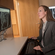
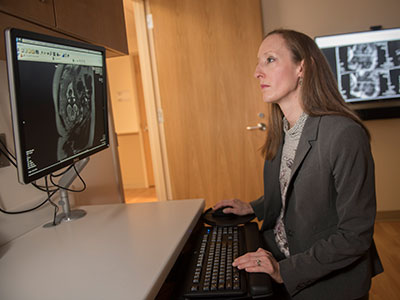
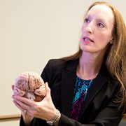
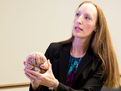
Researchers in the Center for Prenatal, Neonatal & Maternal Health Research are focused on the role of perinatal factors — including maternal stress, anxiety and depression — on the developing brain of the child. Studies also are revealing the impact of congenital anomalies such as heart disease and acquired conditions such as maternal infection with COVID-19 or Zika virus. New approaches to prenatal and postnatal care promise to optimize long-term outcomes of many hospitalized babies.

“Children in the Washington, D.C., area and across the world benefit greatly from the breakthroughs that have emerged from the incredible decades-long partnership between the UAE and Children’s National,” said Michelle Riley-Brown, President and CEO of Children’s National. “I am deeply grateful for the UAE’s most recent gift. The contribution will positively impact children and families and support the teams of researchers and specialists who dedicate their lives to developing innovative medical care.”

Key milestones

The UAE helped to establish the Sheikh Zayed Institute for Pediatric Surgical Innovation at Children’s National in 2009. Today, the Sheikh Zayed Institute (SZI) has grown into a world-class, self-sustaining research center receiving more than 80% of its funding from grants and outside sources.

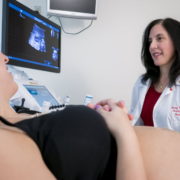
This platform for invention is advancing autonomous, robotic surgery. The institute’s researchers believe pediatric surgical outcomes will improve if the precision and delicacy of a robot are incorporated into procedures such as gallbladder removal. SZI is also propelling the use of artificial intelligence to improve pediatric medicine and expand health equity. One example is a deep learning algorithm that uses hand-held ultrasounds to detect early signs of rheumatic heart disease, which kills nearly 400,000 people worldwide each year.

“The lives and health of countless children and families in the Washington area, in the UAE and around the world have been transformed by our partnership,” said Yousef Al Otaiba, the UAE Ambassador to the United States. “Our continued support promises even more breakthrough innovations in pediatric medicine.”

The UAE also supported the opening of the Children’s National Research & Innovation Campus through a 2019 commitment. The campus represents the first pediatric innovation hub of its kind, where scientists, inventors, caregivers, patients’ families and health authorities come together to advance pediatric health.

The Children’s National Rare Disease Institute and Center for Genetic Medicine Research are two of the teams housed at the campus. Together, they are pioneering care for children in the Washington region and abroad as an international referral site for rare disorders. Two examples of their research endeavors include: next-generation genomic testing to better understand how differences in genetic material can affect human health and identifying biochemical analytes.

The UAE opened a medical office in Washington, D.C., in 1991. Since then, thousands of Emirati patients have visited Children’s National for life-changing care for conditions such as congenital heart disease, neurological disorders and cancer. The hospital is currently treating 40 Emirati patients.

“Having our child treated at Children’s National means accessing specialized pediatric care from a renowned institution dedicated to children’s health,” said Hamad Alnuaimi, an Emirati father of a Children’s National patient. “It provides us with confidence and reassurance that our son is receiving the best possible medical attention from experts who understand and prioritize the unique needs of children. For the UAE to have a strong relationship with Children’s National signifies a valuable connection that enhances pediatric healthcare in our country. This partnership allows us to benefit from advanced treatments, medical innovations, and expertise that might otherwise be inaccessible. It represents a commitment to improving the health and well-being of children through international collaboration.”

School entry neurodevelopmental outcomes of Zika-exposed Colombian children

Members of the Columbia Zika virus research team

The Children’s National Hospital Zika Research Team and collaborators from Biomelab, in Barranquilla, Colombia take a picture after a study visit in Sabanalarga, Colombia following the neurodevelopmental outcomes of children who had in utero exposure to Zika virus. Pictured from Children’s National Hospital: Dr. Sarah Mulkey, Regan Andringa-Seed, Margarita Arroyave-Wessel, and Dr. Madison Berl.

The long-term neurodevelopmental effects of antenatal Zika virus (ZIKV) exposure in children without congenital Zika syndrome (CZS) remain unclear, as few children have been followed to the age of starting primary school.

In a new study published in Pathogens, researchers found children with in utero ZIKV exposure appear to have an overall positive developmental trajectory at 4 to 5 years of age but may experience risks to neurodevelopment in areas of emotional regulation and adaptive mobility.

The hold up in the field

Children who were born during the ZIKV epidemic and who had in utero exposure to ZIKV are only now at the age to start school. Child neurodevelopmental outcome data has not been reported at the age of school entry for children with antenatal ZIKV exposure who do not have the severe birth defects of CZS.

“As these children approach the early school-age years, we aim to examine whether there are neurodevelopmental differences in executive function, motor ability, language development or scholastic skills as compared to a group of unexposed control participants from the same communities in Colombia,” says Sarah Mulkey, M.D., Ph.D., prenatal-neonatal neurologist in The Zickler Family Prenatal Pediatrics Institute at Children’s National Hospital and lead author of the study.

Moving the field forward

Building on previous findings, this study presents the longitudinal outcomes of a well-characterized Colombian cohort of ZIKV-exposed children without CZS at ages 4 to 5 years. These children have been seen for neurodevelopmental follow-up as infants and toddlers at approximately 6 months, 18 months and 3 years of age as part of an international collaboration between researchers in Barranquilla, Colombia and at Children’s National beginning in 2016. The objective of this study was to assess the multi-domain neurodevelopmental outcomes in 4 to 5-year-old children with antenatal ZIKV exposure without CZS compared to unexposed controls in Colombia.

Why we’re excited

Many of the children who had antenatal ZIKV exposure are making good progress in multiple areas of their neurodevelopment. However, the researchers found that children with antenatal ZIKV exposure have differences in areas of emotional regulation, executive function, mood and behavior which may relate to virus exposure during their early brain development.

“These areas of brain function are important for future academic achievement, employment, mental health and social relationships,” says Dr. Mulkey. “So, it will be important to continue to follow these children at older ages when they start school.”

Children’s National leads the way

Children’s National is a leader in conducting outcome studies of children born following antenatal ZIKV exposure. This study follows children in Colombia who are now 5 years old who were first studied while they were in the womb. These children have contributed unique longitudinal understanding to early child neurodevelopment following in utero exposure to ZIKV.

Dr. Mulkey is committed to studying the long-term neurodevelopmental impacts that viruses like Zika and SARS-CoV-2 have on infants born to mothers who were infected during pregnancy through research with the Congenital Infection Program at Children’s National and in collaboration with colleagues in Colombia.

Additional Children’s National authors include Meagan Williams M.S.P.H., C.C.R.C., senior research coordinator; Regan Andringa-Seed, clinical research coordinator, Margarita Arroyave-Wessel, clinical research coordinator; L. Gilbert Vezina, M.D., director, Neuroradiology Program; Dorothy Bulas, M.D., chief, Diagnostic Imaging and Radiology; Robert Podolsky, biostatistician.

Researchers showing paintings of zika virus

Dr. Sarah Mulkey and Children’s National clinical research coordinators in the Prenatal Pediatrics Institute and the Division of Pediatric Infectious Diseases display their paintings of the Zika virus. Pictured from left to right: Manuela Iglesias, Elizabeth Corn, Dr. Sarah Mulkey, Emily Ansusinha and Meagan Williams.

Exposure to Zika in utero may produce neurodevelopmental differences

Sarah Mulkey

“There are still many unanswered questions about the long-term impacts of Zika on children exposed in utero,” says Sarah Mulkey, M.D., Ph.D., a prenatal-neonatal neurologist in the Prenatal Pediatrics Institute at Children’s National Hospital.

Children who are exposed to the Zika virus while in the womb, but who are not subsequently diagnosed with Zika-related birth defects and congenital Zika syndrome (CZS), may still display differences in some aspects of cognitive development, mood and mobility compared to unexposed children, reports a study published in Pediatric Research. These findings suggest that Zika-exposed children may need some additional support and monitoring as they get older.

“There are still many unanswered questions about the long-term impacts of Zika on children exposed in utero,” says Sarah Mulkey, M.D., Ph.D., a prenatal-neonatal neurologist in the Prenatal Pediatrics Institute at Children’s National Hospital and the study’s first author. “These findings are another piece of the puzzle that provides insight into the long-term neurodevelopment of children with prenatal Zika virus exposure. Further evaluation is needed as these children get older.”

It has not been clear how children who were exposed to the Zika virus in the womb during the 2015–2017 epidemic, but who did not develop CZS and serious neurological complications, will develop as they get older.

Dr. Mulkey and colleagues examined the neurodevelopment of 55 children aged 3-5 years who were exposed to Zika in the womb in Sabanalarga, Colombia, and compared them to 70 control children aged 4-5 years who had not been exposed to Zika. Assessments occurred between December 2020 and February 2021. Health professionals tested the children’s motor skills (such as manual dexterity, aiming and catching, and balance) and their readiness for school (including knowledge of colors, letters, numbers and shapes). Parents completed three questionnaires providing information about their child’s cognitive function (such as memory and emotional control), behavioral and physical conditions (such as responsibility and mobility), and their parenting experience (including whether they felt distress).

Parents of Zika-exposed children reported significantly lower levels of mobility and responsibility compared to control children, although differences in cognitive function scores were not significant. Additionally, parents of 6 (11%) Zika-exposed children reported mood problems compared to 1 (1%) of control children, and Zika-exposed parents were significantly more likely to report parental distress.

Professional testing revealed no significant differences in the Zika-exposed children’s manual dexterity, such as their ability to catch an object or post a coin through a slot, compared to the control children. Both Zika-exposed and control children also scored lowly on readiness for school.

The authors highlight that parental responses may have been influenced by the Zika-exposed children’s parents’ perceptions or increased worry about the development of their child. Some differences in results may also have been caused by the age – and therefore developmental – differences between the groups of children.

The authors conclude that while these Zika-exposed children are making progress as they develop, they may need additional support as they prepare to start school.

Dr. Mulkey is committed to studying the long-term neurodevelopmental impacts that viruses like Zika and SARS-CoV-2 have on infants born to mothers who were infected during pregnancy through research with the Congenital Infection Program at Children’s National and in collaboration with colleagues in Colombia.

MRI and ultrasound imaging detect the spectrum of Zika’s impact

Sarah Mulkey

“A combination of prenatal MRI and US was able to detect Zika-related brain abnormalities during pregnancy, giving families timely information to prepare for the potential complex care needs of these infants,” says Sarah B. Mulkey, M.D., Ph.D.

Worldwide, thousands of babies have been born to mothers who were infected during pregnancy with Zika, a virus associated with neurological deficits, impaired vision and neurodevelopmental disabilities, among other birth defects. These birth defects are sometimes severe, causing lifelong disability. But they’re also relatively rare compared with the overall rates of infection.

Predicting how many Zika-exposed babies would experience neurological birth defects has been challenging.

However, an international study led by Children’s faculty suggests that ultrasound (US) imaging performed during pregnancy and after childbirth revealed most Zika-related brain abnormalities experienced by infants exposed to the Zika virus during pregnancy, according to a prospective cohort study published online Nov. 26, 2018, in JAMA Pediatrics. Some Zika-exposed infants whose imaging had been normal during pregnancy had mild brain abnormalities detected by US and magnetic resonance imaging (MRI) after they were born.

“A combination of prenatal MRI and US was able to detect Zika-related brain abnormalities during pregnancy, giving families timely information to prepare for the potential complex care needs of these infants,” says Sarah B. Mulkey, M.D., Ph.D., a fetal-neonatal neurologist at Children’s National Health System and the study’s lead author. “In our study, we detected mild brain abnormalities on postnatal neuroimaging for babies whose imaging was normal during pregnancy. Therefore, it is important for clinicians to continue to monitor brain development for Zika-exposed infants after birth.”

As of Nov. 20 2018, nearly 2,500 pregnant women in the U.S. had laboratory confirmed Zika infection, and about 2,400 of them had given birth, according to the Centers for Disease Control and Prevention (CDC). While more than 100 U.S. infants were born with Zika-associated birth defects, the vast majority of Zika-exposed U.S. infants were apparently normal at birth. The sequential neuroimaging study Dr. Mulkey leads seeks to determine the spectrum of brain findings in infants exposed to Zika in the womb using both US and MRI before and after birth.

The international research team enrolled 82 women in the study from June 15, 2016, through June 27, 2017. All of the women had been exposed to Zika during pregnancy; all but one experienced clinical symptoms by a mean gestational age of 8.2 weeks. Eighty of those women lived in or near Barranquilla, Colombia, and were exposed to Zika there. Two U.S. study participants were exposed to the primarily mosquito-borne illness during travel to Zika hot zones.

All women received fetal MRIs and US during the second and/or third trimester of pregnancy. After their infants were born, the children received brain MRI and cranial US. Blood samples from both mothers and babies were tested for Zika using polymerase chain reaction and serology.

Fetal MRI was able to discern Zika-related brain damage as early as 18 weeks gestation and picked up significant fetal brain abnormalities not fully appreciated in US imaging. In one case, the US remained normal while fetal MRI alone detected brain abnormalities. Three fetuses (4 percent) had severe fetal brain abnormalities consistent with Zika infection, including:

  • Two cases of heterotopias and malformations in cortical development, and
  • One case of parietal encephalocele, Chiari II malformation and microcephaly.

Seventy-five infants were born at term. One pregnancy was terminated at 23 weeks gestation due to the gravity of the fetal brain abnormalities. One fetus with normal imaging died during pregnancy. One newborn who was born with significant fetal brain abnormalities died at age 3 days.

Cranial US and brain MRI was performed on the majority of infants whose prenatal imaging had been normal.  Seven of 53 (13 percent) Zika-exposed infants had mild brain abnormalities detected by MRI after birth. In contrast, postnatal cranial US was better at detecting changes of lenticulostriate vasculopathy, cysts within the brain’s choroid plexus (cells that produce cerebrospinal fluid), germinolytic/subependymal cysts and/or calcifications, which were seen in 21 of 57 (37 percent) infants.

“Sequential neuroimaging revealed that the majority of Zika-exposed fetuses had normal brain development. Tragically, in a small number of pregnancies, Zika-related brain abnormalities were quite severe,” Dr. Mulkey adds. “Our data support the CDC’s recommendation that cranial US be performed after Zika-exposed babies are born. In addition, there is clearly a need to follow these babies over time to gauge whether the brain anomalies we see in imaging affects language, motor and social skills.”

Companion editorial: Revealing the effects of Zika

In addition to Dr. Mulkey, study co-authors include Dorothy I. Bulas, M.D.Gilbert Vezina, M.D., Margarita Arroyave-Wessel, MPH,  Stephanie Russo, B.S, Youssef A. Kousa, D.O, Ph.D.Roberta L. DeBiasi, M.D., MS, Senior Author Adré J. du Plessis, M.B.Ch.B., MPH, all of Children’s National; Christopher Swisher, BS, Georgetown University and Caitlin Cristante, BS, Loyola University, both of  whose contributions included research performed at Children’s National; Yamil Fourzali, M.D., Armando Morales, M.D., both of Sabbag Radiologos; Liliana Encinales, M.D., Allied Research Society; Nelly Pacheco, Bacteriologa, Bio-Nep; Robert S. Lanciotti, Ph.D., Arbovirus Diseases Branch, Centers for Disease Control and Prevention; and Carlos Cure, M.D., BIOMELAB.

Research reported in this news release was supported by the IKARIA fund.

MRI finds novel brain defects in Zika-exposed newborns

Sarah Mulkey

“Imaging is constantly helping us make new discoveries with this virus, and in these two cases we found things that had not been previously described,” says Sarah Mulkey, M.D., Ph.D.

Magnetic resonance imaging (MRI) has identified two brain abnormalities never before reported in newborns with prenatal exposure to the Zika virus. Children’s National Health System researchers reported these findings from a study of more than 70 fetuses or newborns with Zika exposure in utero. The study was published in the January 2018 edition of Pediatric Neurology.

The two novel defects – cranial nerve enhancement and cerebral infarction – may join the growing list of neurological findings associated with congenital Zika infection.

“Imaging is constantly helping us make new discoveries with this virus, and in these two cases we found things that had not been previously described,” says Sarah Mulkey, M.D., Ph.D., the study’s lead author and a fetal-neonatal neurologist at Children’s National. Dr. Mulkey works in Children’s Congenital Zika Virus Program, one of the nation’s first comprehensive, dedicated Zika programs.

The research team recommends that postnatal brain MRI be considered in addition to ultrasound for newborns exposed to Zika in utero. “Brain MRI can be performed in the newborn often without sedation and provides an opportunity to look for brain abnormalities we might not catch otherwise – or might not detect until much later,” says Dr. Mulkey.

Birth defects are seen in 6 to 11 percent of pregnancies affected by Zika, and some of the neurological complications in infants are not apparent until well after birth.

Of the two infants in which the new abnormalities were observed, both had normal head size at birth. Neither had smaller-than-normal head size (microcephaly), one of the more severe effects associated with congenital Zika syndrome.

One infant had a normal neurological evaluation at 2 days of age. However, a brain MRI conducted the following day, using gadolinium contrast due to concern of infection, showed enhancement of multiple cranial nerves. “Nerve root enhancement is very rare in a newborn and had not been described with Zika before,” Dr. Mulkey says. “Yet, there was no neurological deficit that we could identify by physical exam.”

The research team acknowledges that the clinical significance of this finding is not yet known.

In the second patient, brain MRI conducted without contrast at 16 days of age revealed a small area consistent with chronic infarction (ischemic stroke) that likely occurred during the third trimester.

“We followed the mother throughout her pregnancy, and both MRI and ultrasound imaging were normal at 28 weeks gestation,” Dr. Mulkey says. “A postnatal ultrasound was also normal, but the postnatal MRI showed a stroke that had occurred at least one month prior to the MRI and after the last fetal study.”

She adds: “This is the first published report of fetal stroke associated with Zika infection, and it may add to our knowledge of what can occur with congenital Zika infection.”

Unlike most congenital infections, Zika virus does not appear to cause viral-induced placental inflammation, which can lead to fetal stroke. So, the authors say they cannot be sure that congenital Zika contributed to the infarct in this case. However, they write, “Given the relatively low incidence of perinatal ischemic infarct and the lack of other maternal- or birth-related risk factors for this patient, Zika infection is considered a possible etiology.”

In both patients, neonatal brain MRI identified subclinical findings that had not previously been described as part of congenital Zika syndrome. As the body of evidence about the Zika virus has grown, the spectrum of associated brain abnormalities has expanded to include considerably more findings than isolated microcephaly.

Data gathered in 2017 from the Centers for Disease Control and Prevention’s Zika pregnancy and infant registry indicates that 25 percent of eligible U.S. infants receive recommended postnatal imaging. Dr. Mulkey said this represents many possible missed opportunities for earlier identification of brain abnormalities.

“Brain MRI should be considered in all newborns exposed to Zika virus in utero, even in the presence of normal birth head circumference, normal cranial ultrasound and normal fetal imaging,” she says. “In both of these patients, the changes we observed were not evident on cranial ultrasound or on fetal MRI and fetal ultrasound.”

In addition to Dr. Mulkey, Children’s co-authors include L. Gilbert Vezina, M.D., Neuroradiology Program director; Dorothy I. Bulas, M.D., chief of Diagnostic Imaging and Radiology; Zarir Khademian, M.D., radiologist; Anna Blask, M.D., radiologist; Youssef A. Kousa, M.S., D.O., Ph.D., child neurology fellow; Lindsay Pesacreta, FNP; Adré  J. du Plessis, M.B.Ch.B., M.P.H., Fetal Medicine Institute director; and Roberta L. DeBiasi, M.D., M.S., senior author and Pediatric Infectious Disease division chief; and Caitlin Cristante, B.S.

Financial support for this research was provided by the Thrasher Research Fund.

Children’s National experts contribute to new Zika guidelines

Roberta DeBiasi and Sarah Mulkey

Roberta DeBiasi, M.D., M. S., and Sarah B. Mulkey, M.D., Ph.D., members of Children’s multidisciplinary Congenital Zika Virus Program, were among the experts invited to participate in a forum held in Atlanta at CDC headquarters in late August to formulate new Zika recommendations.

The Centers for Disease Control and Prevention (CDC) on Oct. 19, 2017 updated guidelines for evaluation of women, fetuses and infants exposed to the Zika virus during pregnancy. Although only women with symptoms will now be routinely tested, asymptomatic and symptomatic infants born to these women will still be tested for the Zika virus using blood and urine tests.

Infants who appear normal, whose mothers either had negative Zika results or who had not undergone testing, will not undergo Zika testing. These infants still will undergo a standard evaluation, including a detailed physical exam, hearing screen and routine developmental assessments. The revised Zika guidance includes input from practitioners on the front lines of the Zika epidemic, including Children’s National Health System clinicians.

“These changes in the recommendations for Zika testing should not be interpreted as Zika infection risks subsiding for pregnant women and their infants in the United States. It’s simply an acknowledgement of the limitations of current testing methods – which must occur within a narrow window after Zika exposure – and the poor predictive value of Zika testing right now,” says Roberta L. DeBiasi, M.D., M.S., chief of Children’s Division of Pediatric Infectious Diseases. Dr. DeBiasi and Sarah B. Mulkey, M.D., Ph.D., members of Children’s multidisciplinary Congenital Zika Virus Program, were among the experts invited to participate in the Zika forum held in Atlanta at CDC headquarters in late August to formulate the recommendations.

While all infants will receive a standard evaluation, expanded evaluations that include an ophthalmologic assessment, more detailed hearing evaluation and ultrasound of the newborn’s head will be reserved for infants born to mothers confirmed to be Zika positive or Zika probable, or for infants born with abnormalities potentially consistent with congenital Zika syndrome, regardless of maternal status.

The majority of U.S. infants who have been exposed to Zika in the womb appeared normal at birth, according to CDC registries. Now, the next wave of these normal-appearing babies will receive standard evaluations when they are born, including a newborn hearing screening. At each well-child visit, these infants will receive:

  • A comprehensive physical examination
  • An age-appropriate vision screening
  • Developmental monitoring and screening using validated tools

“This is a natural evolution in the diagnosis and screening strategy now that the peak of the first wave of Zika transmission appears to be over,” Dr. DeBiasi says. “While we continue to evaluate new possible cases of Zika infection among pregnant women in our practice, a sizable proportion of Children’s cases are Zika-exposed infants whose physical exam and neuroimaging appeared normal at birth. Through ongoing monitoring, we hope to learn more about these children’s long-term neurodevelopment outcomes.”

Will the Zika epidemic re-emerge in 2017?

Anthony Fauci

Anthony S. Fauci, M.D., director of the National Institute of Allergy and Infectious Diseases at the National Institutes of Health, discussed the possibility of a reemergence of Zika virus at Children’s National Research and Education Week.

Temperatures are rising, swelling the population of Aedes mosquitoes that transmit the Zika virus and prompting an anxious question: Will the Zika epidemic re-emerge in 2017?

Anthony S. Fauci, M.D., director of the National Institute of Allergy and Infectious Diseases at the National Institutes of Health (NIH), sketched out contrasting scenarios. Last year in Puerto Rico, at least 13 percent of residents were infected with Zika, “a huge percentage of the population to get infected in any one outbreak,” Dr. Fauci says. But he quickly adds: “That means that 87 percent of the population” did not get infected. When the chikungunya virus swept through the Caribbean during an earlier outbreak, it did so in multiple waves. “We are bracing for a return of Zika, but we shall see what happens.” Dr. Fauci says.

When it comes to the continental United States, however, previous dengue and chikungunya outbreaks were limited to southern Florida and Texas towns straddling the Mexican border. Domestic Zika transmission last year behaved in much the same fashion.

“Do we think we’re going to get an outbreak [of Zika] that is disseminated throughout the country? The answer is no,” Dr. Fauci adds. “We’re not going to see a major Puerto Rico-type outbreak in the continental United States.”

Dr. Fauci’s remarks were delivered April 24 to a standing-room-only auditorium as part of Research and Education Week, an annual celebration of the cutting-edge research and innovation happening every day at Children’s National. He offered a sweeping, fact-filled summary of Zika’s march across the globe: The virus was first isolated from a primate placed in a treehouse within Uganda’s Zika forest to intentionally become infected; Zika lurked under the radar for the first few decades, causing non-descript febrile illness; it bounced from country to country, causing isolated outbreaks; then, it transformed into an infectious disease of international concern when congenital Zika infection was linked to severe neural consequences for babies born in Brazil.

zika virus

Zika virus lurked under the radar for several decades, causing non-descript febrile illness; it bounced from country to country, resulting in isolated outbreaks; then, it transformed into an infectious disease of international concern.

“I refer to Brazil and Zika as the perfect storm,” Dr. Fauci told attendees. “You have a country that is a large country with a lot of people, some pockets of poverty and economic depression –  such as in the northeastern states –  without good health care there, plenty of Aedes aegypti mosquitoes and, importantly, a totally immunologically naive population. They had never seen Zika before. The right mosquitoes. The right climate. The right people. The right immunological status. And then, you have the explosion in Brazil.”

In Brazil, 139 to 175 babies were born each year with microcephaly – a condition characterized by a smaller than normal skull – from 2010 to 2014. From 2015 through 2016, that sobering statistic soared to 5,549 microcephaly cases, 2,366 of them lab-confirmed as caused by Zika.

Microcephaly “was the showstopper that changed everything,” says Dr. Fauci. “All of a sudden, [Zika] went from a relatively trivial disease to a disease that had dire consequences if a mother was infected, particularly during the first trimester.”

As Zika infections soared, ultimately affecting more than 60 countries, the virus surprised researchers and clinicians a number of times, by:

  • Being spread via sex
  • Being transmitted via blood transfusion, a finding from Brazil that prompted the Food and Drug Administration to recommend testing for all U.S. donated blood and blood products
  • Decimating developing babies’ neural stem cells and causing a constellation of congenital abnormalities, including vision problems and contractions to surviving infants’ arms and legs
  • Causing Guillain-Barré syndrome
  • Triggering transient hearing loss
  • Causing myocarditis, heart failure and arrhythmias

When it comes to the U.S. national response, Dr. Fauci says one of the most crucial variables is how quickly a vaccine becomes available to respond to the emerging outbreak. For Zika, the research community was able to sequence the virus and launch a Phase I trial in about three months, “the quickest time frame from identification to trial in the history of all vaccinology,” he adds.

Zika is a single-stranded, enveloped RNA virus that is closely related to dengue, West Nile, Japanese encephalitis and Yellow fever viruses, which gives the NIH and others racing to produce a Zika vaccine a leg up. The Yellow fever vaccine, at 99 percent effectiveness, is one of the world’s most effective vaccines.

“I think we will wind up with an effective vaccine. I don’t want to be over confident,” Dr. Fauci  says. “The reason I say I believe that we will is because [Zika is] a flavivirus, and we have been able to develop effective flavivirus vaccines. Remember, Yellow fever is not too different from Zika.”

Damage may lurk in “normal” Zika-exposed brains

Sarah Mulkey Columbia Zika Study

An international study that includes Sarah B. Mulkey, M.D., Ph.D., aims to answer one of the most vexing questions about Zika: If babies’ brains appear “normal” at birth, have they survived Zika exposure in the womb with few neurological repercussions? Dr. Mulkey presented preliminary findings at PAS2017.

It has been well established by researchers, including scientists at Children’s National Health System, that the Zika virus is responsible for a slew of birth defects – such as microcephaly, other brain malformations and retinal damage – in babies of infected mothers. But how the virus causes these often devastating effects, and who exactly is affected, has not been explained fully.

Also unknown is whether exposed babies that appear normal at birth are truly unaffected by the virus or have hidden problems that might surface later. The majority of babies born to Zika-infected mothers in the United States appear to have no evidence of Zika-caused birth defects, but that’s no guarantee that the virus has not caused lingering damage.

Recently, Sarah B. Mulkey, M.D., Ph.D., made a trip to Colombia, where Children’s National researchers are collaborating on a clinical study. There, she tested Zika-affected babies’ motor skills as they sat, stood and lay facing upward and downward. The international study aims to answer one of the most vexing questions about Zika: If babies’ brains appear “normal” at birth, have they survived Zika exposure in the womb with few neurological repercussions?

“We don’t know the long-term neurological consequences of having Zika if your brain looks normal,” says Dr. Mulkey, a fetal-neonatal neurologist who is a member of Children’s Congenital Zika Virus Program. “That is what’s so scary, the uncertainty about long-term outcomes.”

According to the Centers for Disease Control and Prevention (CDC), one in 10 pregnancies across the United States with laboratory-confirmed Zika virus infection results in birth defects in the fetus or infant. For the lion’s share of Zika-affected pregnancies, then, babies’ long-term prospects remain a mystery.

“This is a huge number of children to be impacted and the impact, as we understand, has the potential to be pretty significant,” Dr. Mulkey adds.

Dr. Mulkey, the lead author, presented the research group’s preliminary findings during the 2017 annual meeting of the Pediatric Academic Societies (PAS). The presentation was one of several that focused on the Zika virus. Roberta L. DeBiasi, M.D., M.S., chief of the Division of Pediatric Infectious Diseases at Children’s National, organized two invited symposia devoted to the topic of Zika: Clinical perspectives and knowledge gaps; and the science of Zika, including experimental models of disease and vaccines. Dr. DeBiasi’s presentation included an overview of the 68 Zika-exposed or infected women and infants seen thus far by Children’s multidisciplinary Congenital Zika Virus Program.

“As the world’s largest pediatric research meeting, PAS2017 is an ideal setting for panelists to provide comprehensive epidemiologic and clinical updates about the emergence of Congenital Zika Syndrome and to review the pathogenesis of infection as it relates to the fetal brain,” Dr. DeBiasi says. “With temperatures already rising to levels that support spread of the Aedes mosquito, it is imperative for pediatricians around the world to share the latest research findings to identify the most effective interventions.”

As one example, Dr. Mulkey’s research sought to evaluate the utility of using magnetic resonance imaging (MRI) to evaluate fetal brain abnormalities in 48 babies whose mothers had confirmed Zika infection during pregnancy. Forty-six of the women/infant pairs enrolled in the prospective study are Colombian, and two are Washington, D.C. women who were exposed during travel to a Zika hot zone.

The women were infected with Zika during all three trimesters and experienced symptoms at a mean gestational age of 8.4 weeks. The first fetal MRIs were performed as early as 18 weeks’ gestation. Depending upon the gestational age when they were enrolled in the study, the participants had at least one fetal MRI as well as serial ultrasounds. Thirty-six fetuses had a second fetal MRI at about 31.1 gestational weeks. An experienced pediatric neuroradiologist evaluated the images.

Among the 48 study participants, 45 had “normal” fetal MRIs.

Three fetuses exposed to Zika in the first or second trimester had abnormal fetal MRIs:

  • One had heterotopia and an early, abnormal fold on the surface of the brain, indications that neurons did not migrate to their anticipated destination during brain development. This pregnancy was terminated at 23.9 gestational weeks.
  • One had parietal encephalocele, a rare birth defect that results in a sac-like protrusion of the brain through an opening in the skull. According to the CDC, this defect affects one in 12,200 births, or 340 babies, per year. It is not known if this rare finding is related to Zika infection.
  • One had a thin corpus callosum, dysplastic brainstem, heterotopias, significant ventriculomegaly and generalized cerebral/cerebellar atrophy.

“Fetal brain MRI detected early structural brain changes in fetuses exposed to the Zika virus in the first and second trimester,” Dr. Mulkey says. “The vast majority of fetuses exposed to Zika in our study had normal fetal MRI, however. Our ongoing study, underwritten by the Thrasher Research Fund, will evaluate their long-term neurodevelopment.”

Adré J. du Plessis, MB.Ch.B., M.P.H., director of the Fetal Medicine Institute and senior author of the paper, notes that this group “is a very important cohort to follow as long as Dr. Mulkey’s funding permits. We know that microcephaly is among the more devastating side effects caused by Zika exposure in utero. Unanswered questions remain about Zika’s impact on hearing, vision and cognition for a larger group of infants. Definitive answers only will come with long-term follow-up.”

Many of the Colombian families live in Sabanalarga, a relatively rural, impoverished area with frequent rain, leaving pockets of fresh water puddles that the mosquito that spreads Zika prefers, Dr. Mulkey adds. Families rode buses for hours for access to fetal MRI technology, which is not common in Colombia.

“The mothers are worried about their babies. They want to know if their babies are doing OK,” she says.

Zika virus, one year later

Drs. DeBiasi and du Plessis

A multidisciplinary team at Children’s National has consulted on 66 Zika-affected pregnancies and births since May 2016.

The first pregnant patient with worries about a possible Zika virus infection arrived at the Children’s National Health System Fetal Medicine Institute on Jan. 26, 2016, shortly after returning from international travel.

Sixteen months ago, the world was just beginning to learn how devastating the mosquito-borne illness could be to fetuses developing in utero. As the epidemic spread, a growing number of sun-splashed regions that harbor mosquitoes that efficiently spread the virus experienced a ballooning number of Zika-affected pregnancies and began to record sobering birth defects.

The Washington, D.C. patient’s concerns were well-founded. Exposure to Zika virus early in her pregnancy led to significant fetal brain abnormalities, and Zika virus lingered in the woman’s bloodstream months after the initial exposure — longer than the Centers for Disease Control and Prevention (CDC) then thought was possible.

The research paper describing the woman’s lengthy Zika infection, published by The New England Journal of Medicine, was selected as one of the most impactful research papers written by Children’s National authors in 2016.

In the intervening months, a multidisciplinary team at Children National has consulted on 66 pregnancies and infants with confirmed or suspected Zika exposure. Thirty-five of the Zika-related evaluations were prenatal, and 31 postnatal evaluations assessed the impact of in utero Zika exposure after the babies were born.

The continuum of Zika-related injuries includes tragedies, such as a 28-year-old pregnant woman who was referred to Children’s National after imaging hinted at microcephaly. Follow-up with sharper magnetic resonance imaging (MRI) identified severe diffuse thinning of the cerebral cortical mantle, evidence of parenchymal cysts in the white matter and multiple contractures of upper and lower extremities with muscular atrophy.

According to a registry of Zika-affected pregnancies maintained by the CDC, one in 10 pregnancies across the United States with laboratory-confirmed Zika virus infection has resulted in birth defects in the fetus or infant.

“More surprising than that percentage is the fact that just 25 percent of infants underwent neuroimaging after birth – despite the CDC’s recommendation that all Zika-exposed infants undergo postnatal imaging,” says Roberta L. DeBiasi, M.D., M.S., chief of the Division of Pediatric Infectious Diseases and co-director of the Congenital Zika Virus Program at Children’s National. “Clinicians should follow the CDC’s guidance to the letter, asking women about possible exposure to Zika and providing multidisciplinary care to babies after birth. Imaging is an essential tool to accurately monitor the growing baby’s brain development.”

Adré du Plessis, M.B.Ch.B., M.P.H., director of the Fetal Medicine Institute and Congenital Zika Virus Program co-leader, explains the challenges: ”When it comes to understanding the long-term consequences for fetuses exposed to the Zika virus, we are still on the steepest part of the learning curve. Identifying those children at risk for adverse outcomes will require a sustained and concerted multidisciplinary effort from conception well beyond childhood.”

In addition to counseling families in the greater Washington, D.C. region, the Children’s research team is collaborating with international colleagues to conduct a clinical trial that has been recruiting Zika-infected women and their babies in Colombia. Pediatric Resident Youssef A. Kousa, D.O., Ph.D., M.S., and Neurologist Sarah B. Mulkey, M.D., Ph.D., will present preliminary findings during Research and Education Week 2017.

In Colombia as well as the District of Columbia, a growing challenge continues to be assessing Zika’s more subtle effects on pregnancies, developing fetuses and infants, says Radiologist Dorothy Bulas, M.D., another member of Children’s multidisciplinary Congenital Zika Virus Program.

The most severe cases from Brazil were characterized by interrupted fetal brain development, smaller-than-normal infant head circumference, brain calcifications, enlarged ventricles, seizures and limbs folded at odd angles. In the United States and many other Zika-affected regions, Zika-affected cases with such severe birth defects are outnumbered by infants who were exposed to Zika in utero but have imaging that appears normal.

In a darkened room, Dr. Bulas pores over magnified images of the brains of Zika-infected babies, looking for subtle differences in structure that may portend future problems.

“There are some questions we have answered in the past year, but a number of questions remain unanswered,” Dr. Bulas says. “For neonates, that whole area needs assessment. As the fetal brain is developing, the Zika virus seems to affect the progenitor cells. They’re getting hit quite early on. While we may not detect brain damage during the prenatal period, it may appear in postnatal images. And mild side effects that may not be as obvious early on still have the potential to be devastating.”

Fetal medicine update: fetal brain development, zika virus

May 2, 2016 -Impaired global and tissue-specific brain development in the growth-restricted fetus.
A team of researchers applied an advanced imaging technique, three-dimensional MRI, to study brain development in high-risk pregnancies and are the first to report regional, tissue-specific volume delays for the developing fetal brain in FGR-affected pregnancies. The team compared overall fetal brain volume as well as regional brain volumes for a control group of healthy young pregnant women with a group of young women whose pregnancies were complicated by FGR. While fetuses in both groups grew exponentially as pregnancies progressed, the researchers began to see dramatic differences when they compared the volumes of specific regions of the brain, including the cerebellum, which coordinates balance and smooth movement; the deep gray matter, which also is involved in complex functions, such as memory and emotion; and the white matter, which is made up of millions of nerve fibers that connect to neurons in different regions.

March 30, 2016Congenital Zika viral infection linked to significant fetal brain abnormalities, despite ‘normal’ ultrasounds.
Infectious Zika virus was isolated from the brain of a 21-week-old fetus after causing extensive damage to brain tissue – despite ultrasounds that showed no sign of microcephaly at weeks 13, 16, and 17, according to a report published online in The New England Journal of Medicine. “While this is a single case, it poses troubling questions that could inform future research,” says the study’s co-senior author, Adre du Plessis, M.B.Ch.B., Director of the Fetal Medicine Institute and Chief of the Fetal and Transitional Medicine Division at Children’s National Health System. “Evidence is mounting that the Zika virus can persist in pregnant women’s bloodstreams weeks after their initial infection, arguing for changes to how these pregnancies are monitored,” Dr. du Plessis said. Six of the named authors are affiliated with Children’s National, where the pregnant woman sought more thorough assessment after testing positive for the Zika virus herself following international travel.