Tag Archive for: Zika-affected pregnancies

Neonatal baby

Multidisciplinary experts help CDC’s Zika research

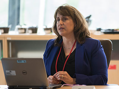
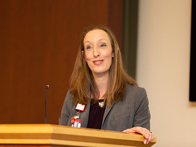
“We are very excited about this next phase in our Zika research,” says Roberta L. DeBiasi, M.D., M.S. “It is a natural extension of our earlier participation as subject matter experts assisting as the CDC developed and published guidelines to inform the care of Zika-exposed and Zika-infected infants across the nation and U.S. territories.”

The Centers for Disease Control and Prevention (CDC) is funding three multidisciplinary experts from the Congenital Zika Virus Program at Children’s National Health System to collaborate on two of the CDC’s longitudinal Zika research projects in Colombia, South America.

“Zika en embarazadas y niños en Colombia” (ZEN) is a research study jointly designed by Colombia’s Instituto Nacional de Salud (INS) and the CDC to evaluate the association between Zika virus infection and adverse maternal, fetal and infant health outcomes. The study is following a large cohort of Colombian women from the first trimester of pregnancy, their male partners and their infants.

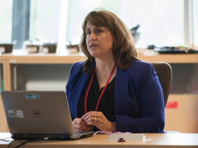
Under the six-month contract, Roberta L. DeBiasi, M.D., M.S., Sarah B. Mulkey, M.D., Ph.D., and Cara Biddle, M.D., M.P.H., will serve as consultants for the ZEN study providing expertise in pediatric infectious diseases, neurology, neurodevelopment and coordination of the complex care needs of Zika-affected infants.

The federal funding will underwrite the consultants’ work effort, as well as travel to the CDC’s headquarters in Atlanta and to research sites in Colombia. To that end, Drs. DeBiasi, Mulkey and Biddle participated in a December 2017 kickoff meeting, joining ZEN team leaders based in the U.S. at the CDC, as well as the INS in Colombia, with whom they will conduct research and collaborate academically.

Cara-Biddle-and-Sarah-Mulkey

Cara Biddle, M.D., M.P.H., and Sarah B. Mulkey, M.D., Ph.D., also will serve as consultants for the ZEN study.

“We are very excited about this next phase in our Zika research,” says Dr. DeBiasi, chief of the Division of Pediatric Infectious Diseases and co-director of the Children’s Zika program. “It is a natural extension of our earlier participation as subject matter experts assisting as the CDC developed and published guidelines to inform the care of Zika-exposed and Zika-infected infants across the nation and U.S. territories.”

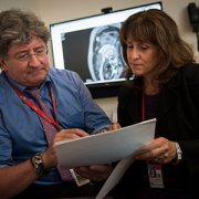
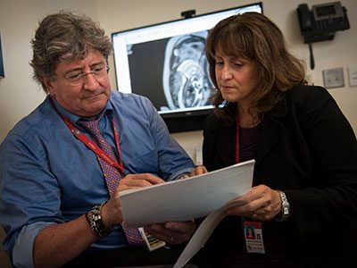
Children’s National is leading its own longitudinal studies in Colombia that explore such questions as whether Zika-exposed infants whose neuroimaging appears normal when they are born experience any longer-term neurological issues and the role of genetics in neurologic injury following congenital Zika virus exposure and infection.

Sarah Mulkey

Fetal MRI plus ultrasound assess Zika-related brain changes

Sarah Mulkey

Magnetic resonance imaging and ultrasound provide complementary data needed to assess ongoing changes to the brains of fetuses exposed to Zika in utero, says Sarah B. Mulkey, M.D., Ph.D.

For Zika-affected pregnancies, fetal magnetic resonance imaging (MRI) used in addition to standard ultrasound (US) imaging can better assess potential brain abnormalities in utero, according to research presented by Children’s National Health System during IDWeek 2017. In cases of abnormal brain structure, fetal MRI can reveal more extensive areas of damage to the developing brain than is seen with US.

“MRI and US provide complementary data needed to assess ongoing changes to the brains of fetuses exposed to Zika in utero,” says Sarah B. Mulkey, M.D., Ph.D., a fetal/neonatal neurologist at Children’s National Health System and lead author of the research paper. “In addition, our study found that relying on ultrasound alone would have given one mother the false assurance that her fetus’ brain was developing normally while the sharper MRI clearly pointed to brain abnormalities.”

As of Sept. 13, the Centers for Disease Control and Prevention (CDC) reported that 1,901 U.S. women were exposed to Zika at some point during their pregnancies but their infants appeared normal at birth. Another 98 U.S. women, however, gave birth to infants with Zika-related birth defects.  And eight more women had pregnancy losses with Zika-related birth defects, according to CDC registries.

The longitudinal neuroimaging study led by Children’s National enrolled 48 pregnant women exposed to the Zika virus in the first or second trimester whose infection was confirmed by reverse transcription polymerase chain reaction, which detects Zika viral fragments shortly after exposure, and/or Immunoglobulin M testing, which reveals antibodies the body produces to neutralize the virus. Forty-six of the study volunteers live in Barranquilla, Colombia, where Zika infection is endemic. Two women live in the Washington region and were exposed to Zika during travel elsewhere.

All of the women underwent at least one diagnostic imaging session while pregnant, receiving an initial MRI or US at 25.1 weeks’ gestational age. Thirty-six women underwent a second MRI/US imaging pair at roughly 31 weeks’ gestation. Children’s National radiologists read every image.

Three of 48 pregnancies, or 6 percent, were marked by abnormal fetal MRIs:

  • One fetus had heterotopias (clumps of grey matter located at the wrong place) and abnormal cortical indent (a deformation at the outer layer of the cerebrum, a brain region involved in consciousness). The US taken at the same gestational age for this fetus showed its brain was developing normally.
  • Another fetus had parietal encephalocele (an uncommon skull defect) and Chiari malformation Type II (a life-threatening structural defect at the base of the skull and the cerebellum, the part of the brain that controls balance). The US for this fetus also detected these brain abnormalities.
  • The third fetus had a thin corpus callosum (bundle of nerves that connects the brain’s left and right hemispheres), an abnormally developed brain stem, temporal cysts, subependymal heterotopias and general cerebral/cerebellar atrophy. This fetal US showed significant ventriculomegaly (fluid-filled structures in the brain that are too large) and a fetal head circumference that decreased sharply from the 32nd to 36th gestational week, a hallmark of microcephaly.

After they were born, infants underwent a follow-up MRI without sedation and US. For nine infants, these ultrasounds revealed cysts in the choroid plexus (cells that produce cerebrospinal fluid) or germinal matrix (the source for neurons and glial cells that migrate during brain development). And one infant’s US after birth showed lenticulostriate vasculopathy (brain lesions).

“Because a number of factors can trigger brain abnormalities, further studies are needed to determine whether the cystic changes to these infants’ brains are attributable to Zika exposure in the womb or whether some other insult caused these troubling results,” Dr. Mulkey says.

What Children’s has learned about congenital Zika infection

Roberta DeBiasi

Roberta DeBiasi, M.D., M.S., outlined lessons learned during a pediatric virology workshop at IDWeek2017, one of three such Zika presentations led by Children’s National research-clinicians during this year’s meeting of pediatric infectious disease specialists.

The Congenital Zika Virus Program at Children’s National Health System provides a range of advanced testing and services for exposed and infected fetuses and newborns. Data that the program has gathered in evaluating and managing Zika-affected pregnancies and births may offer instructive insights to other centers developing similar programs.

The program evaluated 36 pregnant women and their fetuses from January 2016 through May 2017. Another 14 women and their infants were referred to the Zika program for postnatal consultations during that time.

“As the days grow shorter and temperatures drop, we continue to receive referrals to our Zika program, and this is a testament to the critical need it fulfills in the greater metropolitan D.C. region,” says Roberta L. DeBiasi, M.D., M.S., chief of the Division of Pediatric Infectious Diseases and co-leader of the program. “Our multidisciplinary team now has consulted on 90 dyads (mothers and their Zika-affected fetuses/infants). The lessons we learned about when and how these women were infected and how their offspring were affected by Zika may be instructive to institutions considering launching their own programs.”

Dr. DeBiasi outlined lessons learned during a pediatric virology workshop at IDWeek2017, one of three such Zika presentations led by Children’s National research-clinicians during this year’s meeting of pediatric infectious disease specialists.

“The Zika virus continues to circulate in dozens of nations, from Angola to the U.S. Virgin Islands. Clinicians considering a strategic approach to managing pregnancies complicated by Zika may consider enlisting an array of specialists to attend to infants’ complex care needs, including experts in fetal imaging, pediatric infectious disease, physical therapists, audiologists, ophthalmologists and radiologists skilled at reading serial magnetic resonance images as well as ultrasounds,” Dr. DeBiasi says. “At Children’s we have a devoted Zika hotline to triage patient and family concerns. We provide detailed instructions for referring institutions explaining protocols before and after childbirth, and we provide continuing education for health care professionals.”

Of the 36 pregnant women possibly exposed to Zika during pregnancy seen in the program’s first year, 32 lived in the United States and traveled to countries where Zika virus was circulating. Two women had partners who traveled to Zika hot zones. And two moved to the Washington region from places where Zika is endemic. Including the postnatal cases, 89 percent of patients had been bitten by Zika-tainted mosquitoes, while 48 percent of women could have been exposed to Zika via sex with an infected partner.

Twenty percent of the women were exposed before conception; 46 percent were exposed to Zika in the first trimester of pregnancy; 26 percent were exposed in the second trimester; and 8 percent were exposed in the final trimester. In only six of 50 cases (12 percent) did the Zika-infected individual experience symptoms.

Zika infection can be confirmed by detecting viral fragments but only if the test occurs shortly after infection. Twenty-four of the 50 women (nearly 50 percent) arrived for a Zika consultation outside that 12-week testing window. Eleven women (22 percent) had confirmed Zika infection and another 28 percent tested positive for the broader family of flavivirus infections that includes Zika. Another detection method picks up antibodies that the body produces to neutralize Zika virus. For seven women (14 percent), Zika infection was ruled out by either testing method.

“Tragically, four fetuses had severe Zika-related birth defects,” Dr. DeBiasi says. “Due to the gravity of those abnormalities, two pregnancies were not carried to term. The third pregnancy was carried to term, but the infant died immediately after birth. The fourth pregnancy was carried to term, but that infant survived less than one year.”

Drs. DeBiasi and du Plessis

Zika virus, one year later

Drs. DeBiasi and du Plessis

A multidisciplinary team at Children’s National has consulted on 66 Zika-affected pregnancies and births since May 2016.

The first pregnant patient with worries about a possible Zika virus infection arrived at the Children’s National Health System Fetal Medicine Institute on Jan. 26, 2016, shortly after returning from international travel.

Sixteen months ago, the world was just beginning to learn how devastating the mosquito-borne illness could be to fetuses developing in utero. As the epidemic spread, a growing number of sun-splashed regions that harbor mosquitoes that efficiently spread the virus experienced a ballooning number of Zika-affected pregnancies and began to record sobering birth defects.

The Washington, D.C. patient’s concerns were well-founded. Exposure to Zika virus early in her pregnancy led to significant fetal brain abnormalities, and Zika virus lingered in the woman’s bloodstream months after the initial exposure — longer than the Centers for Disease Control and Prevention (CDC) then thought was possible.

The research paper describing the woman’s lengthy Zika infection, published by The New England Journal of Medicine, was selected as one of the most impactful research papers written by Children’s National authors in 2016.

In the intervening months, a multidisciplinary team at Children National has consulted on 66 pregnancies and infants with confirmed or suspected Zika exposure. Thirty-five of the Zika-related evaluations were prenatal, and 31 postnatal evaluations assessed the impact of in utero Zika exposure after the babies were born.

The continuum of Zika-related injuries includes tragedies, such as a 28-year-old pregnant woman who was referred to Children’s National after imaging hinted at microcephaly. Follow-up with sharper magnetic resonance imaging (MRI) identified severe diffuse thinning of the cerebral cortical mantle, evidence of parenchymal cysts in the white matter and multiple contractures of upper and lower extremities with muscular atrophy.

According to a registry of Zika-affected pregnancies maintained by the CDC, one in 10 pregnancies across the United States with laboratory-confirmed Zika virus infection has resulted in birth defects in the fetus or infant.

“More surprising than that percentage is the fact that just 25 percent of infants underwent neuroimaging after birth – despite the CDC’s recommendation that all Zika-exposed infants undergo postnatal imaging,” says Roberta L. DeBiasi, M.D., M.S., chief of the Division of Pediatric Infectious Diseases and co-director of the Congenital Zika Virus Program at Children’s National. “Clinicians should follow the CDC’s guidance to the letter, asking women about possible exposure to Zika and providing multidisciplinary care to babies after birth. Imaging is an essential tool to accurately monitor the growing baby’s brain development.”

Adré du Plessis, M.B.Ch.B., M.P.H., director of the Fetal Medicine Institute and Congenital Zika Virus Program co-leader, explains the challenges: ”When it comes to understanding the long-term consequences for fetuses exposed to the Zika virus, we are still on the steepest part of the learning curve. Identifying those children at risk for adverse outcomes will require a sustained and concerted multidisciplinary effort from conception well beyond childhood.”

In addition to counseling families in the greater Washington, D.C. region, the Children’s research team is collaborating with international colleagues to conduct a clinical trial that has been recruiting Zika-infected women and their babies in Colombia. Pediatric Resident Youssef A. Kousa, D.O., Ph.D., M.S., and Neurologist Sarah B. Mulkey, M.D., Ph.D., will present preliminary findings during Research and Education Week 2017.

In Colombia as well as the District of Columbia, a growing challenge continues to be assessing Zika’s more subtle effects on pregnancies, developing fetuses and infants, says Radiologist Dorothy Bulas, M.D., another member of Children’s multidisciplinary Congenital Zika Virus Program.

The most severe cases from Brazil were characterized by interrupted fetal brain development, smaller-than-normal infant head circumference, brain calcifications, enlarged ventricles, seizures and limbs folded at odd angles. In the United States and many other Zika-affected regions, Zika-affected cases with such severe birth defects are outnumbered by infants who were exposed to Zika in utero but have imaging that appears normal.

In a darkened room, Dr. Bulas pores over magnified images of the brains of Zika-infected babies, looking for subtle differences in structure that may portend future problems.

“There are some questions we have answered in the past year, but a number of questions remain unanswered,” Dr. Bulas says. “For neonates, that whole area needs assessment. As the fetal brain is developing, the Zika virus seems to affect the progenitor cells. They’re getting hit quite early on. While we may not detect brain damage during the prenatal period, it may appear in postnatal images. And mild side effects that may not be as obvious early on still have the potential to be devastating.”