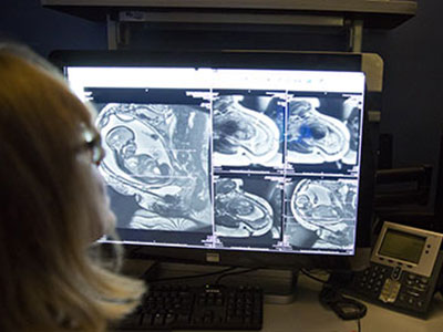
Paving the way toward better understanding and treatment of neonatal brain injuries

The Gallo Lab’s latest research finds reduced expression of Sirt2 in the white matter of premature human infants and characterizes its role in white matter of the brain in normal conditions and during hypoxia.
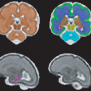
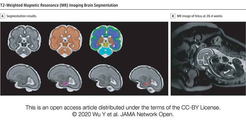
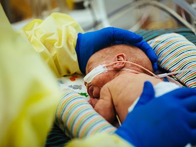
Changes in myelination due to diffuse white matter injury are a common consequence of premature birth and hypoxic-ischemic injury due to asphyxia of sick term-born newborns. Hypoxic damage during the neonatal period can lead to motor disabilities and cognitive deficits with long-term consequences, including cerebral palsy, intellectual disability or epilepsy, which are often due to cellular and functional abnormalities.
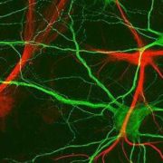
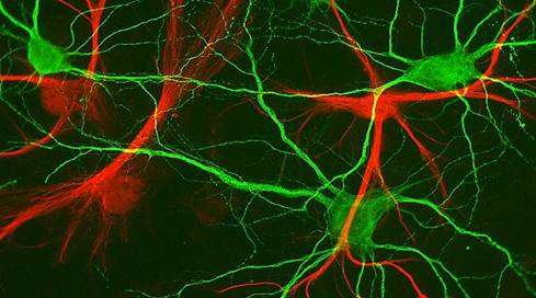
The Gallo Lab, within the Center for Neuroscience Research at Children’s National Hospital, is focused on studying postnatal neural development and the impact of injury and disease on development and regeneration of neurons and glia. Their latest research, published in Nature Communications, finds reduced expression of Sirt2 in the white matter of premature human infants (born earlier than 32 weeks of gestation) and characterizes its role in white matter of the brain in normal conditions as well as during hypoxia.
What it means
The lab previously identified Sirt1 as important for the proliferative regenerative response of oligodendrocyte progenitor cells in response to chronic neonatal hypoxia. This new study characterizes the function of Sirt2 and finds that it acts as a critical promoter of oligodendrocyte differentiation during both normal brain development and after hypoxia.
It’s likely this reduced expression of Sirt2 contributes to the arrest in oligodendrocyte maturation and myelination failure seen in extremely low gestational age neonates. Therefore, targeting Sirt2 may be an opportunity to capture the early and small window of opportunity for therapeutic intervention.
How this moves the field forward
Sirtuins have been shown to play crucial therapeutic roles in various diseases, including aging, neurodegenerative disorders, cardiovascular disease and cancer. Identifying Sirt2 as a major regulator of white matter development and recovery and increasing the understanding of its protein and genomic interactions opens new avenues for Sirt2 as a therapeutic target for white matter injury in premature babies.
Why we’re excited
Interestingly, the team found that overexpression of Sirt2 in oligodendrocyte progenitor cells, but not mature oligodendrocytes, restores oligodendrocyte populations after hypoxia through enhanced proliferation and protection from apoptosis. This is exciting because:
- It tells us that Sirt2 expression is very important for the transition from progenitor to differentiated oligodendrocyte.
- It’s the first report, to the team’s knowledge, of Sirt2 regulating cell survival of oligodendrocytes.
Read more in Nature Communications
- Sirt2 promotes white matter oligodendrogenesis during development and in models of neonatal hypoxia
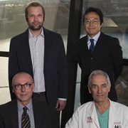
- Researchers on this paper include Beata Jablonska, Ph.D.; Katrina Adams, Ph.D.; Panagiotis Kratimenos, M.D., Ph.D.; Zhen Li, Ph.D.; Emma Strickland, B.S.; Tarik F. Haydar, Ph.D.; and Vittorio Gallo, Ph.D.