Tag Archive for: tumors

tube labeled "CRISPR"

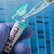
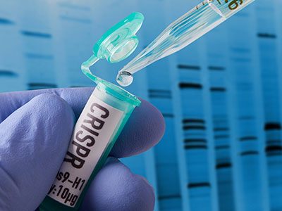
$2M from NIH to extract meaningful data from CRISPR screens

tube labeled "CRISPR"

Protein-coding genes comprise a mere 1% of DNA. While the other 99% of DNA was once derided as “junk,” it has become increasingly apparent that some non-coding genes enable essential cellular functions.

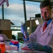
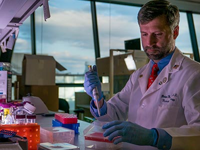
Wei Li, Ph.D., a principal investigator in the Center for Genetic Medicine Research at Children’s National in Washington, D.C., proposes to develop statistical and computational methods that sidestep existing hurdles that currently complicate genome-wide CRISPR/Cas9 screening. The National Institutes of Health has granted him $2.23 million in funding over five years to facilitate the systematic study of genes, non-coding elements and genetic interactions in various biological systems and disease types.

Right now, a large volume of screening data resides in the public domain, however it is difficult to compare data that is stored in one library with data stored at a different library. Over the course of the five-year project, Li aims to:

  • Improve functional gene identification from CRISPR screens.
  • Develop new analyses algorithms for screens targeting non-coding elements.
  • Study genetic interactions from CRISPR screens targeting gene pairs.

Ultimately, Li’s work will examine a range of disease types. Take cancer.

“There is abundant information already available in the public domain, like the Project Achilles  from the Broad Institute. However, no one is looking to see what is going in inside these tumors,” Li says. “Cancer is a disease of uncontrolled cell growth that makes tumors grow faster.”

Li and colleagues are going to ask which genes control this process by looking at genes that hit the brakes on cell growth as well as genes that pump the gas.

“You knock out one gene and then look: Does the cell grow faster or does it grow more slowly? If the cell grows more slowly, you know you are knocking out a gene that has the potential to stop tumor growth. If cells are growing faster, you know that you’re hitting genes that suppress cancer cell growth.”

In a nutshell, CRISPR (clustered regularly interspaced short palindromic repeats) screens knock out different genes and monitor changes in corresponding cell populations. When CRISPR first became popular, Li decided he wanted to do something with the technology. So, as a Postdoc at Harvard, he developed comprehensive computational algorithms for functional screens using CRISPR/Cas9.

To reach as many people as possible, he offered that MAGeCK/MAGeCK-VISPR software free to as many researchers as possible, providing source code and offering internet tutorials.

“So far, I think there are quite a lot of people using this. There have been more than 40,000 software downloads,” he adds. “It’s really exciting and revolutionary technology and, eventually, we hope the outcomes also will be exciting. We hope to find something really helpful for cancer patients.”

Research reported in this publication was supported by the National Human Genome Research Institute of the National Institutes of Health under award number R01HG010753.

Jeffrey Dome

The impact of surveillance imaging to detect relapse in Wilms tumor patients

Jeffrey Dome

Dr. Jeffrey Dome, M.D., Ph.D., vice president, Center for Cancer and Blood Disorders.

The Children’s Oncology Group published an article in the Journal of Clinical Oncology looking at the impact that surveillance imaging has on patients with Wilms tumor (WT), the most common kidney cancer in children.

Despite the risks and costs, the use of computed tomography (CT) for routine surveillance to detect recurrence in patients with WT has increased in recent years. The rationale for using CT scans rather than chest x-rays (CXR) and abdominal ultrasounds (US) is that CT scans are more sensitive, thereby enabling recurrences to be detected earlier.

In this study, led by Jeffrey S. Dome, M.D., Ph.D, vice president of the Center for Cancer and Blood Disorders at Children’s National Health System, researchers conducted a retrospective analysis of patients enrolled in the fifth National Wilms Tumor Study (NWTS-5) who experienced relapse to determine if relapse detection with CT scan correlates with improved overall survival compared with relapse detection by CXR or abdominal US.

A total of 281 patients with favorable-histology WT (FHWT) were included in the analysis. The key findings of the study were that:

  • Among patients with relapse after completion of therapy, outcome was improved in patients whose relapse was detected by surveillance imaging rather after signs and symptoms developed.
  • A higher disease burden at relapse, defined by the diameter of the relapsed tumor and the number of sites of relapse, was associated with inferior survival.
  • Relapses detected by CT scan were detected earlier and were smaller on average than relapses detected by CXR or US.
  • However, there was no difference in survival between patients whose relapse was detected by CT versus CXR or US.

An analysis of radiation exposure levels showed that surveillance regimes including CT scans have about seven times the radiation exposure compared to regimens including only CXR and US. Moreover, the cost to detect each recurrence reduced by 50 percent when CXR and US are used for surveillance.

“The results of this study will be practice changing,” said Dr. Dome, one of the doctors leading the clinical trial. “The extra sensitivity that CT scans provide compared to CXR and US do not translate to improved survival and are associated with the downsides of extra radiation exposure, cost and false-positive results that can lead to unnecessary stress and medical interventions,” he added. “Although counter-intuitive, the more sensitive technology is not necessarily better for patients.”

In conclusion, the doctors found that the elimination of CT scans from surveillance programs for unilateral favorable histology Wilms tumor is unlikely to compromise survival. However, it could result in substantially less radiation exposure and lower health care costs. Overall, the risk-benefit ratio associated with imaging modalities should be considered and formally studied for all pediatric cancers.

Learn more about this research in a podcast from the Journal of Clinical Oncology.

Affiliations

Elizabeth A. Mullen, Dana-Farber Cancer Institute/Boston Children’s Cancer and Blood Disorders Center, Boston, MA; Yueh-Yun Chi and Emily Hibbitts, University of Florida, Gainesville, FL; James R. Anderson, Merck Research Laboratories, North Wales, PA; Katarina J. Steacy, University of Maryland Medical Center, Baltimore, MD; James I. Geller, Cincinnati Children’s Hospital Medical Centre, Cincinnati, OH; Daniel M. Green, St Jude Children’s Research Hospital, Memphis, TN; Geetika Khanna, Washington University School of Medicine, St Louis, MO; Marcio H. Malogolowkin, University of California at Davis Comprehensive Cancer Center, Sacramento, CA; Paul E. Grundy, Stollery Children’s Hospital, University of Alberta, Alberta; Conrad V. Fernandez, University, Halifax, Nova Scotia, Canada; and Jeffrey S. Dome, Children’s National Health System, George Washington University School of Medicine and Health Sciences, Washington, D.C.

Yuan Zhu

The brain tumor field moves forward with new findings and a research grant

Yuan Zhu

Yuan Zhu, Ph.D., and other experts completed new research findings evaluating the effects of manipulating the growth-promoting signaling pathways in brain tumors associated with adults and children.

This month, experts at Children’s National Health System made great strides in brain tumor research, specifically in gliomas, glioblastomas and medulloblastomas. Led by Yuan Zhu, Ph.D., the scientific director and Gilbert Endowed Professor of the Gilbert Family Neurofibromatosis Institute and Center for Cancer and Immunology Research at Children’s National, the team completed new research findings evaluating the effects of manipulating the growth-promoting signaling pathways in brain tumors associated with adults and children. Dr. Zhu’s research was recently published in Cell Reports and he was also awarded a U.S. Department of Defense (DoD) grant to gain a better understanding of how low-grade gliomas form. Together, this work moves the needle on developing more effective treatments for these debilitating and life-threatening tumors.

The study

In his recently published paper, Dr. Zhu and his colleagues, including Drs. Seckin Akgul and Yinghua Li, studied glioblastomas, the most common brain tumor in adults, and medulloblastomas, the most common brain tumor found in children, in genetically engineered experimental models. Dr. Zhu found that when they removed the p53 gene (the most commonly mutated tumor suppressor gene in human cancers) in the experimental model’s brain, most developed malignant gliomas and glioblastomas, while Sonic Hedgehog (SHH)-subtype (SHH) medulloblastomas were also observed. They further suppressed the Rictor/mTorc2 molecular pathway that is known in the regulation of tumor growth. This action greatly reduced the incidence of malignant gliomas and extended the survival of the models, validating the concept that Rictor/mTorc2 could be a viable drug target for this lethal brain cancer in adults.

The study also found that the same Rictor/mTorc2 molecular pathway serves the opposite function in SHH medulloblastoma formation, acting as a tumor suppressor. Findings suggest that if the same drug treatment is used for treating SHH medulloblastoma in children, it could potentially have an adverse effect and promote growth of the tumors.

Ultimately, the study demonstrates that Rictor/mTORC2 has opposing functions in glioblastomas in adults and SHH medulloblastomas in children. While drug therapies targeting Rictor/mTORC2 may be successful in adults, the findings reveal the risks of treating children with pediatric brain tumors when using the same therapies.

The grant

Continuing the study of brain tumors, Dr. Zhu recently received a $575,000 grant from DoD to research benign gliomas, with the hope of gaining a greater understanding of how the tumors form. Low-grade gliomas, or benign brain tumors, are the most common brain tumors in children. While not lethal like their high-grade counterpart, these tumors can lead to significant neurological defects, permanently impacting a child’s quality of life. Most commonly, the tumor can impair vision, often leading to blindness.

Since the tumors only occur in children under the age of eight, Dr. Zhu believes they are linked to neural stem or progenitor cells that exist in the optic nerve only during development, or when children are under eight-years-old. To test if his hypothesis is correct, Dr. Zhu will develop a preclinical model that mimics human brain tumors to study the development of the optic nerve. If his theory proves correct, Dr. Zhu’s long-term goal is to develop a strategy that prevents the tumor formation from ever occurring, ultimately preventing vision loss in children. The grant begins in July and will run for three years.


Brian Rood

Improving the understanding of medulloblastoma

Brian Rood

Brian Rood, M.D., employed quantitative proteomics to tumor samples that led to novel therapeutic targets for Medulloblastoma and other tumors.

In a recently published study, Brian Rood, M.D., a neuro-oncologist at Children’s National Health System, employed quantitative proteomics to tumor samples, a technique that could lead to novel therapeutic targets for medulloblastoma and other tumors in the future.

Currently, many experts use genomic characterization to understand the genetic makeup of cancer cells, which has deepened the field’s collective knowledge of tumor biology. However, it has remained challenging to infer specific information about how the tumors will respond and consequently develop more effective therapies. Medulloblastoma is the most common pediatric, malignant brain tumor. Through Dr. Rood’s research using proteomic analysis, he was able to identify and measure the protein makeup of medulloblastoma, which led to a potential pathway for clinical intervention to treat this life-threatening cancer. The findings were published online June 7, 2018, in Acta Neuropathologica Communications.

“The goal of this research was to find out how these tumor cells function at the protein level, which may ultimately help the field identify drug therapies to stop them,” says Dr. Rood. “The genes of a cancer cell are like a blueprint for a building, but the blueprints aren’t always followed in a cancer cell: Not every active gene will produce its corresponding protein. Proteins do the work of the cell, and understanding them will provide a better overall understanding of a cancer cell’s biology.”

Dr. Rood compared proteomic and genomic data to confirm that genetics do not accurately predict the quantity of proteins. By directly quantitating the proteins and comparing them between different subgroups of the disease, they were able to identify protein-based pathways driving tumor biology. With this information, Dr. Rood was able to demonstrate that medulloblastoma depends on a crucial pathway, the eukaryotic initiation factor 4F protein synthesis pathway, resulting in the identification of a potential target for new treatments in medulloblastoma.

Ultimately, Dr. Rood found that proteomic analysis complements genomic characterization and the two can be used together to create a more complete understanding of tumor biology. Going forward, he hopes proteomic analysis will become common practice for studying all tumors, allowing tumors to be categorized and grouped together by protein makeup to help the field identify more effective therapies for all tumors.