Tag Archive for: therapeutic hypothermia

MRI of the patient's head close-up

Early versus late MRI in newborn brain injury

MRI of the patient's head close-up

A single magnetic resonance imaging (MRI) performed in the first week after birth is adequate to assess brain injury and offer prognostic information in newborn infants with hypoxic ischemic encephalopathy (HIE) treated with therapeutic hypothermia, according to a new study published in The Journal of Pediatrics.

A collaborative team of neonatology, neurology and neuroradiology experts from Children’s National Hospital that included Gilbert Vezina, M.D., Taeun Chang, M.D., and An N. Massaro, M.D., came together to evaluate the agreement in brain injury findings between early and late MRI in newborn infants with hypoxic ischemic encephalopathy (HIE) treated with therapeutic hypothermia. The team then compared the ability of early versus late MRI to predict early neurodevelopmental outcomes.

This was a prospective longitudinal study of 49 patients with HIE who underwent therapeutic hypothermia and had MRI performed at both <7 and ≥7 days of age. MRIs were reviewed by an experienced neuroradiologist and assigned brain injury severity scores according to established systems. Scores for early and late MRIs were assessed for agreement using the kappa statistic. The ability of early and late MRI scores to predict death or developmental delay at 15-30 months of age was assessed by logistic regression analyses.

The results of the study found agreement between the early and late MRI was substantial to near perfect (k>0.75, p<0.001) across MRI scoring systems. In cases of discrepant scoring, early MRI was more likely to identify severe injury when compared with late MRI. Early MRI scores were more consistently predictive of adverse outcomes compared with late MRI.

Read the full study in The Journal of Pediatrics.

newborn in incubator

Working to reduce brain injury in newborns

A new study from Children’s National Health System and Drexel University College of Medicine has identified a promising treatment to reduce or prevent brain injury in newborns who have suffered hypoxia-ischemia.

Research-clinicians at Children’s National Health System and Drexel University College of Medicine led the first study to identify a promising treatment to reduce or prevent brain injury in newborns who have suffered hypoxia-ischemia, a serious complication in which restricted blood flow deprives the brain of oxygen.

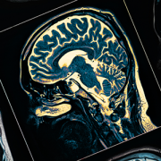
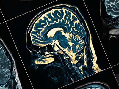
Consequences of brain injury resulting from oxygen deprivation affect the entire lifespan and range from mild (learning disabilities) to severe (inability to breathe, walk, talk or see). This complication can occur during or before birth due to maternal/placental problems, such as placental abruption or cord prolapse, or due to fetal/newborn issues, such as asphyxia due to labor difficulties, infection, fetal-maternal bleeding or twin-to-twin transfusion.

Published in Neonatology on Oct. 13, 2017, the study evaluated newborn experimental models exposed to hypoxia-ischemia. The experimental models were given standard cooling therapy (therapeutic hypothermia) alone and in combination with a selective Src kinase inhibitor, PP2, that blocks a regulatory enzyme of apoptosis (cell death), which intensifies as a result of hypoxia-ischemia. The Food and Drug Administration has approved a Src kinase inhibitor as an oncology treatment. This study is the first to test the benefits of blocking this enzyme in reducing the neurological damage caused by brain hypoxia-ischemia.

“In hypoxia-ischemia, CaM kinase is over-activated, but hypothermia has been shown to decrease this enzyme’s activation. We theorized that a Src kinase inhibitor, in addition to hypothermia, would further attenuate the activation of CaM kinase IV and that the result might be less brain damage,” explains Panagiotis Kratimenos, M.D., Ph.D., the study’s lead author, and a specialist in neonatology and neonatal neurocritical care at Children’s National. “From this study, we were pleased that this seems to be the case.”

The research team assessed neuropathology, adenosine triphosphate and phosphocreatine  concentrations as well as CaM kinase IV activity. The CaM kinase IV activity in cerebral tissue was 2,002 (plus or minus 729) with normal oxygen levels and in normal temperatures, 4,104 (plus or minus 542) in hypoxia with hypothermia treatment, and 2,165 (plus or minus 415) in hypoxia with hypothermia treatment combined with PP2 administration.

The authors conclude that hypothermia alone attenuated the over-activation of CaM kinase IV and improved neuropathology after hypoxia. However, the combination of hypothermia with Src kinase inhibition following hypoxia further attenuated the increased activation of CaM kinase IV, compared with hypothermia alone in the newborn experimental model brain.

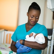
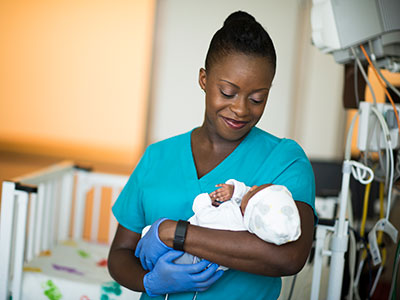
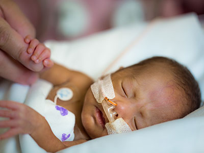
Currently, the only treatment for hypoxia-ischemia is therapeutic hypothermia. Starting in the first six hours of life, doctors in the neonatal intensive care unit lower a baby’s temperature by about 3 degrees Celsius for three days. This therapy is proven to reduce neural defects by up to 30 percent, yet many infants still have poor outcomes even after the therapeutic cooling treatment.

“In oxygen deprivation of the brain, the pathways leading to cell death are over-activated, including the nuclear enzyme CaM kinase IV. We sought to intervene in this pathway to reduce the heightened cell death, which leads to brain damage,” explains Dr. Kratimenos, an assistant professor of pediatrics at The George Washington University School of Medicine and Health Sciences whose research focus is neonatal encephalopathy and therapeutic hypothermia.

To continue preclinical research into this approach, Dr. Kratimenos envisions studying the effect of other types of small molecule inhibitors to target the apoptotic cascade, perhaps in multiple doses, eliminating the potential side effects, and determining the best dose and duration of treatment.

“If confirmed by further studies, this approach─in combination with cooling─may help to further attenuate neurological damage that babies suffer after experiencing hypoxia-ischemia,” says Dr. Kratimenos.

The study co-authors include Ioannis Koutroulis, M.D., Ph.D., a faculty member in Children’s Division of Emergency Medicine; and Amit Jain, M.D.; Shadi Malaeb, M.D.; and the world-renowned neonatologist and pioneer in bioenergetics of the brain, Maria Delivoria-Papadopoulos, M.D., all of the Drexel University College of Medicine.

nurse holding newborn baby

Continuous EEG monitoring better predicts HIE outcomes

nurse holding newborn baby

For newborns who experience a serious complication that deprives their brain of oxygen, continuously monitoring brain activity and examining how the electrical signals evolve may be a more reliable way to identify infants most at risk for brain injury.

For newborns who experience a serious complication that deprives their brain of oxygen, continuously monitoring brain activity and examining how the electrical signals evolve may be a more reliable way to identify infants most at risk for brain injury, compared with doing evaluations at discreet intervals, according to a prospective cohort study led by Children’s National Health System research-clinicians.

Amplitude-integrated electroencephalogram (aEEG) is a bedside tool that permits clinicians to monitor the complex electrical activity of the child’s brain over time. It’s a positive sign when an aEEG shows babies beginning to sleep and wake normally by the time they are 3 days old. Conversely, severely abnormal aEEG readings in the first days of life predict poor outcomes.

The Children’s team used aEEG with infants born with hypoxic-ischemic encephalopathy (HIE), one of the most severe complications that can affect full-term infants. During pregnancy, birth or shortly after birth, a hypoxic-ischemic event can occur that impedes blood flow and oxygen delivery to the brain, resulting in destruction of brain tissue. Cooling (therapeutic hypothermia) is now standard for newborns with HIE in order to stave off life-long consequences, but deaths and neurodevelopmental disability still can occur.

“We know whole-body cooling – or lowering the body’s temperature by about 3 degrees Celsius – can help vulnerable newborns survive and can protect their brains from suffering profound injuries,” says An N. Massaro, M.D., a Children’s National neonatologist and senior author of the study published online Sept. 28, 2017 in the American Journal of Perinatology.  “What we were trying to determine with this study is whether evaluating the pattern of evolution of the aEEG as a whole provides more information compared with looking at snapshots in time.”

Eighty infants undergoing therapeutic cooling who met the inclusion criteria were enrolled in the five-year study, one of the largest such studies to date. The babies weighed more than 1,800 grams and were older than 35 weeks’ gestational age at birth, and either needed prolonged resuscitation after birth or had low APGAR scores – a measure of how well newborns fare outside the womb. Continuous recordings of EEG data occurred from the time of admission up to 12 hours after the infants’ temperatures were raised to normal and aEEG tracings were calculated.

After the therapeutic cooling blankets were removed, the infants underwent at least one magnetic resonance imaging (MRI) scan prior to discharge. During the routine follow-up check at about 18 months of age, the HIE survivors’ cognitive and motor skills were assessed using validated instruments.

Fifty-six of the infants in the study had favorable outcomes. Twenty-four infants had adverse outcomes, including 15 with severe brain injury detected by MRI and nine infants who died. These children had lower APGAR scores at five minutes, and were more likely to have severe HIE and to have experienced more frequent seizures.

“Infants whose aEEG abnormalities do not improve were at increased risk: Infants who do not reach a discontinuous background pattern by 15.5 hours of life, achieve cycling by 45.5 hours after birth and who fail to achieve continuous normal voltage by 78 hours after birth are most at risk for adverse outcomes,” Dr. Massaro says. “In addition to defining worrisome trends, we found that overall assessment of continuous aEEG readings through the course of hypothermia treatment provide the most meaningful predictive power. This means we can speak with families at the bedside with more confidence about their child’s outcomes after the infant undergoes cooling therapy.”

Related Resources