Tag Archive for: seizure

The endovascular embolic hemispherectomy team.

New hemimegalencephaly procedure is all about teamwork

Children’s National experts pioneered a novel approach of inducing strokes to stop seizures and improve neurodevelopmental outcomes in newborns under three months old with hemimegalencephaly (HME). The procedure, called an endovascular embolic hemispherectomy, can be safely used to provide definitive treatment of HME-related epilepsy in neonates and young infants. Monica Pearl, M.D., neurointerventional radiologist, and Panagiotis Kratimenos, M.D., Ph.D., neonatologist, discuss why having a multidisciplinary team skilled at this procedure is the reason we’re the only center in the world capable of providing this treatment.

baby with brain monitor

The history behind the novel hemimegalencephaly procedure

Traditionally, when a baby is diagnosed with hemimegalencephaly (HME), doctors turn to a hemispherectomy at 3 months of age, which involves surgically removing half of a baby’s brain. At Children’s National Hospital, our doctors pioneered the endovascular embolic hemispherectomy, an approach using induced controlled strokes to eliminate the affected part of the brain, halting seizures. Monica Pearl, M.D., neurointerventional radiologist, and Tammy Tsuchida, M.D., Ph.D., neonatal neurologist, talk about this life-changing procedure.

Angelique and family pose in front of their house

Inducing strokes to better treat babies with hemimegalencephaly

When a family from Texas received a shocking diagnosis for their newborn daughter, they knew there was one place they needed to go – Children’s National Hospital in Washington, D.C. At birth, Angelique was diagnosed with a rare and devastating condition known as hemimegalencephaly (HME) which causes uncontrollable and frequent seizures. Monica Pearl, M.D., neurointerventional radiologist, and the team at Children’s National have pioneered an approach to treat HME, where they induce controlled strokes to eliminate the affected part of the brain, halting seizures in their tracks. They’re the only team in the world doing this work. Angelique’s parents knew the clock was ticking — every day they waited meant irreversible damage to their daughter’s developing brain.

Illustration of brain and brainwaves

Risk factors for pharmacoresistant pediatric epilepsy

Illustration of brain and brainwaves

New study evaluates risk factors for the timing and development of drug-resistant pediatric epilepsy.

Focal cortical dysplasia (FCD) is the most common cause of surgically-treatable epilepsy in children. In a new study published in Neurology, researchers evaluated 143 children with confirmed FCD risk factors for the timing and development of pharmacoresistant epilepsy.

What this means

The current definition of pharmacoresistance requires failure of two appropriately-dosed and selected antiseizure medications before being able to be considered for epilepsy surgery.

“We found that the failure of just one antiseizure medication is associated with an enormous increased incidence and earlier development of pharmacoresistance,” says Nathan Cohen, M.D., neurologist at Children’s National and lead author of the study. “Our data supports the redefinition of pharmacoresistant epilepsy to the failure of just one antiseizure medication in this population, which would potentially allow these patients to benefit from earlier curative surgery.”

Why it matters

The findings showed that in children with FCD the failure of just one antiseizure medication is associated with an enormous risk and earlier incidence of pharmacoresistance. Therefore, the authors advocate for its redefinition in FCD-related epilepsy to the failure of just one antiseizure medication.

“This will allow children to be considered much earlier for potentially curative epilepsy surgery,” adds Dr. Cohen. “We find that the majority of FCD patients develop epilepsy and that the majority of those with epilepsy develop pharmacoresistance.”

In a multivariate analysis, the authors show that the FCD cortical lobar location, pathologic subtype, and age of seizure onset are not important factors in the development of pharmacoresistance.

What’s next

This data supports operational re-definition of pharmacoresistance for surgical planning in FCD-related epilepsy to the failure of one antiseizure medication, and support early, potentially curative surgery to improve outcomes in this patient population.

You can read the full study, Prevalence and Risk Factors for Pharmacoresistance in Children With Focal Cortical Dysplasia–Related Epilepsy, in Neurology.

model of the brain

Treating newborns with hemimegalencephaly by inducing strokes

model of the brain

Experts at Children’s National Hospital have pioneered a novel approach using controlled strokes to stop seizures and improve neurodevelopmental outcomes in newborns under three months born with hemimegalencephaly (HME). They now consider it their new standard of care for babies in this age group with HME and refractory epilepsy.

Asking a physician to induce strokes in newborns is asking her to do something contrary to her training. But over the past eight years, experts at Children’s National Hospital have pioneered a novel approach using controlled strokes to stop seizures and improve neurodevelopmental outcomes in newborns under three months born with hemimegalencephaly (HME). They now consider it their new standard of care for babies in this age group with HME and refractory epilepsy.

“We have demonstrated the ability to intervene and stop the intractable seizures during a critical time of neurodevelopment in which no other effective medical or surgical option exists. That is extremely rewarding,” said Monica Pearl, M.D., director of the Neurointerventional Radiology Program at Children’s National. Children’s National is the only center in the world currently offering this treatment. A multi-disciplinary team led by Dr. Pearl; Taeun Chang, M.D., director of the Neonatal Neurology and Neonatal Neurocritical Care Program; neurophysiologist and neonatal neurologist Tammy Tsuchida, M.D., Ph.D.; and other experts has now successfully treated seven patients using this minimally-invasive approach.

“We want patients and providers to understand this is a better alternative to a delayed hemispherectomy, the standard of care currently offered to newborns with HME,” said Dr. Chang.

The best treatment for newborns with hemimegalencephaly

HME, a rare congenital condition occurring in a handful of newborns each year, is characterized by abnormal growth and enlargement of half of the brain which leads to intractable seizures. The seizures often result in severe cognitive delays and hemiparesis. The standard treatment is an anatomic hemispherectomy — surgical removal of the affected half of the brain, allowing the remaining half of the brain to develop and function without constant seizures.

Such a large and complex surgery poses serious risks for infants younger than three months, leaving doctors with the difficult choice to delay surgery until these newborns grow bigger and stronger, even as they are experiencing seizures. These persistent seizures compromise the development of the healthy half of the brain. One study reports as much as a drop of 10 to 20 IQ points with each month’s delay in surgical hemispherectomy.

“I was willing to consider performing these procedures because there is a clear, unmet medical need and these babies are in dire circumstances,” Dr. Pearl said. “Waiting for curative hemispherectomy means more than just lost time; uncontrolled seizures and anti-seizure medications have detrimental effects on the ‘normal,’ unaffected parts of the brain. We needed a better option for these patients.” Dr. Pearl said that complete embolization of the affected hemisphere as both primary and definitive treatment had never been described. They could only find one example in the literature – a paper from 1995 – suggesting embolization as an adjunct to surgery, and nothing suggesting it as a primary modality.

About the care received

Dr. Pearl is one of only a handful of dedicated pediatric neurointerventionalists across the country with neurovascular expertise in people of all ages, in particular neonates and young infants. For these procedures to be performed safely, the neurointerventionalist must be proficient in obtaining femoral arterial access and navigating small caliber cervicocerebral blood vessels that are less than one millimeter in diameter.

Additionally, one needs a neonatal neurocritical care service and NICU that can medically manage large strokes and their potential complications in newborns. Dr. Chang has developed a specialized protocol based on decades of managing strokes and other acute brain injuries in newborns. She created the neonatal neurocritical care service at Children’s National, the only one in the region and the largest in the world.

“Our teams are fortunate in that we each respectively have extensive prior experience in treating and managing neonates and very young infants for various cerebrovascular disorders,” Dr. Pearl said. “We relied on this collective experience to make this hemispheric embolization pathway possible.”

How it happens

To perform the embolizations, Dr. Chang and her team first optimizes control of the seizures using medications. Dr. Pearl places a sheath in the femoral artery using ultrasound guidance – a delicate task in a neonate whose femoral artery diameter is only two to three millimeters. She then navigates a catheter up the aorta and selects the targeted carotid artery using radiographic guidance. What follows is a set of intricate navigations to direct the microcatheter through small blood vessels in the brain, often less than one millimeter.

Using x-ray guidance, Dr. Pearl injects contrast through the microcatheter to visualize the arterial anatomy and advance the microcatheter into position for embolization. She uses glue that hardens when exposed to blood, blocking off the blood supply to the seizure-inducing areas. The process is repeated until the blood supply to the entire affected hemisphere is occluded. Meanwhile, Dr. Chang and her team monitor the brain’s electrical activity using an electroencephalogram (EEG) to watch how the brain responds to each stroke. The surgical epilepsy, neonatal neurocritical care and neonatology teams are all in constant communication throughout the procedure.

Together, they have to contend with the same symptoms patients have immediately following a stroke, most notably brain swelling that can cause bleeding and herniation. The resultant brain swelling is complicated further by the already enlarged hemisphere of the brain. Using neuroprotective strategies learned from treating over a thousand newborns with perinatal brain injury, Dr. Chang and her team and the NICU coordinate to minimize brain swelling and protect the healthy half of the brain by tightly controlling the brain temperature, glucose, sodium levels, and blood pressure. Over the course of a few weeks, Dr. Pearl performs three to four embolization sessions to halt blood supply to the seizing half of the brain.

“The risks of intracranial vasospasm and hemorrhage during embolization are higher in this distinct group of patients compared to other neonates requiring embolization, such as in vein of Galen malformations. These events must be controlled immediately to prevent complications and I know I only have seconds to react,” Dr. Pearl said.

“Here, we have the cultivation of brain-centric neonatal care, a large level IV tertiary NICU with expertise in keeping critically ill babies alive and rare pediatric neurologic subspecialists like Dr. Pearl and myself. All of this is what makes this level of innovation possible,” Dr. Chang said. Now, they wish this minimally invasive approach to be available to all newborns with HME and refractory epilepsy.

“This is not a fluke. This is not a one-time thing. Our team at Children’s National has been perfecting this method for close to a decade,” Dr. Chang said. As for proof, her answer is clear.

Follow our patients: Bella and Trace.

newborn

Predicting risk for infantile spasms after acute symptomatic neonatal seizures

newborn

Infantile spasms (IS) is a severe epilepsy in early childhood. Early treatment of IS provides the best chance of seizure remission and favorable developmental outcome.

Taeun Chang, M.D., director of the Neonatal Neurology and Neurocritical Care Program at Children’s National Hospital, participated in a study with other national pediatric experts which aimed to develop a prediction rule to accurately predict which neonates with acute symptomatic seizures will develop IS.

The group of researchers found that multiple potential predictors were associated with IS, including Apgar scores, EEG features, seizure characteristics, MRI abnormalities and clinical status at hospital discharge. The final model born from this work included three risk factors: (a) severely abnormal EEG or ≥3 days with seizures recorded on EEG, (b) deep gray or brainstem injury on MRI and (c) abnormal tone on discharge exam.

The significance of these findings is that IS risk after acute symptomatic neonatal seizures can be stratified using commonly available clinical data. No child without risk factors, vs >50% of those with all three factors, developed IS. This risk prediction rule may be valuable for clinical counseling as well as for selecting participants for clinical trials to prevent post‐neonatal epilepsy. This tailored approach may lead to earlier diagnosis and treatment and improve outcomes for a devastating early life epilepsy.

Read the full study in Epilepsia.

Jake and Dr. Oluigbo

Doctors at Children’s National give Jake his life back

Jake and Dr. Oluigbo

At the age of 17, Jake underwent surgery led by neurosurgeon Chima Oluigbo, M.D., where he conducted a temporal lobe resection, also called temporal lobectomy, that works to lower the number of seizures, make them less severe or stop them completely. The surgery ended up being successful and it worked to greatly improve his overall quality of life.

Since 1969, November has been considered Epilepsy Awareness Month to highlight the importance of recognizing a seizure and promoting seizure first aid. At Children’s National Hospital, doctors in the division of neurology are committed to finding treatments for epilepsy and have done just that by helping Jacob Yates, an 18-year-old patient, get his life back.

For many families the holidays are meant for spending time with loved ones and enjoying the seasonal festivities. However, the holidays were not always a joyous occasion for Jake and his family. Since he was a baby, many of his holidays were spent in a bed due to a brain disorder that caused him to have developmental delays and, at times, up to 17 seizures a day.

“The holidays were always a tough time for the family because Jake would get so excited around Christmas that it would overwhelm his system and induce seizures that took him days to recover from,” says his mom, Jennifer.

Jake was born a preemie and hours after he was born, doctors at his local hospital had identified that he was having trouble breathing. By coincidence, the Children’s National transport team was on-site to take another patient to Children’s National, but once they looked at Jake they immediately took him instead by SkyBear Air Transport, the hospital’s rapid helicopter transport service.

During his stay at Children’s National, Jake was in the neonatal intensive care unit (NICU) for 11 days and was supported by breathing machines to help with respiratory distress and other issues stemming from him being born prematurely.

“If it wasn’t for the Children’s National transport team coincidentally being at our local hospital, Jake wouldn’t have survived staying at that location,” said Jennifer.

After he was taken care of at Children’s National, he was discharged 11 days later, but at the age of three months Jake was still experiencing respiratory issues and was taken back to his local hospital in Charles County.

“When he first arrived back at the University of Maryland Charles Regional Medical Center, the doctors thought he may have had cystic fibrosis, but it came back that perhaps he was suffering from reflux and they put him on medication,” Jennifer recalls. Unfortunately, this was not the cause and it would not be the family’s last visit to the hospital.

By the age of six months, Jake had his first seizure and he was flown back to Children’s National. Over the next year he was repeatedly admitted to the hospital as his seizures had caused him to stop breathing.

Between the ages of 4 to 6 years old, Jake became a patient of William D. Gaillard, M.D., division chief of epilepsy and neurophysiology and Roger Packer, M.D., senior vice president at the Center of Neuroscience and Behavioral Health at Children’s National. After his visit, both doctors recommended surgery, but Dr. Packer recommended that Jake receive an electroencephalogram (EEG), magnetic resonance imaging (MRI) and go through a sleep study first to identify the specific causes of his seizures.

Now on a new medication, his seizures were maintained for the most part, but doctors were still recommending that it was time for surgery. When Jake was 15, his parents re-evaluated the surgery and learned that their son had a 76% chance of being seizure and medication free.

At the age of 17, Jake underwent surgery led by Chima Oluigbo, M.D., neurosurgeon at Children’s National, where he conducted a temporal lobe resection, also called temporal lobectomy, that works to lower the number of seizures, make them less severe or stop them completely. The surgery ended up being successful and it worked to greatly improve his overall quality of life.

Before the surgery, Jake didn’t speak much, experienced anxiety and had difficulty expressing his emotions. He had never told his mother that he loved her. After the surgery, Jake looked at his mother and said, “I love you babe.”

According to Jennifer, since the surgery her son is a completely different person and states that he has been seizure free for over a year. Equally, Jake and the family can now all look forward to the holidays.

“We’re so excited to have him share the holidays,” Jennifer says. “He feels better and it shows through his attitude and the way he responds to things. Words can’t express the gratitude we have for the doctors at Children’s National Hospital. They gave my son his life back.”

Chima Oluigbo examines a patient

Eradicating epilepsy with Visualase

Chima Oluigbo examines a patient

Chima Oluigbo, M.D., and his team are using Visualase to identify and eliminate seizure foci and provide patients with a minimally invasive procedure for treating epilepsy.

About one in 26 people will be diagnosed with epilepsy in their lifetime. That adds up to about 3.4 million people in the U.S., or about 1 percent of the population nationwide. This condition can have huge consequences on quality of life, affecting whether children will learn well in school, eventually drive a car, hold down a job or even survive into adulthood.

For most of those that develop epilepsy, medications can keep seizures in check. However, for about a third of patients, this strategy doesn’t work, says Chima Oluigbo, M.D., an attending neurosurgeon at Children’s National Health System. That’s when he and his team offer a surgical fix.

Epilepsy surgery has come a long way, Dr. Oluigbo explains. When he first began practicing in the early 2000s, most surgeries were open, he says – they involved making a long incision in the scalp that can span half a foot or more. After drilling out a window of skull that can be as long as five inches, surgeons had to dig through healthy brain to find the abnormal tissue and remove it.

Each part of this “maximally invasive” procedure can be traumatic on a patient, Dr. Oluigbo says. That leads to significant pain after the procedure, extended hospital stays of at least a week followed by a long recovery. There are also significant risks for neurological complications including stroke, weakness, paralysis, speech problems and more.

However, open surgery isn’t the only option for epilepsy surgery anymore. Several new minimally invasive alternatives are now available to patients and the most promising, Dr. Oluigbo says, is called Visualase. He and his team are the only surgeons in the region who perform this procedure.

In Visualase surgeries, Dr. Oluigbo and his colleagues start by making a tiny incision, about 5 millimeters, on the scalp. Through this opening, they bore an even tinier hole into the skull and thread a needle inside that’s about 1.6 millimeters wide. “The brain barely notices that it’s there,” he says.

The tip of this wire holds a laser. Once this tip is placed directly at the seizure foci – the cluster of nerve cells responsible for generating a seizure – the patient is placed in an intraoperative magnetic resonance imaging (MRI) device. There, after checking the tip’s precise placement, the surgeons turn the laser on. Heat from the laser eradicates the foci, which the surgeons can see in real time using MRI thermography technology. The margins of the destroyed tissue are well-defined, largely sparing healthy tissue.

After the wire is removed, the incision is closed with a single stitch, and patients go home the next day. The majority of patients are seizure free, with rates as high as 90 percent for some types of epilepsy, Dr. Oluigbo says. Although seizure-free rates are also high for open procedures, he adds, Visualase spares them many of open surgeries’ painful and difficult consequences.

“Having done both open surgeries and Visualase,” Dr. Oluigbo says, “I can tell you the difference is night and day.”

Although open procedures will still be necessary for some patients with particularly large foci that are close to the surface, Dr. Oluigbo says that Visualase is ideal for treating medication-resistant cases in which the foci are buried deep within the brain. A typical example is a condition called hypothalamic hamartoma, in which tumors on the hypothalamus lead to gelastic seizures, an unusual seizure type characterized by uncontrollable laughing. He also uses Visualase for another condition called tuberous sclerosis, in which waxy growths called tubers develop in the brain, and for cancerous and benign brain tumors.

It’s gratifying to be able to help these children become seizure-free for the rest of their lives, says Dr. Oluigbo – even more so with the numerous updates he receives from families telling him how much this procedure has improved their children’s lifestyle.

“Visualase has completely changed the way that we approach these patients,” Dr. Oluigbo says. “It’s extraordinary to see the effects that this one procedure can have on the quality of life for patients here at Children’s National.”

Expanding awareness of SUDEP

Madison Berl

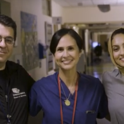
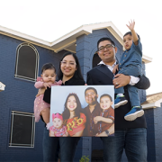
Madison M. Berl, Ph.D., is helping to expand awareness of SUDEP among patients, families and caregivers.

When 4-year-old Henry Lapham died in his sleep just weeks after being diagnosed with epilepsy in 2009, it was a shock to everyone — even his pediatrician and neurologist. Henry’s cause of death was sudden unexpected (or unexplained) death in epilepsy persons (SUDEP), a condition that causes sudden death in about 1 of every 1,000 otherwise healthy patients with epilepsy. Neither health care professional had mentioned this as a possibility, as remote as it was.

“I was desperate to make sense out of our tragedy,” writes Henry’s mother, Gardiner Lapham, R.N., M.P.H., in “Increasing awareness of sudden death in pediatric epilepsy together,” an article published in the February 2017 issue of Pediatrics. After her son’s death, by working with a group called Citizens United for Epilepsy Research, Lapham connected with other families affected by the same heartbreak. “I have met many bereaved family members,” she adds, “and the most consistent thing I hear is that they wish they had known about SUDEP.”

Now, a new collaboration with Children’s National Health System, where Henry received care, University of Virginia Medical Center (UVA) and other academic medical centers is helping to expand awareness of SUDEP among patients, families and caregivers alike. Known as Childhood Epilepsy Risks and Impact on Outcomes (CHERIO), the multiyear effort aims to develop approaches to increase knowledge about SUDEP and other conditions that can accompany epilepsy, such as attention deficit hyperactivity disorder, autism, anxiety, depression and sleep issues, according to co-authors of the Pediatrics article.

CHERIO got its start in 2014 at the American Epilepsy Society annual meeting. There, Lapham met Madison M. Berl, Ph.D., director of research, Division of Pediatric Neuropsychology at Children’s National, who studies epilepsy comorbidities. When Lapham asked what she could do to help raise awareness of SUDEP at Children’s National, she and Berl, along with William Davis Gaillard, M.D., Henry’s neurologist, hatched a plan.

Working with multiple disciplines and stakeholders, including neuropsychologists, psychiatrists, neurologists, epidemiologists, basic scientists, nurses and parent advocates at both Children’s National and UVA, CHERIO plans to assess the level of knowledge about SUDEP and other epilepsy comorbidities among medical providers and parents and to implement ways to increase knowledge. The first item on the agenda, Berl explains, was to conduct a survey to see just how much doctors knew about SUDEP.

“Although many neurologists are aware of this condition, ours was the first to survey pediatricians, and the majority was not aware of SUDEP – despite having children with epilepsy in their practice,” Dr. Gaillard says. “We know that many neurologists do not discuss SUDEP with patients and the reasons for not talking about SUDEP are varied. Thus, CHERIO felt that in addition to educating neurologists about the need to discuss the risk of death associated with epilepsy, increasing pediatricians’ awareness of SUDEP is one approach that could open more opportunities for families to have this discussion.”

To help make it easier to talk about this risk, the CHERIO team is developing strategies for doctors to start the conversation with patients and their families by framing SUDEP in the context of more common epilepsy comorbidities.

“Clinicians walk a fine line in giving information at the right time to make people more aware,” Berl adds, “but also being realistic and giving information that fits with what’s going on in a particular child’s case. By discussing SUDEP along with other, more common epilepsy risks, it brings context to a family so that they’re not unduly concerned about death – which also can paralyze a family and create unnecessary alarm.” The risk of death in most children with epilepsy is very low, slightly higher than the risks faced by healthy children. But parents of children with complicated epilepsy who have more risk factors for sudden death should be especially aware , she says.

Another way to help facilitate discussion may be through a simple tweak in the medical record, Berl adds. The team is currently developing a checklist that pops up annually in a patient’s medical record to remind clinicians of important points to discuss with patients and their families, including SUDEP.

Additionally, they are working on ways that can help families become more empowered to start the discussion themselves. Materials for the waiting room or questionnaires to fill out before appointments could trigger conversations with care providers, Berl says.

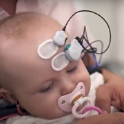
Last, the team also is collaborating with a medical device company that is working on a nighttime monitoring system that could provide an alert if patients with epilepsy experience nighttime seizures, a risk factor for SUDEP. Such technologies have not been proven to prevent SUDEP. Yet, it may help caregivers get help more quickly than if they did not receive the alert.

For each of these efforts, Berl notes, having Lapham as a partner has been key. “She’s part of our meetings and has input into the direction of each project,” Berl explains. “When you have a partner who is so close to the daily work you’re doing, it just heightens those efforts and brings to the forefront the simple message of why this is important.”