Tag Archive for: sandler

In the news: Axios’ Future of Health Summit

“Healthcare is moving very fast. And what often happens in adults, also happens in children. Unfortunately, most of the research is directed initially at adults, and then whittles down to children. At Children’s National, we’re trying to turn that around. We’re trying to do research for children that will expand its way up to adults, turning it on its head.”

Anthony Sandler, MD, senior vice president and surgeon-in-chief, Joseph E. Robert Jr. Center for Surgical Care, and director of the Sheikh Zayed Institute for Pediatric Surgical Innovation highlighted the exciting research and innovation happening at Children’s National – including demonstrating a technology, led by Raj Shekhar, PhD, that uses real-time imaging with augmented reality to project live ultrasound visualization of a patient within the surgeon’s field of view. This enhances surgical precision and ultimately supports positive patient outcomes.

This conversation was a part of Axios’ inaugural Future of Health Summit – an event bringing together the top voices in healthcare, policy and technology to explore the biggest challenges and innovations shaping the future of medicine.

Charging ahead: Researchers develop robotic renal tumor surgery

robotic surgery apparatus

Researchers at Children’s National Hospital are developing supervised autonomous robotic surgery to make expert kidney tumor removal accessible in rural areas, combining robotics, AI and surgeon oversight for safer, more precise outcomes.

Imagine a robot capable of planning and executing the intricate removal of a cancerous kidney tumor — a concept that might sound like science fiction. Yet this groundbreaking work is underway at the Sheikh Zayed Institute (SZI) for Pediatric Surgical Innovation at Children’s National Hospital.

Called Supervised Autonomous Robotic Renal Tumor Surgery (SARRTS), the project aims to prove that a supervised autonomous kidney resection is feasible. Its goal is to enable general surgeons in rural hospitals to oversee robots performing complex resections, democratizing access to specialized surgical care. Backed by a $1 million contract from the Advanced Research Projects Agency for Health (ARPA-H), the initiative represents new opportunities in medical innovation.

“The hope is that, someday, patients will no longer have to travel to major oncology centers to get the best possible surgical outcome when faced with renal tumors,” said Kevin Cleary, PhD, associate director of engineering at SZI. “We hope to combine the precision of robotics with a surgeon’s clinical expertise to create consistently high outcomes.”

The patient benefit

Surgery is a cornerstone of cancer treatment, but access to skilled surgeons remains unevenly distributed nationwide. Autonomous robotic surgery could address this disparity by increasing access to expert-level care, enhancing the precision and consistency of procedures and unlocking new surgical possibilities beyond human surgeons’ capabilities.

Under the initial concept, the SARRTS system will use a combination of CT imaging and 3D mapping from a robot’s RGB-depth camera. While the robot independently plans and executes the incision and tumor resection, the supervising surgeon retains full control, with the ability to approve, modify or halt the procedure at any time — an interplay between human expertise and robotic precision to help ensure safety.

Testing will be conducted on realistic kidney models, called phantoms, which are designed to train and test surgical outcomes. The project aims to validate the feasibility of supervised autonomous tumor resection while advancing technologies that could pave the way for broader applications.

“Robotics and medicine have finally reached a point where we can consider projects requiring this level of complexity,” said Anthony Sandler, MD, senior vice president and surgeon-in-chief at Children’s National and executive director of SZI. By combining autonomous robotics, artificial intelligence and surgical expertise, we can profoundly impact the lives of patients facing life-altering cancer diagnoses.”

Children’s National leads the way

In addition to the kidney surgery initiative, the Children’s National team is pursuing other groundbreaking projects. These include a second ARPA-H contract focused on robotic gallbladder removal and a National Institutes of Health grant to explore robotic hip-pinning, a procedure used to repair fractured hips with pins, screws and plates.

Axel Krieger, PhD, an associate professor of mechanical engineering at Johns Hopkins University, is collaborating closely on the kidney resection and gallbladder projects. The interdisciplinary team believes this state-of-the-art care could be tested and developed within the next decade.

“This particular surgery is complex, and a robot may offer advantages to address difficulties created by patient anatomy and visibility within the surgical field,” said Dr. Sandler. “We can imagine a day – in the not too distant future – when a human and a robotic arm could team up to successfully advance this care.”

This project has been funded in whole with federal funds from ARPA-H under cooperative agreement AY1AX000023.

Children’s National again ranked among the best in the nation by U.S. News & World Report

2024-25 US News BadgesChildren’s National Hospital in Washington, D.C., was ranked as a top hospital in the nation by the U.S. News & World Report 2024-25 Best Children’s Hospitals annual rankings. This marks the eighth straight year Children’s National has made the Honor Roll list. The Honor Roll is a distinction awarded to only 10 children’s hospitals nationwide.

This year, U.S. News ended ordinal rankings on its Honor Roll. Instead of assigning a numerical rank from 1 to 10, all hospitals on the Honor Roll will be recognized as having attained the highest standards of care in the nation.

In addition, Children’s National tied for #1 pediatric hospital in the Mid-Atlantic region, which includes New York, New Jersey, Delaware, Pennsylvania, the District of Columbia, West Virginia and Virginia. It’s also best in the Mid-Atlantic in Neonatology.

For the fourteenth straight year, Children’s National ranked in 10 specialty services. New this year, U.S. News included behavioral health as a service line in the rankings. Since it’s the first year, there are no ordinal rankings for behavioral health, but the Children’s National program was named one of the top 50 programs in the country.

“In my first year here, I witnessed what makes Children’s National so special — our commitment to collaboration, empowering one another, and charting a bold path forward for pediatric care,” said Michelle Riley-Brown, MHA, FACHE, president and chief executive officer of Children’s National. “I’m proud U.S. News again recognized Children’s National as one of the top in the nation and the highest-ranked pediatric hospital in D.C., Maryland and Virginia. Together, we’ll continue to push the boundaries of care, research and innovation to make a difference for those who matter most — the kids.”

The annual rankings are the most comprehensive source of quality-related information on U.S. pediatric hospitals and recognizes the nation’s top 50 pediatric hospitals based on a scoring system developed by U.S. News.

“For nearly two decades, U.S. News has published Best Children’s Hospitals to empower the parents and caregivers of children with complex medical needs,” said Ben Harder, chief of health analysis and managing editor at U.S. News. “Children’s hospitals appearing on the U.S. News Honor Roll have a track record of delivering unparalleled specialized care.”

The bulk of the score for each specialty service is based on quality and outcomes data. The process includes a survey of relevant specialists across the country, who are asked to list hospitals they believe provide the best care for patients with the most complex conditions.

The Children’s National specialty services that U.S. News ranked in the top 10 nationally are:

The other four specialties ranked among the top 50 are Behavioral Health, Cardiology and Heart Surgery, Pulmonology and Lung Surgery, and Urology.

Innovating improved outcomes with robotic gallbladder removal

Patients want to hear they are “in good hands” when choosing a surgeon. A Children’s National Hospital team is investigating whether those hands could be replaced with an autonomous robotic arm during pediatric cholecystectomy procedures.

“The role of autonomous surgery is at a pivot point,” said Anthony Sandler, M.D., senior vice president and surgeon-in-chief at Children’s National and director of the Sheikh Zayed Institute of Pediatric Surgical Innovation (SZI). “Just as it is with autonomously driving cars, we are testing whether a gallbladder removal can be controlled and managed by a robotic arm, rather than a laparoscopic surgeon. In preclinical models, we are evaluating whether we can take the next step forward. We believe we can, and this research will be proof of concept for autonomous surgery.”

The big picture

The rate of cholecystectomy has been on the rise among pediatric patients for the last two decades. It becomes necessary when the gallbladder becomes full of painful gallstones, often caused by obesity and inherited blood disorders like sickle cell disease. Across the country, 99% of gallbladder removals happen without complications.

Yet Children’s National hopes to develop technologies to boost that number even higher. Working with partners, the hospital has embarked on two contracts with the Advanced Research Projects Agency for Health (ARPA-H): a three-year and a two-year contract, worth $3.5 million each (75N91023C00048 & 75N91023C00053, respectively). The Children’s National team is collaborating on solutions with Optosurgical Chief Executive Officer Yoseph Kim, M.S.E., and Axel Krieger, Ph.D., associate professor of mechanical engineering at Johns Hopkins University.

They are tapping into two areas of expertise: robotic surgery and the development of a novel fluorescent dye that – when paired with advanced imaging – can aid surgeons in seeing bleeds during gallbladder removals.

The project also combines the talents of Dr. Sandler and Children’s National optical engineer Richard Cha, Ph.D., principal investigator at SZI, to explore how to integrate these technologies in the operating room.

“Autonomous gallbladder removal involves the identification of the target tissue – the bile duct, the cystic artery and the gallbladder. Our team’s new 3D imaging techniques will help visualize and work through the surgical steps, by locating each target,” Dr. Cha said. “When this technology and related programming come together, it could mark a significant step forward in pediatric surgery.”

The fine print

Given the gallbladder’s small size and accessible location just beneath the liver, cholecystectomy is most often done laparoscopically, using small cameras and incisions. There are three main steps: ligating – or closing off – the cystic artery, ligating the cystic duct while protecting the common bile duct, and removing the gallbladder. Drs. Sandler and Cha believe outcomes will improve if the expertise and delicacy of a robot are incorporated into the procedure.

“If you’re that one patient out of 100 who has significant bleeding post-operatively or, even worse, you are among the 0.5% of patients who have an injury to the bile duct, the impact on your wellbeing is significant after that surgery,” Dr. Sandler said. “Having that extra security of technology and guidance will be an incredible value-add for any patient undergoing this procedure.”

These projects have been funded in whole with federal funds from ARPA-H, National Institutes of Health, Department of Health and Human Services, under Contract No. 75N91023C00053 and Contract No. 75N91023C00048.

Novel dye may improve outcomes for liver surgery

Researchers at Children’s National Hospital and the National Cancer Institute (NCI) have developed a novel, near‐infrared dye that can help surgeons identify structures and detect leakage during liver surgery, offering a promising tool that may someday improve outcomes for patients undergoing gastroenterology procedures.

The problem has vexed the medical community for some time: Despite advances in bile leak detection, only a third of bile duct injuries are found at the time of surgery, extending hospital stays and increasing the risk of liver failure, sepsis and even death.

Why we’re excited

The new dye – known as Bile Label Dye 760 (BL-760) – provided several promising advantages over existing surgical tools during non-clinical testing. When administered into the liver, BL‐760 was excreted and visible in bile ducts within minutes, without significant or prolonged impact on organ tissue. Its fluorescence against the surgical field also provided a superior view of leaks, offering an opportunity to treat the patient while still in the operating room. Details were published recently in Lasers in Surgery and Medicine.

“BL-760 is a promising option for monitoring the health of the liver during surgery, and we are excited to continue our testing and hopefully see first-in-human trials in the future,” said Richard Cha, Ph.D., principal investigator at the Sheikh Zayed Institute of Pediatric Surgical Innovation, part of the NIH-funded team that developed the dye.

doctors doing heart surgery

The new dye – known as Bile Label Dye 760 (BL-760) – provided several promising advantages over existing surgical tools during non-clinical testing.

The big picture

The dye could significantly advance hepatobiliary and pancreatic (HPB) procedures in years to come. More than 40,000 new cases of liver cancer are diagnosed each year, causing more than 30,000 deaths in the U.S. alone. Gallbladder disease is also one of the most common conditions in the U.S., with more than 20 million people affected annually. In pediatrics, gall bladder removal, or cholecystectomy, is on the rise.

Procedures to treat these diseases have many challenges. During minimally invasive surgery, including laparoscopic cholecystectomy or robot-assisted hepatectomy, surgeons can struggle to precisely identify the bile ducts because of a narrow field of view or because they are embedded in fat or other tissues. Existing FDA-approved contrast agents that can enhance the biliary anatomy such as indocyanine green (ICG) aren’t well tailored for HPB surgeries because of the timing of their administration and their inferior ability to highlight biliary structures. In addition, while pre-operative imaging has improved outcomes, it cannot be used to predict leaks from the surgery itself.

What’s ahead

BL-760 was created at Children’s National and NCI by a team of experts in surgery and engineering, led by Anthony Sandler, M.D., senior vice president and surgeon-in-chief. They hope to continue their testing on the dye in the months ahead. The team was encouraged when Michele Saruwatari, M.D., a Joseph E. Robert Fellow in the Sheik Zayed Institute, recently won first place in the resident and fellow abstract presentation competition at the annual meeting of the Society of American Gastrointestinal and Endoscopic Surgeons.

“Having this tool in the operating room will change outcomes for our pediatric patients,” Sandler said.  “This dye has the potential to become an essential step in liver cancer surgery, cholecystectomy and treating other pediatric diseases like biliary atresia. I look forward to the day when we can get it in the hands of surgical teams.”

Pediatric Device Innovators Forum explores state of focused ultrasound

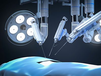
For children living with pediatric tumors, less invasive and less painful treatment with no radiation exposure was not always possible. In recent years, the development of technologies like Magnetic resonance guided high intensity focused ultrasound (MR-HIFU) and Low intensity transcranial focused ultrasound (LIFU) is helping to reverse that trend.

This topic was the focus of the recent Pediatric Device Innovators Forum (PDIF) hosted by the National Capital Consortium for Pediatric Device Innovation (NCC-PDI) in partnership with the U.S. Food and Drug Administration’s (FDA) Pediatric Device Consortia (PDC) grant program. A collaboration between Children’s National Hospital and University of Maryland Fischell Institute for Biomedical Devices, NCC-PDI is one of five PDCs funded by the FDA to support pediatric device innovators in bringing more medical devices to market for children.

The discussion, moderated by Kolaleh Eskandanian, Ph.D., MBA, PMP, vice president and chief innovation officer at Children’s National and principal investigator of NCC-PDI, explored the use of focused ultrasound’s noninvasive therapeutic technology for two pediatric indications, Osteoid Osteoma (OO) and Diffuse Intrinsic Pontine Glioma (DIPG), and the ways it can increase the quality of life for pediatric patients while also decreasing the cost of care.

The discussion also examined the most common barriers preventing more widespread implementation of focused ultrasound technology, specifically small sample size for evidence generation, lack of funding opportunities and reimbursement issues that can make or break a technology’s chances at reaching the patients that need it.

Karun Sharma, M.D., director of Interventional Radiology at Children’s National, emphasized the potential for focused ultrasound to treat localized pain relief and treat other diseases that, like OO, do not have any other therapeutic alternative

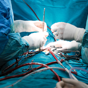
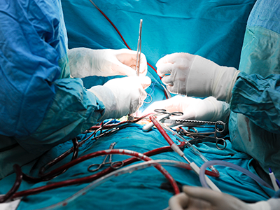
“At Children’s National, we use MR-HIFU to focus an ultrasound beam into lesions, usually tumors of the bone and soft tissues, to heat and destroy the harmful tissue in that region, eliminating the need for incisions,” says Sharma. “In 2015, Children’s National doctors became the first in the U.S. to use MR-HIFU to treat pediatric osteoid osteoma (OO), a painful, but benign, bone tumor that commonly occurs in children and young adults. The trial demonstrated early success in establishing the safety and feasibility of noninvasive MR-HIFU in children as an alternative to current, more invasive approaches to treat these tumors.”

In November 2020, the FDA approved this MR-HIFU system to treat OO in pediatric patients.

Roger Packer, M.D., senior vice president of the Center for Neuroscience and Behavioral Medicine at Children’s National, also discussed how focused ultrasound, specifically LIFU, has also proven to be an attractive modality for its ability to non-invasively, focally and temporarily disrupt the blood brain barrier (BBB) to allow therapies to reach tumors that, until recently, would have been considered unreachable without severe intervention.

“This presents an opportunity in pediatric care to treat conditions like Diffuse Intrinsic Pontine Glioma (DIPG), a highly aggressive brain tumor that typically causes death and morbidity,” says Packer.

Packer is planning a clinical trial protocol to investigate the safety and efficacy of LIFU for this pediatric indication.

The forum also featured insight from Jessica Foley, M.D., chief scientific officer, Focused Ultrasound Foundation; Arjun Desai, M.D., chief strategic innovation officer, Insighttec; Arun Menawat, M.D., chairman and CEO, Profound Medical; Francesca Joseph, M.D., Children’s National; Johannes N. van den Anker, M.D., Ph.D., vice chair of Experimental Therapeutics, Children’s National; Gordon Schatz, president, Schatz Reimbursement Strategies; Mary Daymont, vice president of Revenue Cycle and Care Management, Children’s National; and Michael Anderson, MD, MBA, FAAP, FCCM, FAARC, senior advisor to US Department of Health and Human Services (HHS/ASPR) and Children’s National.

Anthony Sandler, M.D., senior vice president and surgeon-in-chief of the Joseph E. Robert Jr. Center for Surgical Care and director of the Sheikh Zayed Institute for Pediatric Surgical Innovation at Children’s National Hospital, and Sally Allain, regional head of Johnson & Johnson Innovation, JLABS @ Washington, DC, opened the forum by reinforcing both organizations’ commitment to improving pediatric health.

In September 2020, the Focused Ultrasound Foundation designated Children’s National Hospital as the first global pediatric Center of Excellence for using this technology to help patients with specific types of childhood tumors. As a designated COE, Children’s National has the necessary infrastructure to support the ongoing use of this technology, especially for carrying out future pediatric clinical trials. This infrastructure includes an ethics committee familiar with focused ultrasound, a robust clinical trials research support team, a data review committee for ongoing safety monitoring and annual safety reviews, and a scientific review committee for protocol evaluation.

The Pediatric Device Innovators Forum is a recurring collaborative educational experience designed by the FDA-supported pediatric device consortia to connect and foster synergy among innovators across the technology development ecosystem interested in pediatric medical device development. Each forum is hosted by one of the five consortia. This hybrid event took place at the new Children’s National Research and Innovation Campus, the first-of-its-kind focused on pediatric health care innovation, on the former Walter Reed Army Medical Center campus in Washington, D.C.

To view the latest edition of the forum, visit the NCC-PDI website.

Panelists at the Pediatric Device Innovators Forum

The recent Pediatric Device Innovators Forum (PDIF) exploring the state of focused ultrasound was held at the new Children’s National Research and Innovation Campus, a first-of-its-kind focused on pediatric health care innovation.

Novel technique improved nerve visualization in head and neck surgery

In a pre-clinical model, researchers from Children’s National Hospital found that the Mueller polarimetric imaging, a novel technique that improves image contrast, may help identify nerves from other surrounding tissues during neck and head surgical procedures, avoiding accidental nerve damage.

“This technology holds great promise for the possibility of a truly noninvasive imaging approach and may help improve surgical outcomes by potentially reducing inadvertent, ill effects of nerve injuries in head and neck surgery,” said Bo Ning, Ph.D., R&D engineer at Children’s National and lead author of the study.

This pre-clinical study presents the first application of a full-field polarimetric imaging technique in vivo during head and neck surgery to highlight the vagus nerve (VN) and a branch that supplies all the intrinsic muscles to the larynx, known as recurrent laryngeal nerve (RLN).

“Unlike conventional nerve identification devices, this technique is noninvasive and less interruptive to intact tissues without disrupting surgical workflows,” said Ning et al. “Since the technique has an easy mechanism and promising performance in our study, this novel method holds great potential for real-time, noninvasive, and convenient nerve visualization.”

While some promising methods use polarimetric imaging for tissue characterizations, the current literature is still limited to ex vivo conditions due to the system complications and prolonged acquisition speeds.

“Recently, the industry released a new polarimetric camera, which is compact and allows fast and high-definition polarimetric imaging through simple snapshots. Enlightened by this technical advance, we have developed a practical polarimetric imaging method,” said Ning, who also develops compact and practical imaging systems for surgical innovation, including 3D, fluorescent, laser speckle and hyperspectral techniques. “It allows fast polarimetric analysis and can acquire birefringence maps over the whole field of view within 100 milliseconds, which provides an appropriate speed for directly surgical use.”

The new approach proofs that the concept is feasible to set up in live subjects during head and neck surgery, which can also be easily adapted for other surgeries. Among the seven subjects, the VNs and RLNs were successfully differentiated from arteries and other surrounding tissues.

Additional co-authors from Children’s National include Itai Katz, Ph.D., M.S., R&D staff engineer III; Anthony D. Sandler, M.D., Senior Vice President and Surgeon-in-Chief; Richard Jaepyeong Cha, Ph.D., research faculty assistant professor.

schematic of Mueller polarimetric imaging

Researchers at Children’s National used a novel technique that improves image contrast, which may help improve surgical outcomes.

Novel cancer vaccine targets oncogenes known to evade immunity in melanoma and neuroblastoma models

"Neuroblastoma of the Adrenal Gland (2)" by euthman is licensed under CC BY 2.0

Neuroblastoma of the Adrenal Gland (2)” by euthman is licensed under CC BY 2.0.

A personalized tumor cell vaccine strategy targeting Myc oncogenes combined with checkpoint therapy creates an effective immune response that bypasses antigen selection and immune privilege, according to a pre-clinical study for neuroblastoma and melanoma. The neuroblastoma model showed a 75% cure with long-term survival, researchers at Children’s National Hospital found.

Myc is a family of regulator genes and proto-oncogenes that help manage cell growth and differentiation in the body. When Myc mutates to an oncogene, it can promote cancer cell growth. The Myc oncogenes are deregulated in 70% of all human cancers.

Myc mutations, like the amplification of c-MYC and MYCN, are associated with host immune suppression in melanoma and neuroblastoma tumors, according to the study published in The Journal for Immunotherapy of Cancer.

“Paradoxically, from an immunotherapeutic perspective, a lack of an immune response may offer an opportunity to target those tumors [melanoma and neuroblastoma] that would be less resistant to host immunity assuming potent cellular immunity can be generated against the tumor,” said the authors.

The findings suggest that small molecule inhibitors — I-BET726 and JQ1 — suppress Myc’s uncontrolled cellular proliferation and enhance the immune response against tumor cells themselves, enabling their use as a tumor cell vaccine. The combination of cell vaccine and available therapies that keep the immune responses in check, also known as checkpoint inhibitor therapy, can help inform a personalized therapeutic tumor vaccine in the future.

“The work is pre-clinical and although we have seen excellent responses in these models, we need to determine whether this will also be effective in humans,” said Xiaofang Wu, staff scientist III at Sheikh Zayed Institute for Pediatric Surgical Innovation and lead author.  “For this purpose we have started laboratory testing in human cells. Our eventual hope is to translate these basic science findings to clinical application.”

There is a need for more effective therapies for neuroblastoma and melanoma, given the poor outcome of patients experiencing high-risk or advanced disease through traditional chemotherapy methods.  While the field has developed tumor vaccines and immune-based therapies, c-MYC and MYCN seem to protect the tumor against an immune response, so they often evade cure.

The researchers cautioned that both models induced potent immunity but draw different results, which means that this novel therapeutic vaccine is more effective in the neuroblastoma model than in the melanoma model. The neuroblastoma model resulted in a remarkable 75% cure and significantly improved long-term survival despite a larger initial tumor challenge.

“In contrast, the melanoma tumor gained adaptive resistance that is associated with an imbalance between tumor cell growth and cytotoxic killing and thus the vaccine failed to eradicate the tumor,” said the authors. “Despite potent immune effects from the vaccine, other immunosuppressive molecules will need to be targeted to see the full effects of the vaccine protocol in the melanoma model.”

The study proposes a framework that could be translated for therapeutic patient-specific vaccines for MYCN-amplified neuroblastoma tumors resistant to available therapies.

To understand the exact role of c-Myc and MYCN amplification and their association with immune suppression, the researchers examined 21 human neuroblastoma samples — the majority with metastatic disease — and 324 melanoma samples where only 30 were categorized as MYC amplified. Based on the oncogene’s capability to suppress the immune response, the researchers combined checkpoint inhibitors with pharmacologic molecules — I-BET726 and JQ1 — to target Myc oncogenes in mouse neuroblastoma and melanoma models. They also tested for the effects of different doses, drug combinations and incubation times on tumor cell proliferation, differentiation and gene alteration.

Authors on the study from Children’s National Hospital include: Xiaofang Wu, Ph.D., Marie Nelson, M.D., Mousumi Basu, Priya Srinivasan, Ph.D., Christopher Lazarski, Ph.D., and Anthony Sandler, M.D.

Fighting lethal cancer with a one-two punch

The immune system is the ultimate yin and yang, explains Anthony D. Sandler, M.D., senior vice president and surgeon-in-chief of the Joseph E. Robert Jr. Center for Surgical Care at Children’s National in Washington, D.C. With an ineffective immune system, infections such as the flu or diarrheal illness can run unchecked, causing devastating destruction. But on the other hand, excess immune activity leads to autoimmune diseases, such as lupus or multiple sclerosis. Thus, the immune system has “checks and balances” to stay controlled.

Cancer takes advantage of “the checks and balances,” harnessing the natural brakes that the immune system puts in place to avoid overactivity. As the cancer advances, molecular signals from tumor cells themselves turn on these natural checkpoints, allowing cancers to evade immune attack.

Several years ago, a breakthrough in pharmaceutical science led to a new class of drugs called checkpoint inhibitors. These medicines take those proverbial brakes off the immune system, allowing it to vigorously attack malignancies. However, Dr. Sandler says, these drugs have not worked uniformly and in some cancers, they barely work at all against the cancer.

One of these non-responders is high risk neuroblastoma, a common solid tumor found outside the skull in children. About 800 U.S. children are diagnosed with this cancer every year. And kids who have the high-risk form of neuroblastoma have poor prognoses, regardless of which treatments doctors use.

However, new research could lead to promising ways to fight high-risk neuroblastoma by enabling the immune system to recognize these tumors and spark an immune response. Dr. Sandler and colleagues recently reported on these results in the Jan. 29, 2018, PLOS Medicine using an experimental model of the disease.

The researchers created this model by injecting the preclinical models with cancer cells from an experimental version of neuroblastoma. The researchers then waited several days for the tumors to grow. Samples of these tumors showed that they expressed a protein on their cell surfaces known as PD-L1, a protein that is also expressed in many other types of human cancers to evade immune system detection.

To thwart this protective feature, the researchers made a cancer vaccine by removing cells from the experimental model’s tumors and selectively turning off a gene known as Id2. Then, they irradiated them, a treatment that made these cells visible to the immune system but blocked the cells from dividing to avoid new tumors from developing.

They delivered these cells back to the experimental models, along with two different checkpoint inhibitor drugs – antibodies for proteins known as CLTA-4 and PD-L1 – over the course of three treatments, delivered every three days. Although most checkpoint inhibitors are administered over months to years, this treatment was short-term for the experimental models, Dr. Sandler explains. The preclinical models were completely finished with cancer treatment after just three doses.

Over the next few weeks, the researchers witnessed an astounding turnaround: While experimental models that hadn’t received any treatment uniformly died within 20 days, those that received the combined vaccine and checkpoint inhibitors were all cured of their disease. Furthermore, when the researchers challenged these preclinical models with new cancer cells six months later, no new tumors developed. In essence, Dr. Sandler says, the preclinical models had become immune to neuroblastoma.

Further studies on human patient tumors suggest that this could prove to be a promising treatment for children with high-risk neuroblastoma. The patient samples examined show that while tumors with a low risk profile are typically infiltrated with numerous immune cells, tumors that are high-risk are generally barren of immune cells. That means they’re unlikely to respond to checkpoint inhibiting drugs alone, which require a significant immune presence in the tumor microenvironment. However, Dr. Sandler says, activating an immune response with a custom-made vaccine from tumor cells could spur the immune response necessary to make these stubborn cancers respond to checkpoint inhibitors.

Dr. Sandler cautions that the exact vaccine treatment used in the study won’t be feasible for people. The protocol to make the tumor cells immunogenic is cumbersome and may not be applicable to gene targeting in human patients. However, he and his team are currently working on developing more feasible methods for crafting cancer vaccines for kids. They also have discovered a new immune checkpoint molecule that could make this approach even more effective.

“By letting immune cells do all the work we may eventually be able to provide hope for patients where there was little before,” Dr. Sandler says.

In addition to Dr. Sandler, study co-authors include Priya Srinivasan, Xiaofang Wu, Mousumi Basu and Christopher Rossi, all of the Joseph E. Robert Jr. Center for Surgical Care and The Sheikh Zayed Institute for Pediatric Surgical Innovation (SZI), at Children’s National in Washington, D.C.

Financial support for research described in this post was provided by the EVAN Foundation, the Catherine Blair foundation, the Michael Sandler Research Fund and SZI.

ID-KD vaccine induced T-cell cytotoxicity

Mechanism of Id2kd Neuro2a vaccination combined with α-CTLA-4 and α-PD-L1 immunotherapy in a neuroblastoma model. During a vaccine priming phase, CTLA-4 blockade enhances activation and proliferation of T-cells that express programmed cell death 1 (PD1) and migrate to the tumor. Programmed cell death-ligand 1 (PD-L1) is up-regulated on the tumor cells, inducing adaptive resistance. Blocking PD-L1 allows for enhanced cytotoxic effector function of the CD8+ tumor-infiltrating lymphocytes. Artist: Olivia Abbate

Anthony Sandler, M.D., Named Director of Sheikh Zayed Institute

Anthony Sandler

Children’s National Health System is pleased to announce that Anthony Sandler, M.D., current senior vice president and surgeon-in-chief of the Joseph E. Robert Jr. Center for Surgical Care at Children’s National, will now additionally assume the title of director, Sheikh Zayed Institute for Pediatric Surgical Innovation. He will succeed Peter Kim, M.D., the founding vice president of the Sheikh Zayed Institute, who is leaving to pursue other career opportunities after seven years at the helm of our surgical innovation center.

Dr. Sandler will be in a unique position, leading both in the research and clinical enterprises of Children’s National and will help to forge a stronger link between them, especially in the surgical subspecialties.

Internationally known for his work on childhood solid tumors and operative repair of congenital anomalies, Dr. Sandler is the Diane and Norman Bernstein Chair in Pediatric Surgery and is a professor of surgery and pediatrics at the George Washington University School of Medicine & Health Sciences. He is currently on the Board of Examiners for the Pediatric Surgery Qualifying Examination and has served on multiple committees for the American Pediatric Surgical Association and for the Children’s Oncology Group.

Dr. Sandler’s research interests focus on solid tumors of childhood and he’s presently studying tumor immunology and investigating immunotherapeutic vaccine strategies. He has co-developed a surgical polymer sealant that is R01 funded by the National Institutes of Health and is currently in pre-clinical trials. Dr. Sandler has over 120 peer-reviewed publications in clinical and scientific medical journals.

Treatment of neuroblastoma with immunotherapy and vaccine combination shows promise

Anthony Sandler

“Treatment options like these that help the body use its own immune system to fight off cancer are incredibly promising, and we look forward to continuing this work to understand how we can best help our patients and their families,” said Anthony Sandler, M.D.

Despite being the most common extracranial solid tumor found in children and having multiple modes of therapy, neuroblastoma continues to carry a poor prognosis. However, a recent cutting-edge pre-clinical study, PD-L1 checkpoint inhibition and anti-CTLA-4 whole tumor cell vaccination counter adaptive immune resistance: A mouse neuroblastoma model that mimics human disease, published in PLOS Medicine shows the first signs of success in treating high-risk neuroblastoma, a promising step not only for neuroblastoma patients, but potentially for other types of cancer and solid tumors as well. While the research was conducted on mouse models and is in the early stages, the lead author of the study, Anthony Sandler, M.D., senior vice president and surgeon-in-chief of the Joseph E. Robert, Jr., Center for Surgical Care at Children’s National, believes these findings are an encouraging development for the field.

The treatment method combines a novel personalized vaccine and a combination of drugs that target checkpoint inhibitors enabling the immune system to identify and kill cancer cells. When these checkpoints are blocked, it’s similar to taking the brakes off the immune system so that the body’s T cells can be primed by the vaccine, identify the tumor and allow for targeted tumor cell killing. The vaccine then brings in reinforcements to double down on the attack, helping to eradicate the tumor. The vaccine could also be used as a way to prevent recurrence of disease. After a patient has received the vaccine, the T cells would live in the body, remembering the tumor cells, and attack reemerging cancer in a similar way that a flu vaccine helps fight off the flu virus.

“Treatment options like these that help the body use its own immune system to fight off cancer are incredibly promising, and we look forward to continuing this work to understand how we can best help our patients and their families,” said Dr. Sandler.

A vaccine approach to tumor cure

Anthony Sandler

Anthony Sandler, M.D, is trying to understand how cancer cells can change their behavior and activate the immune system – enlisting the patients’ own defenses to fight the tumor.

Building on their groundbreaking research that found a method to cure neuroblastoma tumors in mice, researchers at Children’s National have been working in recent months on a personalized tumor-specific vaccine approach for neuroblastoma and other solid tumors.

The possibility that such a vaccine could non-invasively cure one of the most common childhood cancers is part of Children’s innovative efforts to address some of the most critical medical research challenges facing the field. Anthony Sandler, M.D., Senior Vice President, the Joseph E. Robert, Jr. Center for Surgical Care, and the Diane and Norman Bernstein Professor in Pediatric Surgery, is leading the research that followed an initial publication in PLOS ONE. Sandler’s team seeks to understand how cancer cells can change their behavior and activate the immune system – enlisting the patients’ own defenses to fight the tumor.

Their research is particularly significant because neuroblastoma, most commonly centered in the adrenal glands, is the third most common tumor in childhood, and the most common cancer in babies younger than one year old. It accounts for six percent of all childhood cancers in the United States, with about 700 children younger than 15 diagnosed each year.

“Historically, tumor vaccines held much promise, but demonstrated little clinical success,” Dr. Sandler and his team wrote in their study. “Thus, the task of establishing an effective anti-tumor response in neuroblastoma has been daunting.” However, with this most recent study finding, Dr. Sandler says this failed promise is changing.

The study revealed that “knockdown’” of a DNA-protein inhibitor, known as ID-2, in aggressive high-risk solid tumors resulted in activation of T-cells, which are white blood cells that have figured significantly in immunity research. Gene knockdown refers to a technique in which the expression of one or more of a cell’s genes is reduced.

The research also focused on using “checkpoint blockade,” a therapy in clinical use that allows for expansion of the immune response against tumors. “The combination of selective gene knock-down in tumor cells and checkpoint blockade produced a novel, potent T-cell triggered tumor vaccine strategy,” Dr. Sandler says.

As Children’s researchers examined the impact of the knockdown of ID-2 protein on a tumor, they implanted N2a, a fast growing mouse neuroblastoma cell line, in the mice. Unexpectedly, Sandler said, “Most of the mice rejected the tumor cells and subsequently were protected against further tumor challenges.”

The researchers also noted that a “massive influx” of T-cells infiltrated the shrinking tumor, indicating that T-cells are necessary for antitumor immunity in this vaccination protocol.

The ultimate goal for Sandler’s team is to work toward potential clinical trials to make further progress in neuroblastoma research, with immunotherapy playing a key role.

Dr. Sandler is the Principal Investigator of the Immunology initiative of the Sheikh Zayed Institute for Pediatric Surgical Innovation, and has worked in immunology research related to childhood cancers for more than 20 years.