Tag Archive for: Rood

Drs. Robert Keating, Brian Rood and Catherine Bollard

Children’s National announces new professorships

Drs. Robert Keating, Brian Rood and Catherine Bollard

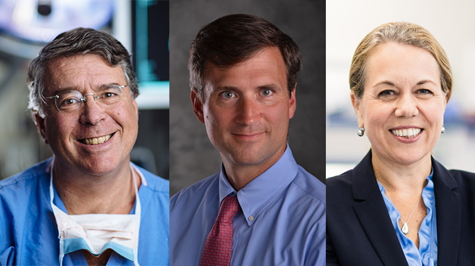
Robert Keating, M.D., Brian Rood, M.D., and Catherine Bollard, M.D., M.B.Ch.B.

Children’s National Hospital named Robert Keating, M.D., as the McCullough Distinguished Professor of Neurosurgery. He serves as the chief of neurosurgery and co-director of the high-intensity focused ultrasound (HIFU) program at Children’s National.

Children’s National Hospital named Brian Rood, M.D., as the Kurt D. Newman, M.D., Professor of Neuro-Oncology. He serves as director of clinical neuro-oncology and medical director of the Brain Tumor Institute at Children’s National.

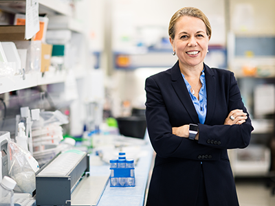
Children’s National Hospital elevated Catherine Bollard, M.D., M.B.Ch.B., to the Dr. Robert J. and Florence T. Bosworth Distinguished Professor of Cancer and Transplantation Biology Research. She is the Interim Executive Vice President and Chief Academic Officer and Interim Director, Children’s National Research Institute. She also serves as the director of the Center for Cancer and Immunology Research and director of the Program for Cell Enhancement and Technologies for Immunotherapy at Children’s National.

About the awards

Professorships at Children’s National support groundbreaking work on behalf of children and their families and foster new discoveries and innovations in pediatric medicine. These appointments carry prestige and honor that reflect the recipient’s achievements and donor’s forethought to advance and sustain knowledge. Children’s National is grateful for its generous donors, who have funded 47 professorships.

Dr. Keating is a longstanding leader in neurosurgery research and care. His areas of expertise include brain tumors, traumatic brain injuries, craniofacial anomalies, Chiari malformations and spinal dysraphism. With Dr. Keating’s leadership, the neurosurgery department is pioneering innovations such as HIFU, a non-invasive therapy using focused ultrasound waves to ablate a focal area of tissue. It can treat tumors located in difficult locations of the brain, movement disorders and epilepsy. Children’s National was one of the first pediatric hospitals in the nation to use HIFU for neuro-oncology patients.

“Our goal is to elevate our top-ranked program to even greater heights,” says Dr. Keating. “We will continue to use cutting-edge technology and non-invasive approaches to make the knife obsolete in pediatric neurosurgery and improve outcomes for children.”

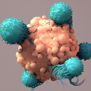
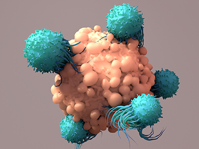
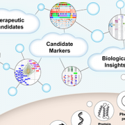
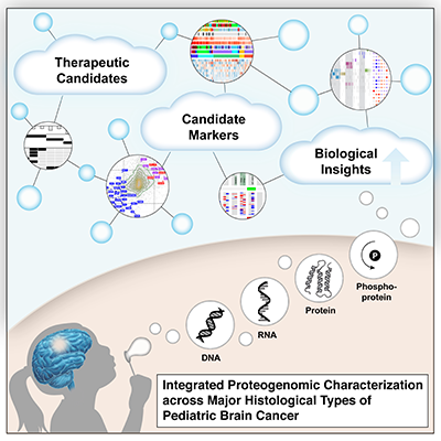
Dr. Rood studies the biology of pediatric brain tumors. He focuses on protein signatures and biomarkers specific to different types of brain cancers. His study of neoantigens is informing the development of T-cell immunotherapies to target a tumor’s unique proteins.

“Immunotherapy is revolutionizing how we treat childhood brain tumors — safely, effectively and with the precision made possible by using a patient’s own cells,” says Dr. Rood. “This professorship enables our team to advance this revolution, which will save lives and improve lifetimes.”

Dr. Bollard received the Dr. Robert J. and Florence T. Bosworth Professor of Cancer and Transplantation Biology Research in 2018 to support her work to develop cell and gene therapies for patients with cancer and underlying immune deficiencies. Her professorship has been elevated to a distinguished professorship to amplify her research and celebrate her accomplishments in the field of immunotherapy.

About the donor

These appointments were made possible through an extraordinary $96 million investment from an anonymous donor family for rare pediatric brain tumor research and care. It is one of the hospital’s largest donations and will transform the hospital’s ability to give patients with rare brain cancer a better chance at healthy lifetimes.

The anonymous family brings a depth of compassion for children facing rare and often challenging diagnoses. Their partnership will immediately advance every aspect of our globally recognized leadership to create new, more effective treatments.

Their investment also endowed the Professorship in Molecular Neuropathology. We look forward to bestowing that honor on a Children’s National pediatric leader.

t cells fighting cancer cell

Personalized T cell immunotherapy for brain tumors closer to becoming reality

t cells fighting cancer cell

Children’s National Hospital experts developed a new approach that discovered unique proteins in an individual tumor’s cells, which then helped scientists generate personalized T cells to target and kill tumors.

Children’s National Hospital experts developed a new approach that discovered unique proteins in an individual tumor’s cells, which then helped scientists generate personalized T cells to target and kill tumors, according to a pre-clinical study published in Nature Communications.

This effort is the first to create a new workflow for neoantigen identification that incorporates both genetic sequencing and protein identification to create a personalized treatment for medulloblastoma in children, a common malignant brain tumor. Given these promising findings, the researchers are now designing a phase I clinical trial slated to open in 12-18 months.

“This work is an incredibly exciting advancement in personalized medicine. It will allow us to treat patients with a novel T cell therapy that is developed for each individual patient to specifically attack and kill their tumor,” said Catherine Bollard, M.D., M.B.Ch.B., director of the Center for Cancer and Immunology Research at Children’s National and co-author on the paper. “This treatment will offer a potential option for children with hard-to-treat brain tumors for which all other therapeutic options have been exhausted.”

Catherine Bollard

Catherine Bollard, M.D., M.B.Ch.B., director of the Center for Cancer and Immunology Research at Children’s National and co-senior author on the paper.

First, the researchers sequenced the DNA of small tissue samples while studying its complete set of proteins that influence cancer biology — also named a “low-input proteogenomic approach” by the authors. After analyzing the empirical data, which shies away from the commonly used predictive models, the researchers developed a T cell immunotherapy that targets the tumor’s unique proteins and allows the T cells to distinguish between healthy cells and tumor cells. This means that Rivero-Hinojosa et al. managed to merge two research fields, proteogenomics and immunotherapy, and lay the groundwork for personalized, targeted T cell therapies to treat children with brain tumors.

“Neoantigen discovery techniques have either been dependent upon in silico prediction algorithms or have required a significant amount of tumor tissue, making them inappropriate for most brain tumors,” said Brian Rood, M.D., medical director of Neuro-oncology and the Brain Tumor Institute at Children’s National. “This neoantigen identification pipeline creates a new opportunity to expand the repertoire of T cell-based immunotherapies.”

Tumor cells have damaged DNA that create mutations during the repair process because they do not do a good job at maintaining their DNA fidelity. The repairs therefore create aberrant DNA that codes for proteins that were never intended by the genetic code and, consequently, they are unique to the individual’s tumor cells.

Brian Rood

Brian Rood, M.D., medical director of Neuro-oncology and the Brain Tumor Institute at Children’s National and co-senior author on the paper.

“We developed a new filtering pipeline to remove non-annotated normal peptides. Targeting antigens that are completely specific to the tumor, and expressed nowhere else in the body, will potentially increase the strength of tumor antigen-specific T cell products while decreasing the toxicity,” said Samuel Rivero-Hinojosa, Ph.D., staff scientist at Children’s National and first author of the study.

Once the experts identified these unique peptides, they used them to select and expand T cells, which showed specificity for the tumor specific neoantigens and the ability to kill tumor cells. The next step is to conduct a clinical trial in which a patient’s own T cells are trained to recognize their tumor’s unique neoantigens and then reinfused back into the patient.

From an immunotherapy standpoint, tumor specificity is important because when clinicians treat patients with T cell therapies, they want to make sure that the T cells directly target and kill the tumor and will not cause devastating harm to healthy cells. This paper demonstrated that it may be possible to create a better efficacy and safety margin with this new approach.

In the past five years, under the leadership of Dr. Bollard, the Center for Cancer and Immunology Research at Children’s National has advanced the scientific knowledge in preclinical and clinical settings. The center discovered a signaling pathway that can be hijacked to prevent brain tumor development, and further advanced translational research with several key first-in-human studies that utilized novel cell therapies to treat cancer and life-threatening viral infections.

graphic abstract for brain tumor paper

First large-scale proteogenomic analysis offers insights into pediatric brain tumor biology

graphic abstract for brain tumor paper

In the first large-scale, multicenter study of its kind, researchers conducted comprehensive analysis yielding a more complete understanding of pediatric brain tumors (PBT), which are the leading cause of cancer-related deaths in children. Researchers from the Clinical Proteomic Tumor Analysis Consortium (CPTAC) and Children’s Brian Tumor Network (CBTN) generated and analyzed proteomic data, which identifies common biological characteristics among different tumor types. The consortia consist of collaborators from the Icahn School of Medicine at Mount Sinai, National Cancer Institute, Fred Hutchinson Cancer Research Center, Children’s National Hospital and Children’s Hospital of Philadelphia. The study, published in Cell on November 25, 2020, provides a clearer understanding of the molecular basis of pediatric brain tumors and proposes new therapeutic avenues.

The molecular characterization of brain tumors has largely hinged upon the presence of unique alterations in the tumor genome ignoring the many layers of regulation that exist between DNA and the functional biology of the tumor cell that is actuated by proteins. The integration of proteomic data identifies common biological themes that span histologic boundaries, suggesting that treatments used for one histologic type may be applied effectively to other tumors sharing similar proteomic features.

Brian Rood, M.D., medical director of the Brain Tumor Institute and associate professor of pediatrics in the Center for Cancer and Blood Disorders at Children’s National Hospital, participated in this study and explains the importance of what the team discovered.

Q: Why was it important that researchers came together to do this work?

A: Comprehensive characterization of the fundamental biology of pediatric brain tumors, including the proteogenomic analysis done in this study, is essential to better understand and treat pediatric brain tumors.

Our study is based on the recognition that proteomics and phosphoproteomics needs to be integrated with other omics data to gain an improved systems biology view of the molecular features of brain tumors. In addition, characterizing biological themes that cross histologic boundaries and cells of origin can suggest extending treatments shown to be effective in one type of tumor to other histologically disparate tumors sharing the same proteomic features.

Proteomic data further reveal the functional impacts of somatic mutations and copy number variations (CNVs) not evident in transcriptomic data alone. Further, kinase-substrate network analyses identify activated biological mechanisms of tumor biology.

This work was only possible because of a unique collaboration between the CPTAC program of the NCI and the CBTN, of which Children’s National is a member.

Q: How will this work advance understanding and treatment of pediatric brain tumors?

A: Pediatric brain tumors have not benefitted from molecularly targeted drugs as much as other tumor types largely because they harbor relatively few gene mutations. Therefore, identifying key pathways to target in these patients’ tumors has been a challenge. The integration of proteomic and phosphoproteomic data with genomic data allows for the construction of a more comprehensive model of brain tumor biology and nominates specific key pathways to be targeted.

Q: What did you find that excites you?

A: Proteomic data revealed a number of findings that were not present in the genomic data. We found evidence to support a molecularly targeted approach to treating craniopharyngioma, a tumor that has previously been unresponsive to chemotherapy. We also found a prognostic marker for high grade gliomas that do not have a mutation in the H3 histone. We were able to identify specific kinases that may dictate the aggressive nature of certain ependymoma tumors. Importantly, we demonstrated the potential of proteomic studies to uncover unique tumor biology, paving the way for more extensive investigations using this approach.

You can find the full study published in Cell. Learn more about the Brain Tumor Institute at Children’s National.


Dr. Rood recently joined a live panel discussion with researchers from the Children’s Brain Tumor Network and the Clinical Proteomic Tumor Analysis Consortium to explore the impact of their landmark study.

pastel colored DNA strands

Germline microsatellite genotypes differentiate children with medulloblastoma

pastel colored DNA strands

A new study suggests that medulloblastoma-specific germline microsatellite variations mark those at-risk for medulloblastoma development.

Brian Rood, M.D., oncologist and medical director at the Brain Tumor Institute, and Harold “Skip” Garner, Ph.D., associate vice provost for research development at Edward Via College of Osteopathic Medicine, published a report in the Society for Neuro-Oncology’s Neuro-Oncology Journal about using a novel approach to identify specific markers in germline (non-tumor) DNA called microsatellites that can differentiate children who have the brain tumor medulloblastoma (MB) from those who don’t.

“Ultimately, the best way to save children from brain tumors and prevent them from bearing long-term side effects from treatment is to prevent those tumors from occurring in the first place,” says Dr. Rood. “New advancements hold the potential to finally realize the dream of cancer prevention, but we must first identify those children at-risk.”

While analyzing germline sequencing data from a training set of 120 MB subjects and 425 controls, the doctors identified 139 individual microsatellites whose genotypes differ significantly between the groups. Using a genetic algorithm, they were able to construct a subset of 43 microsatellites that distinguish MB subjects from controls with a sensitivity and specificity of 92% and 88% respectively.

“We made discoveries in an untapped part of the human genome, enabled by unique bioinformatics data mining approaches combined with clinical insight,” said Dr. Garner. “Our findings establish new genomic directions that can lead to high accuracy diagnostics for predicting susceptibility to medulloblastoma.”

What the doctors discovered and demonstrated in the study was that MB-specific germline microsatellite variations mark those at risk for MB development and suggest that other mechanisms of cancer predisposition beyond heritable mutations exist for MB.

“This work is the first to demonstrate the ability of specific DNA sequences to differentiate children with cancer from their healthy counterparts,” added Dr. Rood.

Contributing Authors to this research study included:  Brian R. Rood, M.D., Harold R. Garner, Ph.D., Samuel Rivero-Hinojosa, Ph.D., and Nicholas Kinney, Ph.D.

Brian Rood

Improving the understanding of medulloblastoma

Brian Rood

Brian Rood, M.D., employed quantitative proteomics to tumor samples that led to novel therapeutic targets for Medulloblastoma and other tumors.

In a recently published study, Brian Rood, M.D., a neuro-oncologist at Children’s National Health System, employed quantitative proteomics to tumor samples, a technique that could lead to novel therapeutic targets for medulloblastoma and other tumors in the future.

Currently, many experts use genomic characterization to understand the genetic makeup of cancer cells, which has deepened the field’s collective knowledge of tumor biology. However, it has remained challenging to infer specific information about how the tumors will respond and consequently develop more effective therapies. Medulloblastoma is the most common pediatric, malignant brain tumor. Through Dr. Rood’s research using proteomic analysis, he was able to identify and measure the protein makeup of medulloblastoma, which led to a potential pathway for clinical intervention to treat this life-threatening cancer. The findings were published online June 7, 2018, in Acta Neuropathologica Communications.

“The goal of this research was to find out how these tumor cells function at the protein level, which may ultimately help the field identify drug therapies to stop them,” says Dr. Rood. “The genes of a cancer cell are like a blueprint for a building, but the blueprints aren’t always followed in a cancer cell: Not every active gene will produce its corresponding protein. Proteins do the work of the cell, and understanding them will provide a better overall understanding of a cancer cell’s biology.”

Dr. Rood compared proteomic and genomic data to confirm that genetics do not accurately predict the quantity of proteins. By directly quantitating the proteins and comparing them between different subgroups of the disease, they were able to identify protein-based pathways driving tumor biology. With this information, Dr. Rood was able to demonstrate that medulloblastoma depends on a crucial pathway, the eukaryotic initiation factor 4F protein synthesis pathway, resulting in the identification of a potential target for new treatments in medulloblastoma.

Ultimately, Dr. Rood found that proteomic analysis complements genomic characterization and the two can be used together to create a more complete understanding of tumor biology. Going forward, he hopes proteomic analysis will become common practice for studying all tumors, allowing tumors to be categorized and grouped together by protein makeup to help the field identify more effective therapies for all tumors.