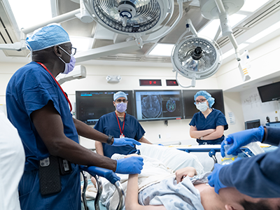
Currently, studies on typical brain and cranium development are limited. One reason for this is that imaging techniques are optimized to best visualize either bone or soft tissue, but not both.
With prevalence of developmental disorders on the rise, the need to understand brain development has never been more critical. Development of the brain is strongly influenced by the cranium, but this relationship has not been adequately studied because of limitations in imaging technology. Now, researchers from Children’s Hospital Los Angeles and Children’s National Hospital are working together to develop techniques that will provide greater insight into this relationship. Their studies will be funded by The National Institute of Dental and Craniofacial Research, which has awarded them $3.5 million.
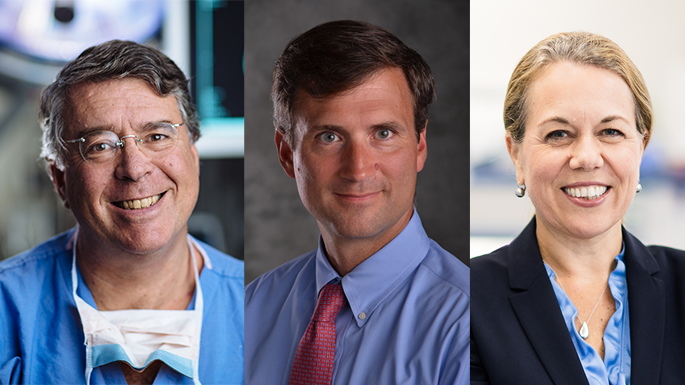
Natasha Leporé, Ph.D., of Children’s Hospital Los Angeles, studies methods to interpret brain imaging data. “There’s a lot of interaction between the skull and the brain,” she says, “and we want to better understand how they grow together.”
Currently, studies on typical brain and cranium development are limited. One reason for this is that imaging techniques are optimized to best visualize either bone or soft tissue, but not both.
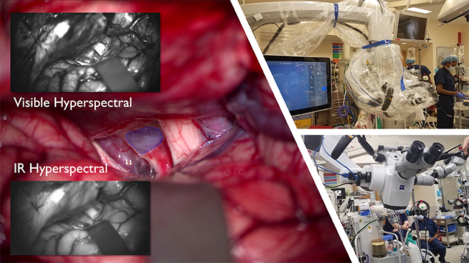
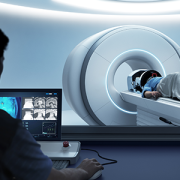
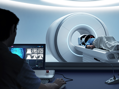
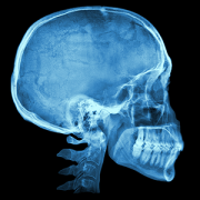
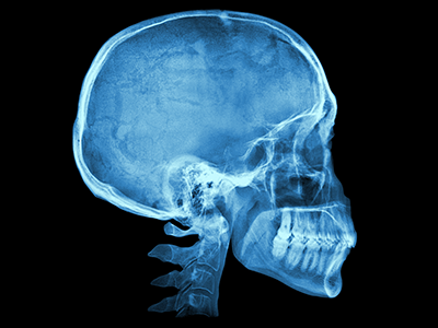
The brain — mostly composed of water, protein and fat — doesn’t show up well on computerized tomography (CT) scans, which use X-ray images. In addition, radiation exposure limits the amount of CT scan data available in children. On the other hand, magnetic resonance imaging (MRI) scans are excellent for brain images but are not optimal for surrounding bone.
This presents researchers with a dilemma if they want to see the brain and the skull together in one image. Fortunately, research barriers like these are often overcome by collaboration.
Leporé will work with Marius George Linguraru, D.Phil, M.A., MS.c., principal investigator in the Sheikh Zayed Institute for Pediatric Surgical Innovation at Children’s National Hospital.
Linguraru works on a set of tools for cranial phenotyping, using existing CT images from typically developing children. In their collaboration, Leporé and Linguraru will extend the tools to MRI scans, allowing the team to analyze the brain and cranium simultaneously. The pair has received a $3.5 million award over 5 years.
“The tools we develop together will help us to better understand the healthy growth of children,” says Linguraru. “We will have the ability to analyze the joint cranial and brain development from large medical image datasets of pediatric patients.”
This, the team says, will be invaluable to the medical community.
“These tools will help clinicians to better assess, diagnose and plan treatment for infants with cranial deformities,” says Linguraru.
Collaborations like this allow expertise to be shared across specialties, ultimately benefiting children in need. Exceptional pediatric care is a result of teamwork; not only doctors, nurses and clinical staff, but also biomedical research, which arms clinicians with the information they depend on.
“We need to have a clear idea of what is expected in normal development,” says Leporé. “This allows doctors to detect and better understand differences in development.”
Other members of the research team include: Vidya Rajagopalan, Ph.D.; Marvin Nelson, M.D.; Alexis Johns, Ph.D.; Niharika Gajawelli, Ph.D. (from Children’s Hospital Los Angeles and University of Southern California); Robert Keating, M.D. (Children’s National Hospital); Yalin Wang, Ph.D. (Arizona State University); Antonio Porras, Ph.D. (University of Colorado); Sean Deoni, Ph.D. (Rhode Island Hospital and Brown University).
A version of this story appeared on the Children’s Hospital Los Angeles newsroom.
 The Divisions of Neurology and Neurosurgery at Children’s National Hospital are proud to host the 35th Annual Pediatric Neurology Update course.