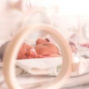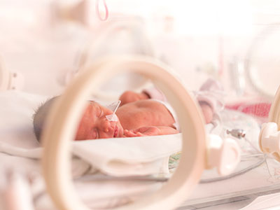
Pandemic stress reshapes the placentas of expectant moms
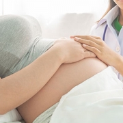
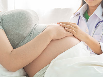
Elevated maternal stress during the COVID-19 pandemic changed the structure, texture and other qualities of the placenta in pregnant mothers.
Elevated maternal stress during the COVID-19 pandemic changed the structure, texture and other qualities of the placenta in pregnant mothers – a critical connection between mothers and their unborn babies – according to new research from the Developing Brain Institute at Children’s National Hospital.
Published in Scientific Reports, the findings spotlight the underappreciated link between the mental health of pregnant mothers and the health of the placenta – a critical organ that develops during pregnancy to nourish and protect babies. The long-term neurodevelopmental impact on their children is under investigation.
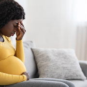
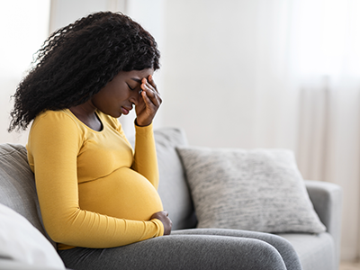
“During the pandemic, mothers were exposed to a litany of negative stressors including social distancing, fear of dying, financial insecurity and more,” said Catherine Limperopoulos, Ph.D., chief and director of the Developing Brain Institute, which led the research. “We now know that this vital organ was changed for many mothers, and it’s essential that we continue to investigate the impact this may have had on children who were born during this global public health crisis.”
The big picture
Dr. Limperopoulos’s team compared magnetic resonance imaging (MRI) of 165 women who were pregnant before March 2020 to 63 women who became pregnant during the pandemic. Those pregnant during the pandemic were not knowingly exposed to COVID-19, and they collectively scored significantly higher on questionnaires measuring stress and depression. They were recruited at Children’s National as part of a clinical trial aimed at reducing pregnant women’s elevated stress levels during the pandemic.
The placenta is a temporary organ that grows during pregnancy to provide oxygen, nutrients and immunological protection to babies, and its health is vital to the well-being of the developing fetus. The data showed key changes in how the placenta grew and developed among women pregnant during the pandemic, especially when compared to placental growth and development among women who were pregnant before the pandemic. Changes in placental development also were associated with the infant’s birth weight at delivery. Importantly, these changes seem to be connected to maternal stress and depression symptoms.
Taken as a whole, the findings suggest that the disturbances measured on placental development in the womb may influence the placenta’s ability to support fetal health and wellness. “We are continuing to follow up on these mother-baby dyads to determine the long-term functional significance of these placental changes in utero,” Dr. Limperopoulos said.
Studies have shown that the placenta adapts to negative changes in the maternal environment and mental health status, and disruptions in placental function impact infant brain development and children’s neurobehavior and temperament.
The patient benefit
Dr. Limperopoulos’s research studying childbirth amid the pandemic builds on her extensive work investigating the impact of maternal stress on unborn children, including its adverse effect on brain structure and biochemistry. She’s also working on treatments and interventions to better support new families. Her program, DC Mother-Baby Wellness, brings together community partners to provide wrap-around care to expectant and new moms with elevated scores for stress, anxiety and depression.
“When identified early, maternal stress is a modifiable risk factor that can be treated with psychotherapy, social support and other personalized, evidence-based interventions,” Dr. Limperopoulos said. “We look forward to continued research in this area to better understand the mechanisms behind these biological changes and the needs of mothers and children who are born during pandemics, natural disasters and other significantly stressful events.”