Tag Archive for: neonatologist

newborn baby

Directly measuring function in tiny hearts

newborn baby

The amount of blood the heart pumps in one minute can be directly measured safely in newborns by monitoring changes in blood velocity after injecting saline, indicates the first clinical study of direct cardiac output measurement in newborns.

The amount of blood that the heart pumps in one minute (cardiac output) can be directly measured safely in newborns by monitoring changes in blood velocity after injecting saline, indicates a paper published online Dec. 17, 2019 in the Journal of Pediatrics and Neonatal Medicine. The research, conducted by Children’s National Hospital faculty, is believed to be the first clinical study of direct cardiac output measurement in newborns.

Right now, cardiac output is measured indirectly in the nation’s neonatal intensive care units (NICU) using newborns’ blood pressure, heart rate, urine output and other indirect measures. However, these techniques can produce imprecise readings in children. And the field lacks a feasible “gold standard” to measure cardiac output in newborns.

The COstatus monitor already uses ultrasound dilution – the expected decrease in the velocity of blood when saline is injected, producing a dilution curve. A Children’s National research team used ultrasound dilution in their small pilot study to gauge the feasibility of directly measuring cardiac output in newborns.

“Infants who stand to benefit most from directly monitoring cardiac hemodynamics are often so sick they already have central venous access,” says Khodayar Rais-Bahrami, M.D., an attending neonatologist at Children’s National and the study’s senior author. “Using the COstatus monitor in these children would enable the clinical team to personalize care based on the newborn’s current hemodynamic status, while introducing minimal fluid during measurements,” Dr. Rais-Bahrami adds.

COstatus monitor

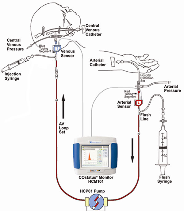
The COstatus Monitor uses an extracorporeal loop attached to arterial and venous lines to measure cardiac output using ultrasound dilution. The research team injected 1mL/kg of body temperature saline into the loop and performed up to two measurement sessions daily.

The research team recruited 12 newborns younger than 2 weeks old who already had central venous and arterial access. The venous line of the arteriovenous AV loop is connected to the umbilical venous catheter while the COstatus monitor’s arterial line is connected to the umbilical arterial catheter. During measurement sessions, two injections of solution are injected into the venous loop, allowing for two measures of cardiac output, cardiac index, active circulating volume index, central blood volume index and systemic vascular resistance index.

Infants enrolled in the pilot study underwent up to two measurement sessions per day for up to four days, for a total of 54 cardiac hemodynamic measurements. The newborns ranged from 720 to 3,740 grams in weight and 24 to 41.3 weeks in gestational age.

The infants’ mean cardiac output was 0.43L/min and increased with gestational age. By contrast, the mean cardiac index was 197mL/kg/min and changed little with infants’ increasing maturity – either by gestational age or postnatal age. Two of the study participants were undergoing therapeutic cooling for hypoxic-ischemic encephalopathy and had their measurements taken during cooling and after rewarming.

“Although this study size is small, it demonstrates that this minimally invasive technique can safely be used in newborns to directly measure cardiac hemodynamics,” says Simranjeet S. Sran, M.D., a Children’s National neonatalogist and the study’s lead author. “This technology may allow for more precise and personalized care of critically ill newborns in a range of disease states – real-world utility in NICUs that serve some of the youngest and sickest newborns,” Dr. Sran adds.

The research team notes that direct measurement by ultrasound dilution revealed a stark increase in cardiac index as infants undergoing therapeutic hypothermia were rewarmed, raising questions about whether indirect measures using other technology, such as echocardiography, underestimate hypothermia’s effect on hemodynamics.

In addition to Drs. Rais-Bahrami and Sran, Mariam Said, M.D., also a Children’s National neonatalogist, was a study co-author.

doctor checking pregnant woman's belly

Novel approach to detect fetal growth restriction

doctor checking pregnant woman's belly

Morphometric and textural analyses of magnetic resonance imaging can point out subtle architectural deviations associated with fetal growth restriction during the second half of pregnancy, a first-time finding that has the promise to lead to earlier intervention.

Morphometric and textural analyses of magnetic resonance imaging (MRI) can point out subtle architectural deviations that are associated with fetal growth restriction (FGR) during the second half of pregnancy. The first-time finding hints at the potential to spot otherwise hidden placental woes earlier and intervene in a more timely fashion, a research team led by Children’s National Hospital faculty reports in Pediatric Research.

“We found reduced placental size, as expected, but also determined that the textural metrics are accelerated in FGR when factoring in gestational age, suggesting premature placental aging in FGR,” says Nickie Andescavage, M.D., a neonatologist at Children’s National and the study’s lead author. “While morphometric and textural features can discriminate placental differences between FGR cases with and without Doppler abnormalities, the pattern of affected features differs between these sub-groups. Of note, placental insufficiency with abnormal Doppler findings have significant differences in the signal-intensity metrics, perhaps related to differences of water content within the placenta.”

The placenta, an organ shared by the pregnant woman and the developing fetus, delivers oxygen and nutrients to the developing fetus and ferries away waste products. Placental insufficiency is characterized by a placenta that develops poorly or is damaged, impairing blood flow, and can result in still birth or death shortly after birth. Surviving infants may be born preterm or suffer early brain injury; later in life, they may experience cardiovascular, metabolic or neuropsychiatric problems.

Because there are no available tools to help clinicians identify small but critical changes in placental architecture during pregnancy, placental insufficiency often is found after some damage is already done. Typically, it is discovered when FGR is diagnosed, when a fetus weighs less than 9 of 10 fetuses of the same gestational age.

“There is a growing appreciation for the prenatal origin of some neuropsychiatric disorders that manifest years to decades later. Those nine months of gestation very much define the breath of who we later become as adults,” says Catherine Limperopoulos, Ph.D., director of MRI Research of the Developing Brain at Children’s National and the study’s senior author. “By identifying better biomarkers of fetal distress at an earlier stage in pregnancy and refining our imaging toolkit to detect them, we set the stage to be able to intervene earlier and improve children’s overall outcomes.”

The research team studied 32 healthy pregnancies and compared them with 34 pregnancies complicated by FGR. These women underwent up to two MRIs between 20 weeks to 40 weeks gestation. They also had abdominal circumference, fetal head circumference and fetal femur length measured as well as fetal weight estimated.

In pregnancies complicated by FGR, placentas were smaller, thinner and shorter than uncomplicated pregnancies and had decreased placental volume. Ten of 13 textural and morphometric features that differed between the two groups were associated with absolute birth weight.

“Interestingly, when FGR is diagnosed in the second trimester, placental volume, elongation and thickness are significantly reduced compared with healthy pregnancies, whereas the late-onset of FGR only affects placental volume,” Limperopoulos adds. “We believe with early-onset FGR there is a more significant reduction in the developing placental units that is detected by gross measures of size and shape. By the third trimester, the overall shape of the placenta seems to have been well defined so that primarily volume is affected in late-onset FGR.”

In addition to Dr. Andescavage and Limperopoulos, study co-authors include Sonia Dahdouh, Sayali Yewale, Dorothy Bulas, M.D., chief of the Division of Diagnostic Imaging and Radiology, and Biostatistician, Marni Jacobs, Ph.D., MPH, all of Children’s National; Sara Iqbal, of MedStar Washington Hospital Center; and Ahmet Baschat, of Johns Hopkins Center for Fetal Therapy.

Financial support for research described in this post was provided by the National Institutes of Health under award number 1U54HD090257, R01-HL116585, UL1TR000075 and KL2TR000076, and the Clinical-Translational Science Institute-Children’s National.

Claire Marie Vacher

Placental function linked to brain injuries associated with autism

Claire Marie Vacher

“We saw long-term cerebellar white matter alterations in male experimental models, and behavioral testing revealed social impairments and increased repetitive behaviors, two hallmark features of ASD,” says Claire-Marie Vacher, Ph.D., lead study author.

Allopregnanolone (ALLO), a hormone made by the placenta late in pregnancy, is such a potent neurosteroid that disrupting its steady supply to the developing fetus can leave it vulnerable to brain injuries associated with autism spectrum disorder (ASD), according to Children’s research presented during the Pediatric Academic Societies 2019 Annual Meeting.

In order to more effectively treat vulnerable babies, the Children’s research team first had to tease out what goes wrong in the careful choreography that is pregnancy. According to the Centers for Disease Control and Prevention, about 1 in 10 babies is born preterm, before 37 weeks of gestation. Premature birth is a major risk factor for ASD.

The placenta is an essential and understudied organ that is shared by the developing fetus and the pregnant mother, delivering oxygen, glucose and nutrients and ferrying out waste products. The placenta also delivers ALLO, a progesterone derivative, needed to ready the developing fetal brain for life outside the womb.

ALLO ramps up late in gestation. When babies are born prematurely, their supply of ALLO stops abruptly. That occurs at the same time the cerebellum – a brain region essential for motor coordination, posture, balance and social cognition– typically undergoes a dramatic growth spurt.

“Our experimental model demonstrates that losing placental ALLO alters cerebellar development, including white matter development,” says Anna Penn, M.D., Ph.D., a neonatologist in the divisions of Neonatology and Fetal Medicine, and a developmental neuroscientist at Children’s National. “Cerebellar white matter development occurs primarily after babies are born, so connecting a change in placental function during pregnancy with lingering impacts on later brain development is a particularly striking result.”

The research team created a novel experimental model in which the gene encoding the enzyme responsible for producing ALLO is deleted in the placenta. They compared these preclinical models with a control group and performed whole brain imaging and RNAseq gene expression analyses for both groups.

“We saw long-term cerebellar white matter alterations in male experimental models, and behavioral testing revealed social impairments and increased repetitive behaviors, two hallmark features of ASD,” says Claire-Marie Vacher, Ph.D., lead study author. “These male-specific outcomes parallel the increased risk of brain injury and ASD we see in human babies born prematurely.”

ALLO binds to specific GABA receptors, which control most inhibitory signaling in the nervous system.

“Our findings provide a new way to frame poor placental function: Subtle but significant changes in utero may set in motion neurodevelopmental disorders that children experience later in life,” adds Dr. Penn, the study’s senior author. “Future directions for our research could include identifying new targets in the placenta or brain that could be amenable to hormone supplementation, opening the potential for earlier treatment for high-risk fetuses.”

Pediatric Academic Societies 2019 Annual Meeting presentation

  • “Placental allopregnanolone loss alters postnatal cerebellar development and function.”
    • Sunday, April 28, 2019, 5:15 p.m. to 5:30 p.m. (EST)

Claire-Marie Vacher, Ph.D., lead author; Jackie Salzbank, co-author; Helene Lacaille, co-author; Dana Bakalar, co-author; Jiaqi O’Reilly, co-author; and Anna Penn, M.D., Ph.D., a neonatologist in the divisions of Neonatology and Fetal Medicine, developmental neuroscientist and senior study author.

Billie Lou Short and Kurt Newman at Research and Education Week

Research and Education Week honors innovative science

Billie Lou Short and Kurt Newman at Research and Education Week

Billie Lou Short, M.D., received the Ninth Annual Mentorship Award in Clinical Science.

People joke that Billie Lou Short, M.D., chief of Children’s Division of Neonatology, invented extracorporeal membrane oxygenation, known as ECMO for short. While Dr. Short did not invent ECMO, under her leadership Children’s National was the first pediatric hospital to use it. And over decades Children’s staff have perfected its use to save the lives of tiny, vulnerable newborns by temporarily taking over for their struggling hearts and lungs. For two consecutive years, Children’s neonatal intensive care unit has been named the nation’s No. 1 for newborns by U.S. News & World Report. “Despite all of these accomplishments, Dr. Short’s best legacy is what she has done as a mentor to countless trainees, nurses and faculty she’s touched during their careers. She touches every type of clinical staff member who has come through our neonatal intensive care unit,” says An Massaro, M.D., director of residency research.

For these achievements, Dr. Short received the Ninth Annual Mentorship Award in Clinical Science.

Anna Penn, M.D., Ph.D., has provided new insights into the central role that the placental hormone allopregnanolone plays in orderly fetal brain development, and her research team has created novel experimental models that mimic some of the brain injuries often seen in very preterm babies – an essential step that informs future neuroprotective strategies. Dr. Penn, a clinical neonatologist and developmental neuroscientist, “has been a primary adviser for 40 mentees throughout their careers and embodies Children’s core values of Compassion, Commitment and Connection,” says Claire-Marie Vacher, Ph.D.

For these achievements, Dr. Penn was selected to receive the Ninth Annual Mentorship Award in Basic and Translational Science.

The mentorship awards for Drs. Short and Penn were among dozens of honors given in conjunction with “Frontiers in Innovation,” the Ninth Annual Research and Education Week (REW) at Children’s National. In addition to seven keynote lectures, more than 350 posters were submitted from researchers – from high-school students to full-time faculty – about basic and translational science, clinical research, community-based research, education, training and quality improvement; five poster presenters were showcased via Facebook Live events hosted by Children’s Hospital Foundation.

Two faculty members won twice: Vicki Freedenberg, Ph.D., APRN, for research about mindfulness-based stress reduction and Adeline (Wei Li) Koay, MBBS, MSc, for research related to HIV. So many women at every stage of their research careers took to the stage to accept honors that Naomi L.C. Luban, M.D., Vice Chair of Academic Affairs, quipped that “this day is power to women.”

Here are the 2019 REW award winners:

2019 Elda Y. Arce Teaching Scholars Award
Barbara Jantausch, M.D.
Lowell Frank, M.D.

Suzanne Feetham, Ph.D., FAA, Nursing Research Support Award
Vicki Freedenberg, Ph.D., APRN, for “Psychosocial and biological effects of mindfulness-based stress reduction intervention in adolescents with CHD/CIEDs: a randomized control trial”
Renee’ Roberts Turner for “Peak and nadir experiences of mid-level nurse leaders”

2019-2020 Global Health Initiative Exploration in Global Health Awards
Nathalie Quion, M.D., for “Latino youth and families need assessment,” conducted in Washington
Sonia Voleti for “Handheld ultrasound machine task shifting,” conducted in Micronesia
Tania Ahluwalia, M.D., for “Simulation curriculum for emergency medicine,” conducted in India
Yvonne Yui for “Designated resuscitation teams in NICUs,” conducted in Ghana
Xiaoyan Song, Ph.D., MBBS, MSc, “Prevention of hospital-onset infections in PICUs,” conducted in China

Ninth Annual Research and Education Week Poster Session Awards

Basic and Translational Science
Faculty:
Adeline (Wei Li) Koay, MBBS, MSc, for “Differences in the gut microbiome of HIV-infected versus HIV-exposed, uninfected infants”
Faculty: Hayk Barseghyan, Ph.D., for “Composite de novo Armenian human genome assembly and haplotyping via optical mapping and ultra-long read sequencing”
Staff: Damon K. McCullough, BS, for “Brain slicer: 3D-printed tissue processing tool for pediatric neuroscience research”
Staff: Antonio R. Porras, Ph.D., for “Integrated deep-learning method for genetic syndrome screening using facial photographs”
Post docs/fellows/residents: Lung Lau, M.D., for “A novel, sprayable and bio-absorbable sealant for wound dressings”
Post docs/fellows/residents:
Kelsey F. Sugrue, Ph.D., for “HECTD1 is required for growth of the myocardium secondary to placental insufficiency”
Graduate students:
Erin R. Bonner, BA, for “Comprehensive mutation profiling of pediatric diffuse midline gliomas using liquid biopsy”
High school/undergraduate students: Ali Sarhan for “Parental somato-gonadal mosaic genetic variants are a source of recurrent risk for de novo disorders and parental health concerns: a systematic review of the literature and meta-analysis”

Clinical Research
Faculty:
Amy Hont, M.D., for “Ex vivo expanded multi-tumor antigen specific T-cells for the treatment of solid tumors”
Faculty: Lauren McLaughlin, M.D., for “EBV/LMP-specific T-cells maintain remissions of T- and B-cell EBV lymphomas after allogeneic bone marrow transplantation”

Staff: Iman A. Abdikarim, BA, for “Timing of allergenic food introduction among African American and Caucasian children with food allergy in the FORWARD study”
Staff: Gelina M. Sani, BS, for “Quantifying hematopoietic stem cells towards in utero gene therapy for treatment of sickle cell disease in fetal cord blood”
Post docs/fellows/residents: Amy H. Jones, M.D., for “To trach or not trach: exploration of parental conflict, regret and impacts on quality of life in tracheostomy decision-making”
Graduate students: Alyssa Dewyer, BS, for “Telemedicine support of cardiac care in Northern Uganda: leveraging hand-held echocardiography and task-shifting”
Graduate students: Natalie Pudalov, BA, “Cortical thickness asymmetries in MRI-abnormal pediatric epilepsy patients: a potential metric for surgery outcome”
High school/undergraduate students:
Kia Yoshinaga for “Time to rhythm detection during pediatric cardiac arrest in a pediatric emergency department”

Community-Based Research
Faculty:
Adeline (Wei Li) Koay, MBBS, MSc, for “Recent trends in the prevention of mother-to-child transmission (PMTCT) of HIV in the Washington, D.C., metropolitan area”
Staff: Gia M. Badolato, MPH, for “STI screening in an urban ED based on chief complaint”
Post docs/fellows/residents:
Christina P. Ho, M.D., for “Pediatric urinary tract infection resistance patterns in the Washington, D.C., metropolitan area”
Graduate students:
Noushine Sadeghi, BS, “Racial/ethnic disparities in receipt of sexual health services among adolescent females”

Education, Training and Program Development
Faculty:
Cara Lichtenstein, M.D., MPH, for “Using a community bus trip to increase knowledge of health disparities”
Staff:
Iana Y. Clarence, MPH, for “TEACHing residents to address child poverty: an innovative multimodal curriculum”
Post docs/fellows/residents:
Johanna Kaufman, M.D., for “Inpatient consultation in pediatrics: a learning tool to improve communication”
High school/undergraduate students:
Brett E. Pearson for “Analysis of unanticipated problems in CNMC human subjects research studies and implications for process improvement”

Quality and Performance Improvement
Faculty:
Vicki Freedenberg, Ph.D., APRN, for “Implementing a mindfulness-based stress reduction curriculum in a congenital heart disease program”
Staff:
Caleb Griffith, MPH, for “Assessing the sustainability of point-of-care HIV screening of adolescents in pediatric emergency departments”
Post docs/fellows/residents:
Rebecca S. Zee, M.D., Ph.D., for “Implementation of the Accelerated Care of Torsion (ACT) pathway: a quality improvement initiative for testicular torsion”
Graduate students:
Alysia Wiener, BS, for “Latency period in image-guided needle bone biopsy in children: a single center experience”

View images from the REW2019 award ceremony.

Robin Steinhorn in the NICU

Coming together as a team for the good of the baby

Robin Steinhorn in the NICU

Children’s National has a new program to care for children who have severe bronchopulmonary dysplasia, a serious complication of preterm birth.

Around the 1-year-old’s crib is a tight circle of smiling adults, and at the foot of his bed is a menagerie of plush animals, each a different color and texture and shape to spark his curiosity and sharpen his intellect.

Gone are the days a newborn with extremely complex medical needs like Elijah would transfer from the neonatal intensive care unit (NICU) to the pediatric intensive care unit and transition through a couple of other hospital units by the time he was discharged. Gone are the days when he’d see a variety of new physician faces at every stop. And gone are the days he’d be confined to his room, divorced from the sights and sounds and scents of the outside world, stimulation that helps little baby’s neural networks grow stronger.

Children’s National has a new program designed to meet the unique needs of children like Elijah who have severe bronchopulmonary dysplasia (BPD), a common complication of preterm birth.

“It’s more forward-thinking – and I mean thinking for the future of each individual baby, and it’s allowing the baby to have one team and one location to take advantage of a deep knowledge of and relationship with that baby and family,” says Robin Steinhorn, M.D. Dr. Steinhorn is senior vice president of the Center for Hospital-Based Specialties and one of Children’s multidisciplinary team members who visited Elijah’s bed twice weekly during his lengthy hospitalization and who continues to see him regularly during outpatient visits.

“The pulmonologist, the neonatologist, the respiratory therapist, the physical therapist, the dietitian, the cardiologist – we all come as a team to work together for the good of the baby,” Dr. Steinhorn adds. “We stick with these babies through thick and thin. We will stick with that baby with this team and this location until they are ready to go home – and beyond.”

BPD, a serious lung condition, mostly affects extremely low birthweight preterm babies whose lungs were designed to continue developing inside the womb until the pregnancy reaches full term. Often born months before their due dates, these extremely vulnerable newborns have immature organs, including the lungs, which are not ready for the task of breathing air. Children’s program targets infants who experience respiratory failure from BPD. The respiratory support required for these infants ranges from oxygen delivered through a nasal cannula to mechanical ventilators.

Robin Steinhorn and Colleague

“It’s more forward-thinking – and I mean thinking for the future of each individual baby, and it’s allowing the baby to have one team and one location to take advantage of a deep knowledge of and relationship with that baby and family,” says Robin Steinhorn, M.D.

About 1 percent of all preterm births are extremely low birthweight, or less than 1,500 grams. Within that group, up to 40 percent will develop BPD. While they represent a small percentage of overall births, these very sick babies need comprehensive, focused care for the first few years of their lives. And some infants with severe BPD also have pulmonary hypertension which, at Children’s National, is co-managed by cardiology and pulmonary specialists.

Children’s BPD team not only focuses on the child’s survival and medical care, they focus on the neurodevelopmental and social care that a baby needs to thrive. From enhanced nutrition to occupational and physical therapy to a regular sleep cycle, the goal is to help these babies achieve their full potential.

“These babies are at tremendous risk for long-term developmental issues. Everything we do is geared to alleviate that,” adds John T. Berger III, M.D., director of Children’s Pulmonary Hypertension Program.

“Our NICU care is more focused, comprehensive and consistent,” agrees Mariam Said, M.D., a neonatologist on the team. “We’re also optimizing the timing of care and diagnostic testing that will directly impact health outcomes.”

Leaving no detail overlooked, the team also ensures that infants have age-appropriate developmental stimuli, like toys, and push for early mobility by getting children up and out of bed and into a chair or riding in a wagon.

“The standard approach is to keep the baby in a room with limited physical or occupational therapy and a lack of appropriate stimulation,” says Geovanny Perez, M.D., a pulmonologist on the team. “A normal baby interacts with their environment inside the home and outside the home. We aim to mimic that within the hospital environment.”

Dr. Steinhorn, who had long dreamed of creating this comprehensive team care approach adds that “it’s been so gratifying to see it adopted and embraced so quickly by Children’s NICU caregivers.”