Tag Archive for: Jaiswal

illustration of laser damaging the plasma membrane

The microscopic world of cell healing: A window into future therapies

illustration of laser damaging the plasma membrane from Advanced Science coverUnraveling how cells mend after injury serves as a key to unlocking potential therapies. Recent findings from the Center for Genetic Medicine Research at Children’s National Hospital offered surprising insights into the cell’s healing mechanisms by illuminating the intricate cellular responses to various types of injuries.

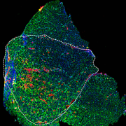
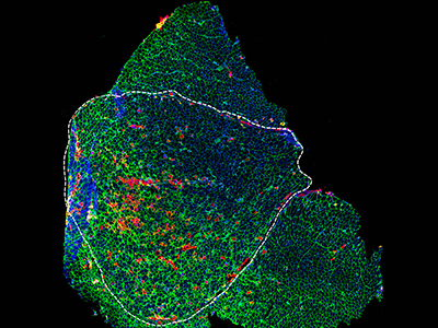
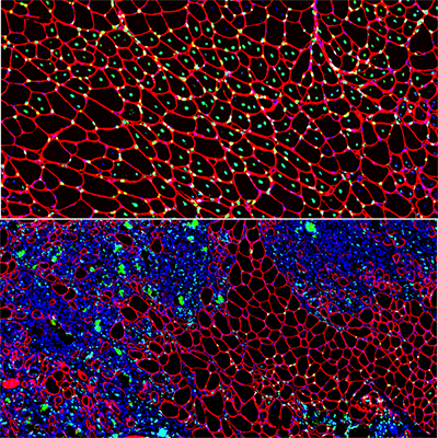
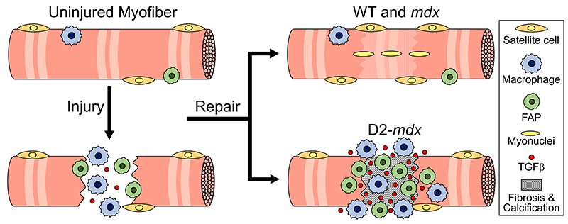
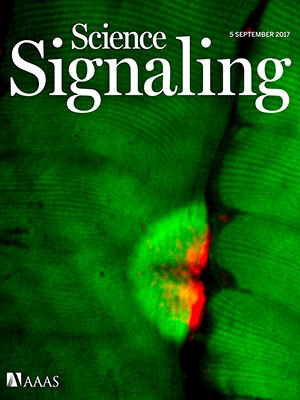
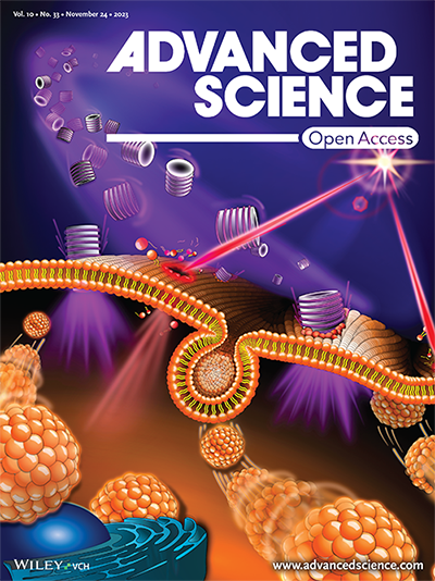
The study, featured on the back cover of the latest issue of Advanced Science, found that cells respond in distinct ways depending on the type of injury, such as a traumatic muscle tear that creates a large injury or tiny holes in the cell membrane caused by pathogenic proteins. Daniel Bittel, DPT, Ph.D., a research postdoctoral fellow at the Center for Genetic Medicine Research, said that cells are routinely injured from even everyday activities, such as walking up a flight of stairs.

“Injuries often involve damage to the plasma membrane,” Bittel said. “We wanted to investigate how healing happens at the subcellular level to better understand diseases and develop targeted therapies. We were especially curious about muscle cells because, interestingly, healthy ones get stronger the more that they are injured.”

The fine print

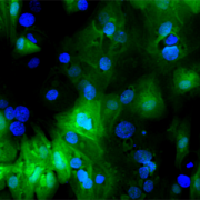
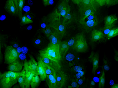
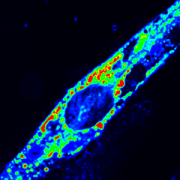
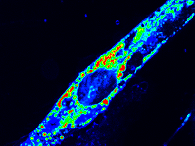
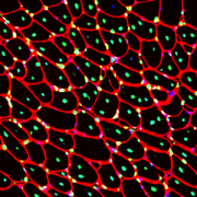
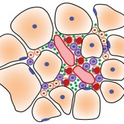
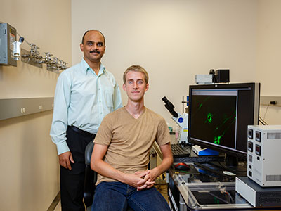
Using the center’s unique, custom-built microscope, the research team zoomed in on the process of cellular healing to watch how cells activate repair after injuries. Using a laser to damage the plasma membrane, they mimicked mechanically induced trauma. They also used a pathogen-derived protein to create nanoscale pinprick injuries in a cell’s plasma membrane that resemble those that are seen after strenuous muscle exertion.

Then, they watched as cells went to work within seconds, engaging healing mechanisms tailored to the type of injury. In the case of a cell facing numerous pinpricks along the cell membrane, it immediately deployed the endocytic pathway used by the cells to eat and drink. This process helped remove the injurious agents and the tiny holes they made. However, with a larger mechanical injury, the cells demonstrated patience, allowing the plasma membrane to seal before clearing up the damage by the same endocytic pathway.

 The big picture

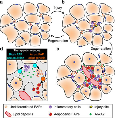
The paper is part of an ongoing body of research on cell injury that will inform future investigations into a wide range of pediatric health issues including muscular dystrophies, injuries to neurons, orthopedic injuries from sports and other mechanical damage to tissues.

Jyoti Jaiswal, M.Sc., Ph.D., senior investigator at the Center for Genetic Medicine Research, said this work is foundational in the development of new therapies. “Knowing where the problem lies will help us figure out what therapy will work best and target the therapy to address the specific deficit,” he said. “This work will pave the way to help tailor therapies and tackle diseases more effectively.”

Microscopic visual of a diseased muscle section

Gene therapy offers potential long-term treatment for limb-girdle muscular dystrophy 2B

Microscopic visual of a diseased muscle section

Microscopic visual of a diseased muscle section. Credit: Daniel Bittel.

Children’s National Hospital experts developed a new pre-clinical gene therapy for a rare disorder, known as limb-girdle muscular dystrophy (LGMD) 2B, that addresses the primary cellular deficit associated with this disease. Using a single injection of a low dose gene therapy vector, researchers restored the ability of injured muscle fibers to repair in a way that reduced muscle degeneration and enhanced the functioning of the diseased muscle. The treatment was safe, attenuated fibro-fatty muscle degeneration, and restored myofiber size and muscle strength, according to the study published in the Journal of Clinical Investigation.

With an incidence of less than 1 in 100,000, LGMD2B is a rare disorder caused by a genetic mutation in a large gene called dysferlin. This faulty gene leads to muscle weakness in the arms, legs, shoulder and pelvic girdle. Affected children and adults face trouble walking, climbing stairs and getting out of chairs. Individuals typically lose the ability to walk within years after the onset of symptoms, and often need assistance with everyday tasks such as showering, dressing and transferring.

This study described a new approach that avoids the need for packaging a large gene, like dysferlin, or giving a large vector dose to target the muscles, which are bottlenecks faced in ongoing gene therapy efforts aimed at muscular dystrophies.

“Currently, patients with LGMD2B have no gene or drug-based therapies available to them, and we are amongst the few centers developing therapeutic approaches for this disease,” said Jyoti K. Jaiswal, M.Sc. Ph.D., senior investigator of the Center for Genetic Medicine Research at Children’s National. “We are working to further enhance the efficacy of this approach and perform a longer-term safety and efficacy study to enable the clinical translation of this therapy.”

The genetic defect in dysferlin that is associated with LGMD2B causes the encoded protein to be truncated or degraded. This hinders the muscle fiber’s ability to heal, which is required for healthy muscles. In recessive genetic disorders, like LGMD2B, common pre-clinical gene therapy approaches usually target the mutated gene in the muscle, making them capable of producing the missing proteins.

“The large size of the gene mutated in this disease, and impediments in body-wide delivery of gene therapy vectors to reach all the muscles, pose significant challenges for developing gene therapies to treat this disease,” said Jaiswal.

To overcome these challenges, the researchers found another way to slow down the disease’s progression. The authors built upon their previous discovery that acid sphingomyelinase (hASM) protein is required to repair injured muscle cells. In this current work, the research team administered a single in vivo dose of an Adeno-associated virus (AAV) vector that produces a secreted version of hASM in the liver, which then was delivered to the muscles via blood circulation at a level determined to be efficacious in repairing LGMD2B patient’s injured muscle cells.

“Increased muscle degeneration necessitates greater muscle regeneration, and we found that improved repair of dysferlin-deficient myofibers by hASM-AAV reduces the need for regeneration, causing a 2-fold decrease in the number of regenerated myofibers,” said Daniel Bittel, D.P.T., PhD., research postdoctoral fellow of the Center for Genetic Medicine Research at Children’s National and a lead author of this study.

Sreetama Sen Chandra, Ph.D., who was a research postdoctoral fellow at Children’s National at the time of this study and served as co-lead author, also added that “these findings are also of interest to patients with Niemann-Pick disease type A since the pre-clinical model for this disease also manifests poor sarcolemma repair.”

Children’s National researchers of the Center for Genetic Medicine Research and the Rare Disease Institute (RDI) are constantly pursuing high-impact opportunities in pediatric genomic and precision medicine. Both centers combine its strengths with public and private partners, including industry, universities, federal agencies, start-up companies and academic medical centers. They also serve as an international referral site for rare disorders.

Gene therapy Schematic

Gene therapy Schematic. Credit: Daniel Bittel.

RSV infected infant cells

$2.13M grant accelerates treatments for kids with Down syndrome experiencing respiratory viruses

RSV infected infant cells

Children’s National Hospital received a combined $2.13 million award from the National Institutes of Health’s (NIH) National Heart, Lung and Blood Institute to better understand the mechanisms of severe viral respiratory infections in patients with Down syndrome and to develop new diagnostic tools and innovative precision medicine approaches for this vulnerable population.

“We have a unique opportunity to discover novel targets that can treat severe viral respiratory infections, including SARS-CoV-2,” said Gustavo Nino, M.D., M.S.H.S., D’A.B.S.M., principal investigator in the Center for Genetic Medicine at Children’s National. “Part of the award will help us accelerate the development of these novel approaches to prevent severe respiratory infections caused by SARS-CoV-2 and other viruses like respiratory syncytial virus infection (RSV) in children and adults with Down syndrome.”

Lower respiratory tract infections are a leading cause of hospitalization and death in children with Down syndrome. Those children have a nine times higher risk for hospitalization and mortality due to respiratory viruses that cause lower respiratory tract infections.

Chromosome 21, which is an extra chromosome copy found in patients with Down syndrome, encodes four of the six known interferon receptors, leading to hyperactivation of interferon response in Down syndrome. With the central role of interferons focused on antiviral defense, it remains puzzling how interferon hyperactivation contributes to severe viral lower respiratory tract infections in children with Down syndrome. This is an area that the researchers will explore to better manage and treat viral lower respiratory tract infections in these patients, with the support of NIH’s INCLUDE initiative. INCLUDE provides institutions with grants to help clinical research and therapeutics to understand and diminish risk factors that influence the overall health, longevity, and quality of life for people with Down syndrome related to respiratory viruses.

“While many of the other studies focus on intellectual and other disabilities, we are exploring a novel viral respiratory infectious disease mechanism and are doing so by working directly with patients and patient-derived samples,” said Jyoti Jaiswal, M.Sc., Ph.D., senior investigator in the Center for Genetic Medicine Research at Children’s National.

Children with Down syndrome have historically been excluded in research related to airway antiviral immunity, which is a focus of this human-based transformative study to improve the health and survival of patients with Down syndrome. There is a critical need for studies that define targetable molecular and cellular mechanisms to address dysregulated antiviral responses in this patient population.

“The clinical expertise at Children’s National in studying Down syndrome and the work of our team in caring for these patients with respiratory and sleep disorders positions us well to pursue this work,” said Jaiswal. “This is further supplemented by our initial studies that have identified a novel mechanism of impaired airway antiviral responses in these patients.”

Congresswoman Eleanor Holmes Norton (D-DC) also celebrated Children’s National and its NIH research funding benefitting people with Down syndrome.

“I am pleased to congratulate Dr. Nino and staff on being the recipients of the National Heart, Lung, & Blood Institute grant. You were chosen from a competitive group of applicants and should be proud of this notable achievement,” said Norton in a letter. “By receiving this grant, you have demonstrated outstanding promise in your field. It is my hope that this grant will enable you to better the local and global community.”

Injury triggered change in ER calcium of a muscle cell

ER maintains ion balance needed for muscle repair

Injury triggered change in ER calcium of a muscle cell

A new study led by Jyoti Jaiswal, M.Sc., Ph.D., principal investigator at Children’s National Hospital, identifies that an essential requirement for the repair of injured cells is to cope with the extracellular calcium influx caused by injury to the cell’s membrane. Credit: Goutam Chandra, Ph.D.

Physical activity can injure our muscle cells, so their ability to efficiently repair is crucial for maintaining muscle health. Understanding how healthy muscle cells respond to injury is required to understand and treat diseases caused by poor muscle cell repair.

A new study led by Jyoti Jaiswal, M.Sc., Ph.D., principal investigator at Children’s National Hospital, identifies that an essential requirement for the repair of injured cells is to cope with the extracellular calcium influx caused by injury to the cell’s membrane.

This study, published in the Journal of Cell Biology, identifies endoplasmic reticulum (ER) – a network of membranous tubules in the cell – as the site where the calcium entering the injured cell is sequestered. Using limb girdle muscular dystrophy 2L (LGMD2L) patient cells and a model for this genetic disease, the study shows impaired ability of diseased muscle cells to cope with this calcium excess. It also shows that a drug to sequester excess calcium counters this ion imbalance and reverses the diseased cell’s repair deficit.

“The study provides a novel insight into how injured cells in our body cope with calcium ion imbalance during injury,” Dr. Jaiswal explained. “This work also addresses how calcium homeostasis is compromised by a genetic defect that leads to LGMD2L. It also offers a proof of principle approach to restore calcium homeostasis, paving the path for future work to develop therapies targeting this disease.”

According to Dr. Jaiswal, this work also addresses the current lack of understanding of the basis for exercise intolerance and other symptoms faced by LGMD2L patients.

“This study opens the path for developing targeted therapies for LGMD2L and provides a fundamental cellular insight into a process crucial for cell survival,” said Goutam Chandra, Ph.D., research fellow and lead author of this study.

The Center for Genetic Medicine Research at Children’s National is among only a handful across the world to study this rare disease. These findings are unprecedented in providing the mechanistic insights needed to develop treatment for it.

In addition to Dr. Jaiswal and Chandra, the study co-authors include Sreetama Sen Chandra, Ph.D., Davi Mazala, Ph.D., and Jack VanderMeulen, Ph.D., from Children’s National, and Karine Charton, Ph.D., and Isabelle Richard, Ph.D., from Université Paris-Saclay.

muscle cells

Experimental model mimics early-stage myogenic deficit in boys with DMD

muscle cells

Muscle regeneration marked by incorporation of muscle stem cell nuclei (green) in the myofibers (red) in dystrophic muscles with low TGFβ level (upper image), but not with high TGFβ level (lower image). Inflammatory and other nuclei are labeled blue.

Boys with Duchenne muscular dystrophy (DMD) experience poor muscle regeneration, but the precise reasons for this remain under investigation. An experimental model of severe DMD that experiences a large spike in transforming growth factor-beta (TGFβ) activity after muscle injury shows that high TGFβ activity suppresses muscle regeneration and promotes fibroadipogenic progenitors (FAPs). This leads to replacement of the damaged muscle fibers by calcified and connective tissue, compromising muscle structure and function. While blocking FAP buildup provides a partial solution, a Children’s National Hospital study team identifies correcting the muscle micro-environment caused by high TGFβ as a ripe therapeutic target.

The team’s study was published online March 26, 2020, in JCI Insight.

DMD is a chronic muscle disease that affects 1 in 6,200 young men in the prime of their lives. The disorder, caused by genetic mutations leading to the inability to produce dystrophin protein, leads to ongoing muscle damage, chronic inflammation and poor regeneration of lost muscle tissue. The patients experience progressive muscle wasting, lose the ability to walk by the time they’re teenagers and die prematurely due to cardiorespiratory failure.

The Children’s National team finds for the first time that as early as preadolescence (3 to 4 weeks of age), their experimental model of severe DMD disease showed clear signs of the type of spontaneous muscle damage, regenerative failure and muscle fiber loss seen in preadolescent boys who have DMD.

“In boys, the challenge due to muscle loss exists from early in their lives, but had not been mimicked previously in experimental models,” says Jyoti K. Jaiswal, MSc, Ph.D., principal investigator in the Center for Genetic Medicine Research at Children’s National, and the study’s co-senior author. “TGFβ is widely associated with muscle fibrosis in DMD, when, in fact, our work shows its role in this disease process is far more significant.”

Research teams have searched for experimental models that replicate the sudden onset of symptoms in boys who have DMD as well as its complex progression.

“Our work not only offers insight into the delicate balance needed for regeneration of skeletal muscle, but it also provides quantitative information about muscle stem cell activity when this balanced is disturbed,” says Terence A. Partridge, Ph.D., principal investigator in the Center for Genetic Medicine Research at Children’s National, and the study’s co-senior author.

This schematic depicts the fate of injured myofibers in healthy or dystrophic muscle

This schematic depicts the fate of injured myofibers in healthy or dystrophic muscle (WT or mdx experimental models) that maintain low TGFβ level, compared with D2-mdx experimental models that experience a large increase in TGFβ level. As the legend shows, various cells are involved in this regenerative response.

“The D2-mdx experimental model is a relevant one to use to investigate the interplay between inflammation and muscle degeneration that is seen in humans with DMD,” adds Davi A.G. Mázala, co-lead study author.  “This model faithfully recapitulates many features of the complex disease process seen in humans.”

Between 3 to 4 weeks of age in the experimental models of severe DMD disease, the level of active TGFβ spiked up to 10-fold compared with models with milder disease. Intramuscular injections of an off-the-shelf drug that inhibits TGFβ signaling tamped down the number of FAPs, improving the muscle environment by lowering TGFβ activity.

“This work lays the foundation for studies that could lead to future therapeutic strategies to improve patients’ outcomes and lessen disease severity,” says James S. Novak, Ph.D., principal investigator in Children’s Center for Genetic Medicine Research, and co-lead study author. “Ultimately, our goal is to improve the ability of patients to continue to maintain muscle mass and regenerate muscle.”

In addition to Mázala, Novak, Jaiswal and Partridge, Children’s National study co-authors include Marshall W. Hogarth; Marie Nearing; Prabhat Adusumalli; Christopher B. Tully; Nayab F. Habib; Heather Gordish-Dressman, M.D.; and Yi-Wen Chen, Ph.D.

Financial support for the research described in this post was provided by the National Institutes of Health under award Nos. T32AR056993, R01AR055686 and U54HD090257; Foundation to Eradicate Duchenne; Muscular Dystrophy Association under award Nos. MDA295203, MDA480160 and MDA 477331; Parent Project Muscular Dystrophy; and Duchenne Parent Project – Netherlands.

mitochondria

Molecular gatekeepers that regulate calcium ions key to muscle function

mitochondria

Controlled entry of calcium ions into the mitochondria, the cell’s energy powerhouses, makes the difference between whether muscles grow strong or easily tire and perish from injury, according to research published in Cell Reports.

Calcium ions are essential to how muscles work effectively, playing a starring role in how and when muscles contract, tap energy stores to keep working and self-repair damage. Not only are calcium ions vital for the repair of injured muscle fibers, their controlled entry into the mitochondria, the cell’s energy powerhouses, spells the difference between whether muscles will be healthy or if they will easily tire and perish following an injury, according to research published Oct. 29, 2019, in Cell Reports.

“Lack of the protein mitochondrial calcium uptake1 (MICU1) lowers the activation threshold for calcium uptake mediated by the mitochondrial calcium uniporter in both, muscle fibers from an experimental model and fibroblast of  a patient lacking MICU1,” says Jyoti K. Jaiswal, MSc, Ph.D., a principal investigator in the Center for Genetic Medicine Research at Children’s National Hospital and one of the paper’s corresponding authors. “Missing MICU1 also tips the calcium ion balance in the mitochondria when muscles contract or are injured, leading to more pronounced muscle weakness and myofiber death.”

Five years ago, patients with a very rare disease linked to mutations in the mitochondrial gene MICU1 were described to suffer from a neuromuscular disease with signs of muscle weakness and damage that could not be fully explained.

To determine what was going awry, the multi-institutional research team used a comprehensive approach that included fibroblasts donated by a patient lacking MICU1 and an experimental model whose MICU1 gene was deleted in the muscles.

Loss of MICU1 in skeletal muscle fibers leads to less contractile force, increased fatigue and diminished capacity to repair damage to their cell membrane, called the sarcolemma. Just like human patients, the experimental model suffers more pronounced muscle weakness, increased numbers of dead myofibers, with greater loss of muscle mass in certain muscles, like the quadriceps and triceps, the research team writes.

“What was happening to the patient’s muscles was a big riddle that our research addressed,” Jaiswal adds. “Lacking this protein is not supposed to make the muscle fiber die, like we see in patients with this rare disease. The missing protein is just supposed to cause atrophy and weakness.”

Patients with this rare disease show early muscle weakness, fluctuating levels of fatigue and lethargy, muscle aches after exercise, and elevated creatine kinase in their bloodstream, an indication of cell damage due to physical stress.

“One by one, we investigated these specific features in experimental models that look normal and have normal body weight, but also show lost muscle mass in the quadriceps and triceps,” explains Adam Horn, Ph.D., the lead researcher in Jaiswal’s lab who conducted this study. “Our experimental model lacking MICU1 only in skeletal muscles responded to muscle deficits so similar to humans that it suggests that some of the symptoms we see in patients can be attributed to MICU1 loss in skeletal muscles.”

Future research will aim to explore the details of how the impact of MICU1 deficit in muscles may be addressed therapeutically and possible implications of lacking MICU1 or its paralog in other organs.

In addition to Jaiswal and Horn, Children’s National Hospital Center for Genetic Medicine Research co-authors include Marshall W. Hogarth and Davi A. Mazala. Additional co-authors include Lead Author Valentina Debattisti, Raghavendra Singh, Erin L. Seifert, Kai Ting Huang, and Senior Author György Hajnóczky, all from Thomas Jefferson University; and Rita Horvath, from Newcastle University.

Financial support for research described in this post was provided by the National Institutes of Health under award numbers R01AR55686, U54HD090257 and RO1 GM102724; National Institute of Arthritis and Musculoskeletal and Skin Diseases under award number T32AR056993; and Foundation Leducq.

sketch of muscle cells

Losing muscle to fat: misdirected fate of a multipotent stem cell drives LGMD2B

Fibro/adipogenic precursors (FAPs) control the onset and severity of disease in limb-girdle muscular dystrophy type 2 (LGMD2B)

Fibro/adipogenic precursors (FAPs) control the onset and severity of disease in limb-girdle muscular dystrophy type 2 (LGMD2B). a) Healthy and/or pre-symptomatic LGMD2B muscle contains resident FAPs. b) After myofiber injury, inflammatory cells invade and trigger FAP proliferation. c) In symptomatic LGMD2B muscle, there is a gradual accumulation of extracellular AnxA2, which prolongs the pro-inflammatory environment, causing excessive FAP proliferation. d) Blocking aberrant signaling due to AnxA2 buildup blocks FAP accumulation and thus preventing adipogenic loss of dysferlinopathic muscle. Credit: “Fibroadipogenic progenitors are responsible for muscle loss in limb girdle muscular dystrophy 2B.” Published online June 3, 2019, in Nature Communications. Marshall W. Hogarth, Aurelia Defour, Christopher Lazarski, Eduard Gallardo, Jordi Diaz Manera, Terence A. Partridge, Kanneboyina Nagaraju and Jyoti K. Jaiswal. https://rdcu.be/bFu9U.

Research led by faculty at Children’s National published online June 3, 2019, in Nature Communications shows that the sudden appearance of symptoms in limb-girdle muscular dystrophy type 2 (LGMD2B) is a result of impaired communication between different cell types that facilitate repair in healthy muscle. Of particular interest are the fibro/adipogenic precursors (FAPs), cells that typically play a helpful role in regenerating muscle after injury by removing debris and enhancing the fusion of muscle cells into new myofibers.

LGMD2B is caused by mutations in the DYSF gene that impair the function of dysferlin, a protein essential for repairing injured muscle fibers. Symptoms, like difficulty climbing or running, do not appear in patients until young adulthood. This late onset has long puzzled researchers, as the cellular consequences of dysferlin’s absence are present from birth and continue through development, but do not impact patients until later in life.

The study found that in the absence of dysferlin, muscle gradually increases the expression of the protein Annexin A2 which, like dysferlin, facilitates repair of injured muscle fiber. However, increasing Annexin A2 accumulates outside the muscle fiber and drives an increase in FAPs within the muscle as well as encourages these FAPs to differentiate into adipocytes, forming fatty deposits. Shutting down Annexin A2 or blocking the adipocyte fate of FAPs using an off-the-shelf medicine arrests the fatty replacement of dysferlinopathic muscle.

“We propose a feed-forward loop in which repeated myofiber injury triggers chronic inflammation which, over time, creates an environment that promotes FAPs to accumulate and differentiate into fat. This, in turn, contributes to more myofiber damage,” says Jyoti K. Jaiswal, MSc, Ph.D., a principal investigator in the Center for Genetic Medicine Research at Children’s National and the study’s senior author.

“Adipogenic accumulation becomes the nucleating event that results in an abrupt decline in muscle function in patients. This new view of LGMD2B disease opens previously unrealized avenues to intervene,” adds Marshall Hogarth, Ph.D., the study’s lead author.

Joyti Jaiswal

“We propose a feed-forward loop in which repeated myofiber injury triggers chronic inflammation which, over time, creates an environment that promotes FAPs to accumulate and differentiate into fat. This, in turn, contributes to more myofiber damage,” says Jyoti K. Jaiswal, MSc, Ph.D.

A research team led by Jaiswal collaborated with Eduard Gallardo and Jordi Diaz Manera, of Hospital de la Santa Creu in Barcelona, Spain, to examine muscle biopsies from people with LGMD2B who had mild to severe symptoms. They found that adipogenic deposits originate in the extracellular matrix space between muscle fibers, with the degree of accumulation tied to disease severity. They found a similar progressive increase in lipid accumulation between myofibers predicted disease severity in dysferlin-deficient experimental models. What’s more, this process can be accelerated by muscle injury, triggering increased adipogenic replacement in areas that otherwise would be occupied by muscle cells.

“Accumulation and adipogenic differentiation of FAPs is responsible for the decline in function for dysferlinopathic muscle. Reversing this could provide a therapy for LGMD2B, a devastating disease with no effective treatment,” predicts Jaiswal as the team continues research in this field.

Promising off-the-shelf drugs include batimastat, an anti-cancer drug that inhibits the extracellular matrix enzyme matrix metalloproteinase. This drug reduces FAP adipogenesis in vitro and lessens injury-triggered lipid formation in vivo. In experimental models, batimastat also increases muscle function.

In addition to Jaiswal, Hogarth, Gallardo and Diaz Manera, other study co-authors include Aurelia Defour, Christopher Lazarski, Terence A. Partridge and Kanneboyina Nagaraju, all of Children’s National.

Financial support for research described in this post was provided by the Muscular Dystrophy Association under awards MDA477331 and MDA277389, the National Institute of Arthritis and Musculoskeletal and Skin Diseases under award R01AR055686 and the National Institutes of Health under awards K26OD011171, R24HD050846 and P50AR060836.

Sen Chandra Sreetama and Jyoti K Jaiswal

Modified glucocorticoid stabilizes dysferlin-deficient muscle cell membrane in experimental models

Sen Chandra Sreetama and Jyoti K Jaiswal

Limb girdle muscular dystrophy type 2B (LGMD2B) – a disease so rare that researchers aren’t even sure how many people it affects – is characterized by chronic muscle inflammation and progressively weakened muscles in the pelvis and shoulder girdle. It can affect able-bodied people during their childbearing years and makes it difficult to tiptoe, walk, run or rise unaided from a squat. Ultimately, many with the muscle-wasting condition require wheelchair assistance. There is no therapy approved by the Food and Drug Administration for this condition.

In a head-to-head trial between the conventional glucocorticoid, prednisolone, and a modified glucocorticoid, vamorolone, in experimental models of LGMD2B, vamorolone improved dysferlin-deficient muscle cell membrane stability and repair. This correlated with increased muscle strength and decreased muscle degeneration, according to a Children’s-led study published online Aug. 27, 2018, in Molecular Therapy. By contrast, prednisolone worsened muscle weakness, impaired muscle repair and increased myofiber atrophy.

“These two steroids differ by only two chemical groups,” says Jyoti K. Jaiswal, MSC, Ph.D., a principal investigator at Children’s National Health System and senior study author. “One made muscle repair better. The other made muscle repair worse or about the same as untreated experimental models. This matches experience in the clinic as patients with LGMD2B experienced increased muscle weakness after being prescribed conventional glucocorticoids, such as prednisolone.”

Healthy muscle cells rely on the protein dysferlin to properly repair the sarcolemmal membrane, a cell membrane specialized for muscle cells that serves a vital role in ensuring that muscle fibers are strong enough and have the necessary resources to contract. Mutations in the DYSF gene that produces this essential protein causes LGMD2B.

Jaiswal likens the plasma membrane to a balloon that sits atop the myofiber, a long cell that when healthy can flex and contract. If, in the process of myofiber contraction, the plasma membrane experiences anything out of sync or overly stressful, it develops a tear that needs to be quickly sealed. An intact balloon keeps air inside; tear it, and air escapes. When the plasma membrane tears, calcium from the outside leaks in, causing the muscle cell to collapse into a ball and die. The body contends with the dead cell by breaking it up into fragments and sending in inflammatory cells to clear the debris.

Lack of dysferlin is associated with increased lipid mobility in the LGMD2B cell membrane

Lack of dysferlin is associated with increased lipid mobility in the limb girdle muscular dystrophy type 2B (LGMD2B) cell membrane, which is further increased by injury and prednisolone treatment, causing failure of these cells to undergo repair. By contrast, vamorolone treatment stabilizes the LGMD2B muscle cell membrane to near healthy cell level, enabling repair of injured cells.

The study team got the idea for the current research project during a previous study of the experimental treatment vamorolone for a different type of muscular dystrophy. “In Duchenne muscular dystrophy (DMD), treatment with vamorolone not only reduced inflammation, but the membranes of muscle fibers were stabilized. That was the team’s ah-hah moment,” he says.

Three different doses of vamorolone were tested on cells derived from patients with LGMD2B with higher cell membrane repair efficacy seen with rising treatment dose. The dysferlinopathic experimental models were treated for three months with daily doses of cherry syrup laced with either 30 mg/kg of vamorolone or prednisolone or cherry syrup alone as the placebo arm.

“Right now there are zero treatments,” he says. People with LGMD2B turn to rehabilitative therapies and movement aids to cope with loss of mobility. Doctors are cautioned not to prescribe steroids. Jaiswal says many patients with LGMD2B grew up doing strenuous exercise, former athletes whose first indication of a problem was muscle cramping and pain. How this progresses to muscle weakness and loss is an area of active research in Jaiswal’s lab. “While additional research is needed, our findings here suggest that modified steroids such as vamorlone may be an option for some patients,” Jaiswal says.

“There is a nuance here: In addition to genomic effects, steroids also have physical effects on the cell membrane which may make some of the approved steroids ‘good’ steroids for dysferlinopathy that could selectively be used for this disease,” adds Sen Chandra Sreetama, lead study author.  Further research could indicate whether vamorolone, which is in Phase II human clinical trials for DMD, or any off-the-shelf drug could slow decline in muscle function for patients with LGMD2B.

Additional Children’s study authors include Goutam Chandra; Jack H. Van der Meulen; Mohammad Mahad Ahmad; Peter Suzuki; Shivaprasad Bhuvanendran; and Kanneboyina Nagaraju and Eric P. Hoffman, both of ReveraGen BioPharma.

Research reported in this news release was supported by the Clark Charitable Foundation; Muscular Dystrophy Association, under award number MDA277389; National Institute of Arthritis and Musculoskeletal and Skin Diseases, under award number R01AR055686; National Institutes of Health (NIH), under award numbers K26OD011171 and R24HD050846; and the District of Columbia Intellectual and Developmental Disabilities Research Center under NIH award number 1U54HD090257.

Jyoti Jaiswal and Adam Horn

Antioxidants could thwart muscle repair

Science Signaling cover image 05Sept17

Science Signaling features a Research Article that describes the pathway by which mitochondria transduce the increase in cytosolic Ca2+ caused by plasma membrane injury into a ROS-dependent repair response. The image shows ROS production and actin polymerization as detected by fluorescent reporters near a plasma membrane injury site in a skeletal myofiber in an intact bicep of an experimental model. Credit: Adam Horn and Jyoti Jaiswal, M.S.C., Ph.D. Children’s National Health System and The George Washington University School of Medicine and Health Sciences

Reactive oxygen species (ROS) are a biological double-edged sword. These atoms, molecules or molecular fragments containing oxygen that is poised for chemical reactions, are a key part of the immune response, used by immune cells to kill potentially dangerous invaders such as bacteria. However, too much ROS – which also are produced as a normal part of cellular metabolism – can cause extreme damage to normal, healthy cells.

Because oxidative damage has been linked with cancer, many people make a concerted effort to consume antioxidants in food and as concentrated supplements. These compounds can neutralize ROS, stemming cellular damage. Taking antioxidants also has been thought to stem the muscle soreness from exercise since ROS are produced in excess during hard physical activity.

However, a new study led by researchers from Children’s National Health System finds that taking antioxidants could thwart the processes that repair muscle fibers. According to the study published Sept. 5, 2017 in Science Signaling and featured on the journal’s cover, oxidative species are crucial signals that start the process of repairing muscle fibers.

Cellular powerhouses known as mitochondria help injured muscle cells (myofibers) repair by soaking up calcium that enters from the site of injury and using it to trigger increased production of reactive oxygen species. Loading up mitochondria with excess antioxidants inhibits this signaling process, blocking muscle repair, exacerbating myofiber damage and diminishing muscle strength.

“Our results suggest a physiological role for mitochondria in plasma membrane repair in injured muscle cells, a role that highlights a beneficial effect of reactive oxygen species,” says Jyoti K. Jaiswal, M.S.C., Ph.D., principal investigator in the Center for Genetic Medicine Research at Children’s National Health System, associate professor of genomics and precision medicine at The George Washington University School of Medicine and Health Sciences and senior study author. “Our work highlights the need to take a nuanced view of the role of reactive oxygen species, as they are necessary when they are present at the right place and right time. Indiscriminate use of antioxidants actually could harm an adult with healthy muscles as well as a child with diseased muscle.”

Antioxidants are widely used by Baby Boomers with muscles that ache from a grueling workout or newborns diagnosed with muscular dystrophy. Jaiswal and Children’s National colleagues understand that their results buck conventional wisdom that antioxidants generally benefit muscle recovery.

“It is still a common belief within the fitness community that taking antioxidant supplements after a workout will help your muscles recover better. That’s what people think; that’s what I thought,” says Adam Horn, lead study author, a graduate student at The George Washington University who works with Jaiswal at Children’s National. “What we’ve done is figure out that mitochondria need to produce a very specific oxidative signal in response to muscle damage in order to help injured muscles repair.”Jyoti Jaiswal and Adam Horn

The oxidative signals produced by mitochondria are delicately balanced by the antioxidant defenses in healthy cells. This balance can be disrupted in diseases such as Duchenne muscular dystrophy, which is caused by the lack of a muscle-specific protein, dystrophin. Lack of dystrophin makes the muscle cell plasma membrane more vulnerable to injury. In an experimental model of Duchenne muscular dystrophy, the muscles at birth are seemingly normal but, within weeks, show obvious muscle damage and progressive weakness.

“What changes? One of the things that changes in the third and fourth week of life of this experimental model is mitochondrial functionality,” Jaiswal adds. “They end up with many dysfunctional mitochondria, which compromise repair of injured myofibers. This permits chronic and excessive oxidation of the myofibers and disruption of the proper oxidant-antioxidant balance.”

In this case, a dose of antioxidants may restore that proper balance and help to reverse muscle damage and progressive weakness.

As a next step, the research team is examining oxidation in healthy and diseased muscle to understand how the oxidant-antioxidant balance is disrupted and how it could be restored efficiently by using existing supplements. In one such study funded by the National Institutes of Health, the team is looking at the potential benefit of vitamin E supplements for patients with muscular dystrophy.

“Antioxidant supplements are made from extracts of bark, sap, chocolate and other compounds so they’re all different,” Jaiswal says. “Knowing which ones can restore balance under a specific circumstance has the potential to help the body maintain proper cellular signaling ability, which will keep muscles healthy and working properly.”


The response of actin protein following injury to a pair of muscle fibers in an intact biceps muscle.

mitochondria

Mitochondria key for repairing cell damage in DMD

mitochondria

A research team led by Jyoti K. Jaiswal, M.S.C., Ph.D., found that dysfunctional mitochondria prevent repair of muscle cells in Duchenne muscular dystrophy.

PDF Version

What’s known

Duchenne muscular dystrophy (DMD), one of the most severe forms of muscular dystrophy, is caused by a defect in the dystrophin gene. The protein that this gene encodes is responsible for anchoring muscle cells’ inner frameworks, or cytoskeletons, to proteins and other molecules outside these cells, the extracellular matrix. Without functional dystrophin protein, the cell membranes of muscle cells become damaged, and the cells eventually die. This cell death leads to the progressive muscle loss that characterizes this disease. Why these cells are unable to repair this progressive damage has been unknown.

What’s new

A research team led by Jyoti K. Jaiswal, M.S.C., Ph.D., a principal investigator in the Center for Genetic Medicine Research at Children’s National Health System, investigated this question in two experimental models of DMD that carry different mutations of the dystrophin gene. The researchers monitored the effects of the lack of functional dystrophin protein in these preclinical models on the level and function of muscle cell. They found that mitochondria – organelles that act as powerhouses to supply the chemical energy to drive cellular activities – are among the first to be affected. They found that the decline in mitochondrial level and activity over time in these experimental models preceded the onset of symptoms. The research team also looked at the ability of the experimental models’ muscle cells to repair damage. As the muscle cell mitochondria lost function, the cells’ ability to repair damage also declined. Efforts to increase mitochondrial activity after these organelles became dysfunctional did not improve muscle repair. This suggests that poor muscle repair may not be caused by a deficit in energy production by mitochondria.

Questions for future research

Q: Does similar mitochondrial dysfunction occur in human patients with DMD?
Q: How can the mitochondrial dysfunction be prevented?
Q: Is there a way to reverse mitochondrial dysfunction to better preserve the ability of muscle cells to repair from DMD-related damage?

Source: “Mitochondria mediate cell membrane repair and contribute to Duchenne muscular dystrophy.” Vila, M.C., S. Rayavarapu, M.W. Hogarth, J.H. Van der Meulen, A. Horn, A. Defour, S. Takeda, K.J. Brown, Y. Hathout, K. Nagaraju and J.K. Jaiswal. Published by Cell Death and Differentiation February 2017.