Tag Archive for: inflammation

muscle cells

Experimental model mimics early-stage myogenic deficit in boys with DMD

muscle cells

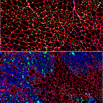
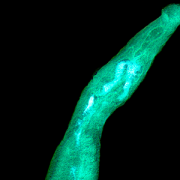
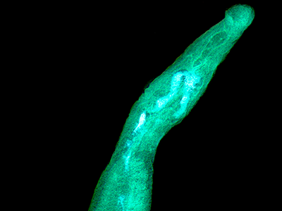
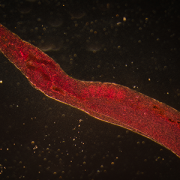
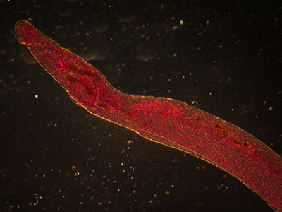
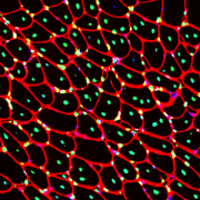
Muscle regeneration marked by incorporation of muscle stem cell nuclei (green) in the myofibers (red) in dystrophic muscles with low TGFβ level (upper image), but not with high TGFβ level (lower image). Inflammatory and other nuclei are labeled blue.

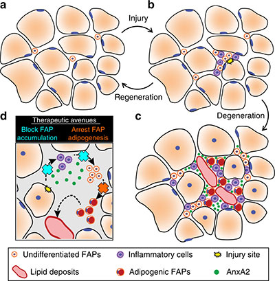
Boys with Duchenne muscular dystrophy (DMD) experience poor muscle regeneration, but the precise reasons for this remain under investigation. An experimental model of severe DMD that experiences a large spike in transforming growth factor-beta (TGFβ) activity after muscle injury shows that high TGFβ activity suppresses muscle regeneration and promotes fibroadipogenic progenitors (FAPs). This leads to replacement of the damaged muscle fibers by calcified and connective tissue, compromising muscle structure and function. While blocking FAP buildup provides a partial solution, a Children’s National Hospital study team identifies correcting the muscle micro-environment caused by high TGFβ as a ripe therapeutic target.

The team’s study was published online March 26, 2020, in JCI Insight.

DMD is a chronic muscle disease that affects 1 in 6,200 young men in the prime of their lives. The disorder, caused by genetic mutations leading to the inability to produce dystrophin protein, leads to ongoing muscle damage, chronic inflammation and poor regeneration of lost muscle tissue. The patients experience progressive muscle wasting, lose the ability to walk by the time they’re teenagers and die prematurely due to cardiorespiratory failure.

The Children’s National team finds for the first time that as early as preadolescence (3 to 4 weeks of age), their experimental model of severe DMD disease showed clear signs of the type of spontaneous muscle damage, regenerative failure and muscle fiber loss seen in preadolescent boys who have DMD.

“In boys, the challenge due to muscle loss exists from early in their lives, but had not been mimicked previously in experimental models,” says Jyoti K. Jaiswal, MSc, Ph.D., principal investigator in the Center for Genetic Medicine Research at Children’s National, and the study’s co-senior author. “TGFβ is widely associated with muscle fibrosis in DMD, when, in fact, our work shows its role in this disease process is far more significant.”

Research teams have searched for experimental models that replicate the sudden onset of symptoms in boys who have DMD as well as its complex progression.

“Our work not only offers insight into the delicate balance needed for regeneration of skeletal muscle, but it also provides quantitative information about muscle stem cell activity when this balanced is disturbed,” says Terence A. Partridge, Ph.D., principal investigator in the Center for Genetic Medicine Research at Children’s National, and the study’s co-senior author.

This schematic depicts the fate of injured myofibers in healthy or dystrophic muscle

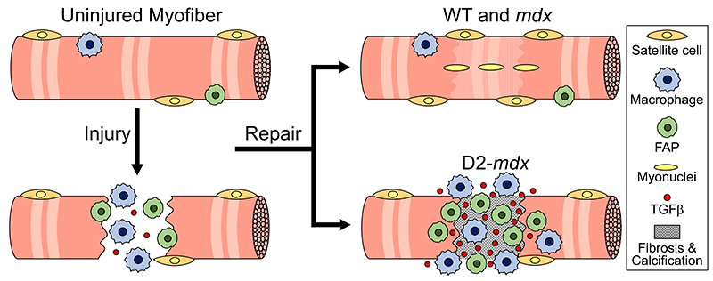
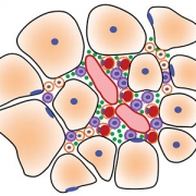
This schematic depicts the fate of injured myofibers in healthy or dystrophic muscle (WT or mdx experimental models) that maintain low TGFβ level, compared with D2-mdx experimental models that experience a large increase in TGFβ level. As the legend shows, various cells are involved in this regenerative response.

“The D2-mdx experimental model is a relevant one to use to investigate the interplay between inflammation and muscle degeneration that is seen in humans with DMD,” adds Davi A.G. Mázala, co-lead study author.  “This model faithfully recapitulates many features of the complex disease process seen in humans.”

Between 3 to 4 weeks of age in the experimental models of severe DMD disease, the level of active TGFβ spiked up to 10-fold compared with models with milder disease. Intramuscular injections of an off-the-shelf drug that inhibits TGFβ signaling tamped down the number of FAPs, improving the muscle environment by lowering TGFβ activity.

“This work lays the foundation for studies that could lead to future therapeutic strategies to improve patients’ outcomes and lessen disease severity,” says James S. Novak, Ph.D., principal investigator in Children’s Center for Genetic Medicine Research, and co-lead study author. “Ultimately, our goal is to improve the ability of patients to continue to maintain muscle mass and regenerate muscle.”

In addition to Mázala, Novak, Jaiswal and Partridge, Children’s National study co-authors include Marshall W. Hogarth; Marie Nearing; Prabhat Adusumalli; Christopher B. Tully; Nayab F. Habib; Heather Gordish-Dressman, M.D.; and Yi-Wen Chen, Ph.D.

Financial support for the research described in this post was provided by the National Institutes of Health under award Nos. T32AR056993, R01AR055686 and U54HD090257; Foundation to Eradicate Duchenne; Muscular Dystrophy Association under award Nos. MDA295203, MDA480160 and MDA 477331; Parent Project Muscular Dystrophy; and Duchenne Parent Project – Netherlands.

electronic cigarette dispenser with different flavors of nicotine

Extreme difficulty breathing and swallowing linked to teen’s vaping?

electronic cigarette dispenser with different flavors of nicotine

After a teen was transferred to Children’s National Hospital suffering from severe difficulty breathing and swallowing, a multidisciplinary team continued the detective work and surmises that vaping was to blame for her unusual symptoms.

A teenage girl with no hint of prior asthma or respiratory illness began to feel hoarseness in her throat and a feeling that she needed to clear her throat frequently. Within a few weeks, her hoarseness and throat-clearing worsened with early morning voice loss and feeling as if food were lodged in her throat. She started having trouble swallowing and began to avoid food all together.

Her pediatrician prescribed loratadine for suspected allergies to no avail. Days later, an urgent care center prescribed a three-day course of prednisone. For a few days, she felt a little better, but went back to feeling like she was breathing “through a straw.” After going to an emergency room with acute respiratory distress and severe difficulty swallowing, staff tried intravenous dexamethasone, ampicillin/sulbactam, and inhaled racemic epinephrine and arranged for transfer.

When she arrived at Children’s National Hospital, a multidisciplinary team continued the detective work with additional testing, imaging and bloodwork.

Examining her throat confirmed moderate swelling and a partially obstructed airway draped with thick chartreuse-colored mucus. The teen had no history of an autoimmune disorder, no international travel and no exposure to animals. She had no fever and had received all her scheduled immunizations.

“With epiglottitis – an inflammation of the flap found at the base of the tongue that prevents food from entering the trachea – our first concern is that an underlying infection is to blame,” says Michael Jason Bozzella, D.O., MS, a third-year infectious diseases fellow and lead author of the case report published Feb. 5, 2020, in Pediatrics. “We tested her specimens in a number of ways for a host of respiratory pathogens, including human rhino/enterovirus, respiratory syncytial virus, influenza, Epstein-Barr virus, Streptococcus and more. All negative. We also looked for more atypical infections with bacteria, like Arcanobacterium, Mycoplasma and Gonorrhea. Those were all negative as well,” Dr. Bozzella adds.

She slowly improved during a seven-day initial hospital stay, though soon returned for another six-day hospital stay after it again became excruciatingly painful for her to swallow.

Every throat culture and biopsy result showed no evidence of fungal, bacterial or viral infection, acid-fast bacilli or other malignancy. But in speaking with doctors, the teen had admitted to using candy-and fruit-flavored e-cigarettes three to five times with her friends over the two months preceding her symptoms. The last time she vaped was two weeks before her unusual symptoms began.

According to the Centers for Disease Control and Prevention, 2,668 people in the U.S. have been hospitalized for e-cigarette or vaping product use-associated lung injury, as of Jan. 14, 2020. The Children’s National case report’s authors say the increasing use of vaping products by teenagers highlights the potential for unknown health risks to continue to grow.

“This teenager’s use of e-cigarettes is the most plausible reason for this subacute epiglottitis diagnosis, a condition that can become life-threatening,” says Kathleen Ferrer, M.D., a hospitalist at Children’s National and the case report’s senior author. “This unusual case adds to a growing list of toxic effects attributable to vaping. While we normally investigate infectious triggers, like Streptococci, Staphylococci and Haemophilus, we and other health care providers should also consider e-cigarettes as we evaluate oro-respiratory complaints.”

In addition to Drs. Bozzella and Ferrer, Children’s National case report co-authors include Matthew Allen Magyar, M.D., a hospitalist; and Roberta L. DeBiasi, M.D., MS, chief of the Division of Pediatric Infectious Diseases.

Schistosoma

Parasitic eggs trigger upregulation in genes associated with inflammation

Schistosoma

Of the 200 million people around the globe infected with Schistosomiasis, about 100 million of them were sickened by the parasite Schistosoma haematobium.

Of the 200 million people around the globe infected with Schistosomiasis, about 100 million of them were sickened by the parasite Schistosoma haematobium. As the body reacts to millions of eggs laid by the blood flukes, people can develop fever, cough and abdominal pain, according to the Centers for Disease Control and Prevention. Schistosomiasis triggered by S. haematobium can also include hematuria, bladder calcification and bladder cancer.

Despite the prevalence of this disease, there are few experimental models specifically designed to study it, and some tried-and-true preclinical models don’t display the full array of symptoms seen in humans. It’s also unclear how S. haematobium eggs deposited in the host bladder modulate local tissue gene expression.

To better understand the interplay between the parasite and its human host, a team led by Children’s National Hospital injected 6,000 S. haematobium eggs into the bladder wall of seven-week-old experimental models.

After four days, they isolated RNA for analysis, comparing differences in gene expression in various treatment groups, including those that had received the egg injection and experimental models whose bladders were not exposed to surgical intervention.

Using the Database for Annotation, Visualization and Integrated Discovery (DAVID) – a tool that helps researchers understand the biological meaning of a long list of genes – the team identified commonalities with other pathways, including malaria, rheumatoid arthritis and the p53 signaling pathway, the team recently presented during the American Society of Tropical Medicine and Hygiene 2019 annual meeting. Some 325 genes were differentially expressed, including 34 genes in common with previous microarray data.

“Of particular importance, we found upregulation in genes associated with inflammation and fibrosis. We also now know that the body may send it strongest response on the first day it encounters a bolus of eggs,” says Michael Hsieh, M.D., Ph.D., director of transitional urology at Children’s National, and the research project’s senior author. “Next, we need to repeat these experiments and further narrow the list of candidate genes to key genes associated with immunomodulation and bladder cancer.”

In addition to Dr. Hsieh, presentation co-authors include Lead Author Kenji Ishida, Children’s National; Evaristus Mbanefo and Nirad Banskota, National Institutes of Health; James Cody, Vigene Biosciences; Loc Le, Texas Tech University; and Neil Young, University of Melbourne.

Financial support for research described in this post was provided by the National Institutes of Health under award No. R01-DK113504.

brain network illustration

$2.5M to protect the brain from metabolic insult

brain network illustration

The brain comprises only 2% of the body’s volume, but it uses more than 20% of its energy, which makes this organ particularly vulnerable to changes in metabolism.

More than 30 million Americans have diabetes, with the vast majority having Type 2 disease. Characterized by insulin resistance and persistently high blood sugar levels, poorly controlled Type 2 diabetes has a host of well-recognized complications: compared with the general population, a greatly increased risk of kidney disease, vision loss, heart attacks and strokes and lower limb amputations.

But more recently, says Nathan A. Smith, MS, Ph.D., a principal investigator in Children’s National Research Institute’s Center for Neuroscience Research, another consequence has become increasingly apparent. With increasing insulin resistance comes cognitive damage, a factor that contributes significantly to dementia diagnoses as patients age.

The brain comprises only 2% of the body’s volume, but it uses more than 20% of its energy, Smith explains – which makes this organ particularly vulnerable to changes in metabolism. Type 2 diabetes and even prediabetic changes in glucose metabolism inflict damage upon this organ in mechanisms with dangerous synergy, he adds. Insulin resistance itself stresses brain cells, slowly depriving them of fuel. As blood sugar rises, it also increases inflammation and blocks nitric oxide, which together narrow the brain’s blood vessels while also increasing blood viscosity.

When the brain’s neurons slowly starve, they become increasingly inefficient at doing their job, eventually succumbing to this deprivation. These hits don’t just affect individual cells, Smith adds. They also affect connectivity that spans across the brain, neural networks that are a major focus of his research.

While it’s well established that Type 2 diabetes significantly boosts the risk of cognitive decline, Smith says, it’s been unclear whether this process might be halted or even reversed. It’s this question that forms the basis of a collaborative Frontiers grant, $2.5 million from the National Science Foundation split between his laboratory; the lead institution, Stony Brook University; and Massachusetts General Hospital/Harvard Medical School.

Smith and colleagues at the three institutions are testing whether changing the brain’s fuel source from glucose to ketones – byproducts from fat metabolism – could potentially save neurons and neural networks over time. Ketones already have shown promise for decades in treating some types of epilepsy, a disease that sometimes stems from an imbalance in neuronal excitation and inhibition. When some patients start on a ketogenic diet – an extreme version of a popular fat-based diet – many can significantly decrease or even stop their seizures, bringing their misfiring brain cells back to health.

Principal Investigator Smith and his laboratory at the Children’s National Research Institute are using experimental models to test whether ketones could protect the brain against the ravages of insulin resistance. They’re looking specifically at interneurons, the inhibitory cells of the brain and the most energy demanding. The team is using a technique known as patch clamping to determine how either insulin resistance or insulin resistance in the presence of ketones affect these cells’ ability to fire.

They’re also looking at how calcium ions migrate in and out of the cells’ membranes, a necessary prerequisite for neurons’ electrical activity. Finally, they’re evaluating whether these potential changes to the cells’ electrophysiological properties in turn change how different parts of the brain communicate with each other, potentially restructuring the networks that are vital to every action this organ performs.

Colleagues at Athinoula A. Martinos Center for Biomedical Imaging at Massachusetts General Hospital and Harvard Medical School, led by Principal Investigator Eva-Maria Ratai, Ph.D.,  will perform parallel work in human subjects. They will use imaging to determine how these two fuel types, glucose or ketones, affect how the brain uses energy and produces the communication molecules known as neurotransmitters. They’re also investigating how these factors might affect the stability of neural networks using techniques that investigate the performance of these networks both while study subjects are at rest and performing a task.

Finally, colleagues at the Laufer Center for Physical and Quantitative Biology at Stony Brook University, led by Principal Investigator Lilianne R. Mujica-Parodi, Ph.D., will use results generated at the other two institutions to construct computational models that can accurately predict how the brain will behave under metabolic stress: how it copes when deprived of fuel and whether it might be able to retain healthy function when its cells receive ketones instead of glucose.

Collectively, Smith says, these results could help retain brain function even under glucose restraints. (For this, the research team owes a special thanks to Mujica-Parodi, who assembled the group to answer this important question, thus underscoring the importance of team science, he adds.)

“By supplying an alternate fuel source, we may eventually be able to preserve the brain even in the face of insulin resistance,” Smith says.

little boy using asthma inhaler

Searching for the molecular underpinnings of asthma exacerbations

little boy using asthma inhaler

It’s long been known that colds, flu and other respiratory illnesses are major triggers for asthma exacerbations, says asthma expert Stephen J. Teach, M.D., MPH. Consequently, a significant body of research has focused on trying to figure out what’s happening on the cellular or molecular level as these illnesses progress to exacerbations.

People with asthma can be indistinguishable from people who don’t have this chronic airway disease – until they have an asthma attack, also known as an exacerbation. During these events, their airways become inflamed and swollen and produce an abundance of mucus, causing dangerous narrowing of the bronchial tubes that leads to coughing, wheezing and trouble breathing. These events are a major cause of morbidity and mortality, leading to the deaths of 10 U.S. residents every day, according to the Centers for Disease Control and Prevention.

It’s long been known that colds, flu and other respiratory illnesses are major triggers for asthma exacerbations, says Children’s National in Washington, D.C., asthma expert Stephen J. Teach, M.D., MPH. Consequently, a significant body of research has focused on trying to figure out what’s happening on the cellular or molecular level as these illnesses progress to exacerbations. Targeted searches have identified several different molecular pathways that appear to be key players in this phenomenon. However, Dr. Teach says researchers have been missing a complete and unbiased snapshot of all the important pathways in illness-triggered exacerbations and how they interrelate.

To develop this big picture view, Dr. Teach and  Inner-City Asthma Consortium colleagues recruited 208 children ages 6-17 years old with severe asthma – marked by the need for daily doses of inhaled corticosteroids, two hospitalizations or systemic corticosteroid treatments over the past year, and a high concentration of asthma-associated immune cells – from nine pediatric medical centers across the country, including Children’s National. (Inhaled corticosteroids are a class of medicine that calms inflamed airways.) The researchers collected samples of nasal secretions and blood from these patients at baseline, when all of them were healthy.

Then, they waited for these children to show symptoms of respiratory illnesses. Within six days of cold symptoms, the researchers took two more samples of nasal secretions and blood. They also administered breathing tests to determine whether these respiratory illnesses led to asthma exacerbations and recorded whether these patients were treated with systemic corticosteroids to stem the associated respiratory inflammation.

The researchers examined nasal fluid samples for evidence of viral infection during illness and used analytical methods to identify the causative virus. They analyzed all the samples they collected for changes in concentrations of various immune cells. They also looked globally in these samples for changes in gene expression compared with baseline and between the two collection periods during respiratory illness.

Together, this information told the molecular story about what took place after these children got sick and after some of them developed exacerbations. Of the 208 patients recruited, 106 got respiratory illnesses during the six-month study period, leading to a total of 154 illness events. Of those, 47 caused exacerbations, and 107 didn’t.

About half the exacerbations appeared to have been triggered by a rhinovirus, a cause of common colds, the research team reports in a study published online April 8, 2019, in Nature Immunology. The other children’s cold-like symptoms could have been triggered by pollution, allergens or other irritants.

In most exacerbations, virally triggered or not, the researchers saw early activation of a network of genes that appeared to be associated with SMAD3, a signaling molecule already known to be involved in airway inflammation. At the same time, genes that control a set of immune cells known as lymphocytes were turned down. However, as the exacerbation progressed and worsened, the researchers saw gene networks turned on that related to airway narrowing, mucus hypersecretion and activation of other immune cells.

Exacerbations triggered by viruses were associated with multiple inflammatory pathways, in contrast to those in which viruses weren’t found, which were associated with molecular pathways that affected cells in the airway lining.

The researchers validated these findings in 19 patients who each got respiratory illnesses at least twice during the study period but only developed an exacerbation during one of these episodes, finding the same upregulated and downregulated molecular pathways in these patients as in the study population as a whole. They also identified a set of molecular risk factors in patients at baseline – signatures of gene activation that appeared to put patients at risk for exacerbations when they got sick. When patients were treated with systemic corticosteroids during exacerbations, these medicines appeared to restore only some of the affected molecular pathways to normal, healthy levels. Other molecular pathways remained markedly changed.

Each finding could represent a new target for drugs that could prevent or more effectively treat exacerbations, keeping more patients with asthma healthy and out of the hospital.

“Our consortium study found increased gene expression of enzymes that produce molecules that contribute to narrowed airways and dilated blood vessels,” Dr. Teach adds. “This is especially intriguing because drugs that target kallikreins or bradykinin may help treat asthma attacks that aren’t caused by viruses.”

In addition to Dr. Teach, study co-authors include Lead Author Matthew C. Altman, University of Washington; Michelle A. Gill, Baomei Shao and Rebecca S. Gruchalla, all of University of Texas Southwestern Medical Center; Elizabeth Whalen and Scott Presnell of Benaroya Research Institute; Denise C. Babineau and Brett Jepson of Rho, Inc.; Andrew H. Liu, Children’s Hospital Colorado; George T. O’Connor, Boston University School of Medicine; Jacqueline A. Pongracic, Ann Robert H. Lurie Children’s Hospital of Chicago; Carolyn M. Kercsmar and Gurjit K. Khurana Hershey, , Cincinnati Children’s Hospital; Edward M. Zoratti and Christine C. Johnson, Henry Ford Health System; Meyer Kattan, Columbia University College of Physicians and Surgeons; Leonard B. Bacharier and Avraham Beigelman, Washington University, St. Louis; Steve M. Sigelman, Peter J. Gergen, Lisa M. Wheatley and Alkis Togias, National Institute of Allergy and Infectious Diseases; and James E. Gern, William W. Busse and Senior author Daniel J. Jackson, University of Wisconsin School of Medicine and Public Health.

Funding for research described in this post was provided by the National Institute of Allergy and Infectious Diseases under award numbers 1UM1AI114271 and UM2AI117870; CTSA under award numbers UL1TR000150, UL1TR001422 and 5UL1TR001425; the National Institutes of Health under award number UL1TR000451;  CTSI under award number 1UL1TR001430; CCTSI under award numbers UL1TR001082 and 5UM1AI114271; and NCATS under award numbers UL1 TR001876 and UL1TR002345.

sketch of muscle cells

Losing muscle to fat: misdirected fate of a multipotent stem cell drives LGMD2B

Fibro/adipogenic precursors (FAPs) control the onset and severity of disease in limb-girdle muscular dystrophy type 2 (LGMD2B)

Fibro/adipogenic precursors (FAPs) control the onset and severity of disease in limb-girdle muscular dystrophy type 2 (LGMD2B). a) Healthy and/or pre-symptomatic LGMD2B muscle contains resident FAPs. b) After myofiber injury, inflammatory cells invade and trigger FAP proliferation. c) In symptomatic LGMD2B muscle, there is a gradual accumulation of extracellular AnxA2, which prolongs the pro-inflammatory environment, causing excessive FAP proliferation. d) Blocking aberrant signaling due to AnxA2 buildup blocks FAP accumulation and thus preventing adipogenic loss of dysferlinopathic muscle. Credit: “Fibroadipogenic progenitors are responsible for muscle loss in limb girdle muscular dystrophy 2B.” Published online June 3, 2019, in Nature Communications. Marshall W. Hogarth, Aurelia Defour, Christopher Lazarski, Eduard Gallardo, Jordi Diaz Manera, Terence A. Partridge, Kanneboyina Nagaraju and Jyoti K. Jaiswal. https://rdcu.be/bFu9U.

Research led by faculty at Children’s National published online June 3, 2019, in Nature Communications shows that the sudden appearance of symptoms in limb-girdle muscular dystrophy type 2 (LGMD2B) is a result of impaired communication between different cell types that facilitate repair in healthy muscle. Of particular interest are the fibro/adipogenic precursors (FAPs), cells that typically play a helpful role in regenerating muscle after injury by removing debris and enhancing the fusion of muscle cells into new myofibers.

LGMD2B is caused by mutations in the DYSF gene that impair the function of dysferlin, a protein essential for repairing injured muscle fibers. Symptoms, like difficulty climbing or running, do not appear in patients until young adulthood. This late onset has long puzzled researchers, as the cellular consequences of dysferlin’s absence are present from birth and continue through development, but do not impact patients until later in life.

The study found that in the absence of dysferlin, muscle gradually increases the expression of the protein Annexin A2 which, like dysferlin, facilitates repair of injured muscle fiber. However, increasing Annexin A2 accumulates outside the muscle fiber and drives an increase in FAPs within the muscle as well as encourages these FAPs to differentiate into adipocytes, forming fatty deposits. Shutting down Annexin A2 or blocking the adipocyte fate of FAPs using an off-the-shelf medicine arrests the fatty replacement of dysferlinopathic muscle.

“We propose a feed-forward loop in which repeated myofiber injury triggers chronic inflammation which, over time, creates an environment that promotes FAPs to accumulate and differentiate into fat. This, in turn, contributes to more myofiber damage,” says Jyoti K. Jaiswal, MSc, Ph.D., a principal investigator in the Center for Genetic Medicine Research at Children’s National and the study’s senior author.

“Adipogenic accumulation becomes the nucleating event that results in an abrupt decline in muscle function in patients. This new view of LGMD2B disease opens previously unrealized avenues to intervene,” adds Marshall Hogarth, Ph.D., the study’s lead author.

Joyti Jaiswal

“We propose a feed-forward loop in which repeated myofiber injury triggers chronic inflammation which, over time, creates an environment that promotes FAPs to accumulate and differentiate into fat. This, in turn, contributes to more myofiber damage,” says Jyoti K. Jaiswal, MSc, Ph.D.

A research team led by Jaiswal collaborated with Eduard Gallardo and Jordi Diaz Manera, of Hospital de la Santa Creu in Barcelona, Spain, to examine muscle biopsies from people with LGMD2B who had mild to severe symptoms. They found that adipogenic deposits originate in the extracellular matrix space between muscle fibers, with the degree of accumulation tied to disease severity. They found a similar progressive increase in lipid accumulation between myofibers predicted disease severity in dysferlin-deficient experimental models. What’s more, this process can be accelerated by muscle injury, triggering increased adipogenic replacement in areas that otherwise would be occupied by muscle cells.

“Accumulation and adipogenic differentiation of FAPs is responsible for the decline in function for dysferlinopathic muscle. Reversing this could provide a therapy for LGMD2B, a devastating disease with no effective treatment,” predicts Jaiswal as the team continues research in this field.

Promising off-the-shelf drugs include batimastat, an anti-cancer drug that inhibits the extracellular matrix enzyme matrix metalloproteinase. This drug reduces FAP adipogenesis in vitro and lessens injury-triggered lipid formation in vivo. In experimental models, batimastat also increases muscle function.

In addition to Jaiswal, Hogarth, Gallardo and Diaz Manera, other study co-authors include Aurelia Defour, Christopher Lazarski, Terence A. Partridge and Kanneboyina Nagaraju, all of Children’s National.

Financial support for research described in this post was provided by the Muscular Dystrophy Association under awards MDA477331 and MDA277389, the National Institute of Arthritis and Musculoskeletal and Skin Diseases under award R01AR055686 and the National Institutes of Health under awards K26OD011171, R24HD050846 and P50AR060836.

An-Massaro

Looking for ‘help’ signals in the blood of newborns with HIE

An Massaro

“This data support our hypothesis that a panel of biomarkers – not a one-time test for a single biomarker – is needed to adequately determine the risk and timing of brain injury for babies with HIE,” says An N. Massaro, M.D.

Measuring a number of biomarkers over time that are produced as the body responds to inflammation and injury may help to pinpoint newborns who are more vulnerable to suffering lasting brain injury due to disrupted oxygen delivery and blood flow, according to research presented during the Pediatric Academic Societies 2019 Annual Meeting.

Hypoxic-ischemic encephalopathy (HIE) happens when blood and oxygen flow are disrupted around the time of birth and is a serious birth complication for full-term infants. To lessen the chance of these newborns suffering permanent brain injury, affected infants undergo therapeutic cooling, which temporarily lowers their body temperatures.

“Several candidate blood biomarkers have been investigated in HIE but we still don’t have one in clinical use.  We need to understand how these markers change over time before we can use them to direct care in patients,” says An N. Massaro, M.D., co-director of the Neonatal Neurocritical Care Program at Children’s National and the study’s senior author. “The newborns’ bodies sent out different ‘help’ signals that we detected in their bloodstream, and the markers had strikingly different time courses. A panel of plasma biomarkers has the potential to help us identify infants most in need of additional interventions, and to help us understand the most optimal timing for those interventions.”

Past research has keyed in on inflammatory cytokines and Tau protein as potential biomarkers of brain injury for infants with HIE who are undergoing therapeutic cooling. The research team led by Children’s faculty wanted to gauge which time periods to measure such biomarkers circulating in newborns’ bloodstreams. They enrolled 85 infants with moderate or severe HIE and tapped unused blood specimens that had been collected as cooling began, as well as 12, 24, 72 and 96 hours later. The infants’ mean gestational age was 38.7 weeks, their mean birth weight was about 7 pounds (3.2 kilograms), and 19% had severe brain disease (encephalopathy).

Cytokines – chemicals like Interleukin (IL) 6, 8 and 10 that regulate how the body responds to infection, inflammation and trauma – peaked in the first 24 hours of cooling for most of the newborns. However, the highest measure of Tau protein for the majority of newborns was during or after the baby’s temperature was restored to normal.

“After adjusting for clinical severity of encephalopathy and five-minute Apgar scores, IL-6, IL-8 and IL-10 predicted adverse outcomes, like severe brain injury or death, as therapeutic hypothermia began. By contrast, Tau protein measurements predicted adverse outcomes during and after the infants were rewarmed,” Dr. Massaro says.

IL-6 and IL-8 proteins are pro-inflammatory cytokines while IL-10 is considered anti-inflammatory.  These chemicals are released as a part of the immune response to brain injury. Tau proteins are abundant in nerve cells and stabilize microtubules.

“This data support our hypothesis that a panel of biomarkers – not a one-time test for a single biomarker – is needed to adequately determine the risk and timing of brain injury for babies with HIE,” she adds.

Pediatric Academic Societies 2019 Annual Meeting presentation

  • “Serial plasma biomarkers of brain injury in infants with hypoxic ischemic encephalopathy (HIE) treated with therapeutic hypothermia (TH).”
    • Saturday, April 27, 2019, 6 p.m. (EST)

Meaghan McGowan, lead author; Alexandra C. O’Kane, co-author; Gilbert Vezina, M.D.,  director, Neuroradiology Program and co-author; Tae Chang, M.D., director, Neonatal Neurology Program and co-author; and An N. Massaro, M.D., co-director of the Neonatal Neurocritical Care Program and senior author; all of Children’s National; and co-author Allen Everett, of Johns Hopkins School of Medicine.

dystrophin protein

Experimental drug shows promise for slowing cardiac disease and inflammation

dystrophin protein

Duchenne muscular dystrophy (DMD) is caused by mutations in the DMD gene, which provides instructions for making dystrophin, a protein found mostly in skeletal, respiratory and heart muscles.

Vamorolone, an experimental medicine under development, appears to combine the beneficial effects of prednisone and eplerenone – standard treatments for Duchenne muscular dystrophy (DMD) – in the heart and muscles, while also showing improved safety in experimental models. The drug does so by simultaneously targeting two nuclear receptors important in regulating inflammation and cardiomyopathy, indicates a small study published online Feb. 11, 2019, in Life Science Alliance.

DMD is a progressive X-linked disease that occurs mostly in males. It is characterized by muscle weakness that worsens over time, and most kids with DMD will use wheelchairs by the time they’re teenagers. DMD is caused by mutations in the DMD gene, which provides instructions for making dystrophin, a protein found mostly in skeletal, respiratory and heart muscles.

Cardiomyopathy, an umbrella term for diseases that weaken the heart, is a leading cause of death for young adults with DMD, causing up to 50 percent of deaths in patients who lack dystrophin. A collaborative research team co-led by Christopher R. Heier, Ph.D., and Christopher F. Spurney, M.D., of Children’s National Health System, is investigating cardiomyopathy in DMD. They find genetic dystrophin loss provides “a second hit” for a specific pathway that worsens cardiomyopathy in experimental models of DMD.

“Some drugs can interact with both the mineralocorticoid receptor (MR) and glucocorticoid receptor (GR) since these two drug targets evolved from a common ancestor. However, we find these two drug targets can play distinctly different roles in heart and skeletal muscle. The GR regulates muscle inflammation, while the MR plays a key role in heart health,” says Heier, an assistant professor at Children’s National and lead study author. “In our study, the experimental drug vamorolone safely targets both the GR to treat chronic inflammation and the MR to treat the heart.”

After gauging the efficacy of various treatments in test tubes, the study team looked at whether any could mitigate negative impacts of the MR on heart health. Wild type and mdx experimental models were implanted with pumps that activated the MR. These models also received a daily oral MR antagonist (or inhibitor) drug, and either eplerenone, spironolactone or vamorolone. Of note:

  • MR activation increased kidney size and caused elevated blood pressure (hypertension).
  • Treatment with vamorolone maintained normal kidney size and prevented hypertension.
  • MR activation increased mdx heart mass and fibrosis. Vamorolone mitigated these changes.
  • MR activation decreased mdx heart function, while vamorolone prevented declines in function.
  • Daily prednisone caused negative MR- and GR-mediated side effects, such as hyperinsulinemia, whereas vamorolone safely improved heart function without these side effects.

“These findings have the potential to help current and future patients,” Heier says. “Clinicians already prescribe several of these drugs. Our new data support the use of MR antagonists such as eplerenone in protecting DMD hearts, particularly if patients take prednisone. The experimental drug vamorolone is currently in Phase IIb clinical trials and is particularly exciting for its unique potential to simultaneously treat chronic inflammation and heart pathology with improved safety.”

In addition to Heier and senior author Spurney, study co-authors include Qing Yu, Alyson A. Fiorillo, Christopher B. Tully, Asya Tucker and Davi A. Mazala, all of Children’s National; Kitipong Uaesoontrachoon and Sadish Srinivassane, AGADA Biosciences Inc.; and Jesse M. Damsker, Eric P. Hoffman and Kanneboyina Nagaraju, ReveraGen BioPharma.

Financial support for research described in this report was provided by Action Duchenne; the Clark Charitable Foundation; the Department of Defense under award W81XWH-17-1-047; the Foundation to Eradicate Duchenne; the Intellectual and Developmental Disabilities Research Center under award U54HD090257 (through the National Institutes of Health’s (NIH) Eunice Kennedy Shriver National Institute of Child Health and Human Development); and the NIH under awards K99HL130035, R00HL130035, L40AR068727 and T32AR056993.

Financial disclosure:  Co-authors employed by ReveraGen BioPharma were involved in creating this news release.

schistosome blood fluke

Therapy derived from parasitic worms downregulates proinflammatory pathways

schistosome blood fluke

A therapy derived from the eggs of the parasitic Schistosoma helps to protect against one of chemotherapy’s debilitating side effects by significantly downregulating major proinflammatory pathways, reducing inflammation.

A therapy derived from the eggs of parasitic worms helps to protect against one of chemotherapy’s debilitating side effects by significantly downregulating major proinflammatory pathways and reducing inflammation, indicates the first transcriptome-wide profiling of the bladder during ifosfamide-induced hemorrhagic cystitis.

The experimental model study findings were published online Feb. 7, 2019, in Scientific Reports.

With hemorrhagic cystitis, a condition that can be triggered by anti-cancer therapies like the chemotherapy drug ifosfamide and other oxazaphosphorines, the lining of the bladder becomes inflamed and begins to bleed. Existing treatments on the market carry their own side effects, and the leading therapy does not treat established hemorrhagic cystitis.

Around the world, people can become exposed to parasitic Schistosoma eggs through contaminated freshwater. Once inside the body, the parasitic worms mate and produce eggs; these eggs are the trigger for symptoms like inflammation. To keep their human hosts alive, the parasitic worms tamp down excess inflammation by secreting a binding protein with anti-inflammatory properties.

With that biological knowledge in mind, a research team led by Michael H. Hsieh, M.D., Ph.D., tested a single dose of IPSE, an Interleukin-4 inducing, Schistosoma parasite-derived anti-inflammatory molecule and found that it reduced inflammation, bleeding and urothelial sloughing that occurs with ifosfamide-related hemorrhagic cystitis.

In this follow-up project, experimental models were treated with ifosfamide to learn more about IPSE’s protective powers.

The preclinical models were given either saline or IPSE before the ifosfamide challenge. The bladders of the experimental models treated with ifosfamide had classic symptoms, including marked swelling (edema), dysregulated contraction, bleeding and urothelial sloughing. In contrast, experimental models “pre-treated” with IPSE were shielded from urothelial sloughing and inflammation, the study team found.

Transcriptional profiling of the experimental models’ bladders found the IL-1-B TNFa-IL-6 proinflammatory cascade via NFkB and STAT3 pathways serving as the key driver of inflammation. Pretreatment with IPSE slashed the overexpression of Il-1b, Tnfa and Il6 by 50 percent. IPSE drove significant downregulation of major proinflammatory pathways, including the IL-1-B TNFa-IL-6 pathways, interferon signaling and reduced (but did not eliminate) oxidative stress.

“Taken together, we have identified signatures of acute-phase inflammation and oxidative stress in ifosfamide-injured bladder, which are reversed by pretreatment with IPSE,” says Dr. Hsieh, a urologist at Children’s National Health System and the study’s senior author. “These preliminary findings reveal several pathways that could be therapeutically targeted to prevent ifosfamide-induced hemorrhagic cystitis in humans.”

When certain chemotherapy drugs are metabolized by the body, the toxin acrolein is produced and builds up in urine. 2-mercaptoethane sulfonate Na (MESNA) binds to acrolein to prevent urotoxicity. By contrast, IPSE targets inflammation at the source, reversing inflammatory changes that damage the bladder.

“Our work demonstrates that there may be therapeutic potential for naturally occurring anti-inflammatory molecules, including pathogen-derived factors, as alternative or complementary therapies for ifosfamide-induced hemorrhagic cystitis,” Dr. Hsieh adds.

In addition to Dr. Hsieh, study co-authors include Lead Author Evaristus C. Mbanefo and Rebecca Zee, Children’s National; Loc Le, Nirad Banskota and Kenji Ishida, Biomedical Research Institute; Luke F. Pennington and Theodore S. Jardetzky, Stanford University; Justin I. Odegaard, Guardant Health; Abdulaziz Alouffi, King Abdulaziz City for Science & Technology; and Franco H. Falcone, University of Nottingham.

Financial support for the research described in this report was provided by the Margaret A. Stirewalt Endowment, the National Institute of Diabetes and Digestive and Kidney Diseases under award R01DK113504, the National Institute of Allergy and Infectious Diseases under award R56AI119168 and a Urology Care Foundation Research Scholar Award.

Dr. Michael Hsieh's clay shield

Innovative urologist Michael Hsieh takes unbeaten path

Dr. Michael Hsieh's clay shield

For an elementary school art project, Michael H. Hsieh, M.D., Ph.D., was instructed to fashion a coat of arms out of clay. In addition to panels for truth, justice and Taiwan, in the shield’s M.D. panel, a snake twists around a rod, like the staff for Asclepius, a Greek god associated with healing.

Children’s urologist Michael H. Hsieh, M.D., Ph.D., knew from age 10 that he would become a doctor. Proof is at his parents’ home. For an elementary school art project, students were instructed to fashion a coat of arms out of clay. In addition to panels for truth, justice and Taiwan, in the shield’s M.D. panel, a snake twists around a rod, like the staff for Asclepius, a Greek god associated with healing.

“I liked science. When I can use it to help patients, that is very rewarding,” says Dr. Hsieh, the first doctor in his family.

These days, Dr. Hsieh’s Twitter profile serves as a digital coat of arms, describing him as “tinker, tailor,” #UTI #biologist, epithelial #immunologist, helminthologist and #urologist.

Tinker/tailor is shorthand for the mystery drama, “Tinker Tailor Solider Spy,” he explains, adding that the “tinker” part also refers “to the fact that I am always questioning things, and science is about experimentation, trying to seek answers to questions.”

While still in medical school during a rotation Dr. Hsieh saw a bladder operation on a young child and thought it was “amazing.” That experience in part inspired Dr. Hsieh to become a urologist and bladder scientist. His training in immunology and study of the bladder naturally led him to study urinary tract infections and parasitic worms that affect the urinary tract. In addition, thanks to R01 funding from the National Institutes of Health (NIH), Dr. Hsieh is co-principal investigator with Axel Krieger, University of Maryland, and Jin U. Kang, Johns Hopkins, on a project to develop imaging robots for supervised autonomous surgery on soft tissue.

The $1 million in NIH funding pushes the boundaries on amazing by using multi-spectral imaging technology and improved techniques to reduce surgical complications.

Anastomosis is a technique used by surgeons to join one thing to another, whether it’s a vascular surgeon suturing blood vessels, an orthopedic surgeon joining muscles or a urologist stitching healthy parts of the urinary tract back together. Complications can set in if their stitching is too tight, prompting scar tissue to form, or too loose, letting fluid seep out.

“The human eye can see a narrow spectrum of electromagnetic radiation. These multi-spectral imaging cameras would see across greater set of wavelengths,” he says.

The project has three aims: figuring out the best way to place sutures using multi-spectral imaging, accurately tracking soft tissue as they model suturing and comparing the handicraft of a robot against anastomosis hand-sewn by surgeons.

“I like challenges, and I like new things. I am definitely not interested in doing permutations of other people’s work,” Dr. Hsieh explains. “I would much rather go on a path that hasn’t been tread. It is more difficult in some ways, but on a day-to-day basis, I know I am making a contribution.”

In another innovative research project, Dr. Hsieh leveraged a protein secreted by a parasitic worm, Schistosoma haematobium, that suppresses inflammation in hosts as a new therapeutic approach for chemotherapy-induced hemorrhagic cystitis, a form of inflammation of the bladder.

Watching his first surgery nearly 30 years ago, he had no idea robots might one day vie to take over some part of that complicated procedure, or that parasite proteins could be harnessed as drugs. However, he has a clear idea which innovations could be on the horizon for urology in the next three decades.

“My hope is 30 years from now, we will have a solid UTI vaccine and more non-antibiotic therapies. UTIs are the second-most common bacterial infection in childhood and, in severe cases, can contribute to kidney failure,” he says.

Globally, parasitic worms pose an ongoing challenge, affecting more than 1 billion worldwide – second only to malaria. People persistently infected by schistosome worms fail to reach their growth potential, struggle academically and lack sufficient energy for exercise or work.


“There is a feeling that the infection prevalence might be decreasing globally, but not as quickly as everyone hopes. In 30 years perhaps with more mass drug administration and additional drugs – including a vaccine – we’ll have it close to eliminated globally. It would become more like polio, casting a slim shadow with small pockets of infection here or there, rather than consigning millions to perpetual poverty.”

newborn in incubator

How EPO saves babies’ brains

newborn in incubator

Researchers have discovered that treating premature infants with erythropoietin can help protect and repair their vulnerable brains.

The drug erythropoietin (EPO) has a long history. First used more than three decades ago to treat anemia, it’s now a mainstay for treating several types of this blood-depleting disorder, including anemia caused by chronic kidney disease, myelodysplasia and cancer chemotherapy.

More recently, researchers discovered a new use for this old drug: Treating premature infants to protect and repair their vulnerable brains. However, how EPO accomplishes this feat has remained unknown. New genetic analyses presented at the Pediatric Academic Societies 2018 annual meeting that was conducted by a multi-institutional team that includes researchers from Children’s National show that this drug may work its neuroprotective magic by modifying genes essential for regulating growth and development of nervous tissue as well as genes that respond to inflammation and hypoxia.

“During the last trimester of pregnancy, the fetal brain undergoes tremendous growth. When infants are born weeks before their due dates, these newborns’ developing brains are vulnerable to many potential insults as they are supported in the neonatal intensive care unit during this critical time,” says An Massaro, M.D., an attending neonatologist at Children’s National Health System and lead author of the research. “EPO, a cytokine that protects and repairs neurons, is a very promising therapeutic approach to support the developing brains of extremely low gestational age neonates.”

The research team investigated whether micro-preemies treated with EPO had distinct DNA methylation profiles and related changes in expression of genes that regulate how the body responds to such environmental stressors as inflammation, hypoxia and oxidative stress.  They also investigated changes in genes involved in glial differentiation and myelination, production of an insulating layer essential for a properly functioning nervous system. The genetic analyses are an offshoot of a large, randomized clinical trial of EPO to treat preterm infants born between 24 and 27 gestational weeks.

The DNA of 18 newborns enrolled in the clinical trial was isolated from specimens drawn within 24 hours of birth and at day 14 of life. Eleven newborns were treated with EPO; a seven-infant control group received placebo.

DNA methylation and whole transcriptome analyses identified 240 candidate differentially methylated regions and more than 50 associated genes that were expressed differentially in infants treated with EPO compared with the control group. Gene ontology testing further narrowed the list to five candidate genes that are essential for normal neurodevelopment and for repairing brain injury:

“These findings suggest that EPO’s neuroprotective effect may be mediated by epigenetic regulation of genes involved in the development of the nervous system and that play pivotal roles in how the body responds to inflammation and hypoxia,” Dr. Massaro says.

In addition to Dr. Massaro, study co-authors include Theo K. Bammler, James W. MacDonald, biostatistician, Bryan Comstock, senior research scientist, and Sandra “Sunny” Juul, M.D., Ph.D., study principal investigator, all of University of Washington.

Human Rhinovirus

Finding the root cause of bronchiolitis symptoms

Human Rhinovirus

A new study shows that steroids might work for rhinovirus but not for respiratory syncytial virus.

Every winter, doctors’ offices and hospital emergency rooms fill with children who have bronchiolitis, an inflammation of the small airways in the lung. It’s responsible for about 130,000 admissions each year. Sometimes these young patients have symptoms reminiscent of a bad cold with a fever, cough and runny nose. Other times, bronchiolitis causes breathing troubles so severe that these children end up in the intensive care unit.

“The reality is that we don’t have anything to treat these patients aside from supportive care, such as intravenous fluids or respiratory support,” says Robert J. Freishtat, M.D., M.P.H., chief of emergency medicine at Children’s National Health System. “That’s really unacceptable because some kids get very, very sick.”

Several years ago, Dr. Freishtat says a clinical trial tested using steroids as a potential treatment for bronchiolitis. The thinking was that these drugs might reduce the inflammation that’s a hallmark of this condition. However, he says, the results weren’t a slam-dunk for steroids: The drugs didn’t seem to improve outcomes any better than a placebo.

But the trial had a critical flaw, he explains. Rather than having one homogenous cause, bronchiolitis is an umbrella term for a set of symptoms that can be caused by a number of different viruses. The most common ones are respiratory syncytial virus (RSV) and rhinovirus, the latter itself being an assortment of more than 100 different but related viruses. By treating bronchiolitis as a single disease, Dr. Freishtat says researchers might be ignoring the subtleties of each virus that influence whether a particular medication is useful.

“By treating all bronchiolitis patients with a single agent, we could be comparing apples with oranges,” he says. “The treatment may be completely different depending on the underlying cause.”

To test this idea, Dr. Freishtat and colleagues examined nasal secretions from 32 infants who had been hospitalized with bronchiolitis from 2011 to 2014 at 17 medical centers across the country that participate in a consortium called the 35th Multicenter Airway Research Collaboration. In half of these patients, lab tests confirmed that their bronchiolitis was caused by RSV; in the other half, the cause was rhinovirus.

From these nasal secretions, the researchers extracted nucleic acids called microRNAs. These molecules regulate the effects of different genes through a variety of different mechanisms, usually resulting in the effects of target genes being silenced. A single microRNA typically targets multiple genes by affecting messenger RNA, a molecule that’s key for producing proteins.

Comparing results between patients with RSV or rhinovirus, the researchers found 386 microRNAs that differed in concentration. Using bioinformatic software, they traced these microRNAs to thousands of messenger RNAs, looking for any interesting clues to important mechanisms of illness that might vary between the two viruses.

Their findings eventually turned up important differences between the two viruses in the NF-kB (nuclear factor kappa-light-chain-enhancer of activated B cells) pathway, a protein cascade that’s intimately involved in the inflammatory response and is a target for many types of steroids. Rhinovirus appears to upregulate the expression of many members of this protein family, driving cells to make more of them, and downregulate inhibitors of this cascade. On the other hand, RSV didn’t seem to have much of an effect on this critical pathway.

To see if these effects translated into cells making more inflammatory molecules in this pathway, the researchers searched for various members of this protein cascade in the nasal secretions. They found an increase in two, known as RelA and NFkB2.

Based on these findings, published online Jan. 17, 2018, in Pediatric Research, steroids might work for rhinovirus but not for RSV, notes Dr. Freishtat the study’s senior author.

“We’re pretty close to saying that you’d need to conduct a clinical trial with respect to the virus, rather than the symptoms, to measure any effect from a given drug,” he says.

Future clinical trials might test the arsenal of currently available medicines to see if any has an effect on bronchiolitis caused by either of these two viruses. Further research into the mechanisms of each type of illness also might turn up new targets that researchers could develop new medicines to hit.

“Instead of determining the disease based on symptoms,” he says, “we can eventually treat the root cause.”

Study co-authors include Kohei Hasegawa, study lead author, and Carlos A. Camargo Jr., Massachusetts General Hospital; Marcos Pérez-Losada, The George Washington University School of Medicine and Health Sciences; Claire E. Hoptay, Samuel Epstein and Stephen J. Teach, M.D., M.P.H., Children’s National; Jonathan M. Mansbach, Boston Children’s Hospital; and Pedro A. Piedra, Baylor College of Medicine.

macrophage

Improving treatment success for Duchenne muscular dystrophy

macrophage

Macrophages, white blood cells involved in inflammation, readily take up a new medicine for Duchenne muscular dystrophy and promote its sustained delivery to regenerating muscle fibers long after the drug has disappeared from circulation.

Chronic inflammation plays a crucial role in the sustained delivery of a new type of muscular dystrophy drug, according to an experimental model study led by Children’s National Health System.

The study, published online Oct. 16, 2017 in Nature Communications, details the cellular mechanisms of morpholino antisense drug delivery to muscles. Macrophages, white blood cells involved in inflammation, readily take up a new medicine for Duchenne muscular dystrophy (DMD) and promote its sustained delivery to regenerating muscle fibers long after the drug has disappeared from circulation.

Until recently, the only approved medicines for DMD targeted its symptoms, rather than the root genetic cause. However, in 2016 the Food and Drug Administration approved the first exon-skipping medicine to restore dystrophin protein expression in muscle: Eteplirsen, an antisense phosphorodiamidate morpholino oligomer (PMO). The drug had shown promise in preclinical studies but had variable and sporadic results in clinical trials.

The Children’s National study adds to the understanding of how this type of medicine targets muscle tissue and suggests a path to improve treatments for DMD, which is the most common and severe form of muscular dystrophy and currently has no cure, explains study co-leader James S. Novak, Ph.D., a principal investigator in Children’s Center for Genetic Medicine Research.

Because the medication vanishes from the blood circulation within hours after administration, Children’s research efforts have focused on the mechanism of delivery to muscle and on ways to increase its cellular uptake – and, by extension, its effectiveness. However, researchers understand little about how this medication actually gets delivered to muscle fibers or how the disease pathology impacts this process, knowledge that could offer new ways of boosting both its delivery and effectiveness, says Terence Partridge, Ph.D., study co-leader and principal investigator in Children’s Center for Genetic Medicine Research.

To investigate this question, Novak, Partridge and colleagues used an experimental model of DMD that carries a version of the faulty DMD gene that, like its human counterparts, destroys dystrophin expression. To track the route of the PMO into muscle fibers, they labeled it with a fluorescent tag. The medicine traveled to the muscle but only localized to patches of regenerating muscle where it accumulated within the infiltrating macrophages, immune cells involved in the inflammatory response that accompanies this process. While PMO is rapidly cleared from the blood, the medication remained in these immune cells for up to one week and later entered muscle stem cells, allowing direct transport into regenerating muscle fibers. By co-administering the PMO with a traceable DNA nucleotide analog, the research team was able to define the stage during the regeneration process that promotes heightened uptake by muscle stem cells and efficient dystrophin expression in muscle fibers.

“These macrophages appear to extend the period of availability of this medication to the satellite cells and muscle fibers at these sites,” Partridge explains. “Since the macrophages are acting as long-term storage reservoirs for prolonged delivery to muscle fibers, they could possibly represent new therapeutic targets for improving the uptake and delivery of this medicine to muscle.”

Future research for this group will focus on testing whether macrophages might be used as efficient delivery vectors to transport eteplirsen to the muscle, which would avert the rapid clearance currently associated with intravenous delivery.

“Understanding exactly how different classes of exon-skipping drugs are delivered to muscle could open entirely new possibilities for improving future therapeutics and enhancing the clinical benefit for patients,” Novak adds.