Tag Archive for: immune response

ID-KD vaccine induced T-cell cytotoxicity

Fighting lethal cancer with a one-two punch

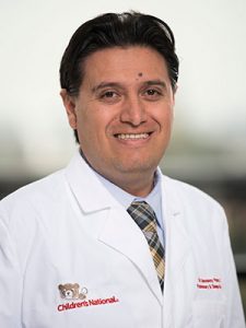
The immune system is the ultimate yin and yang, explains Anthony D. Sandler, M.D., senior vice president and surgeon-in-chief of the Joseph E. Robert Jr. Center for Surgical Care at Children’s National in Washington, D.C. With an ineffective immune system, infections such as the flu or diarrheal illness can run unchecked, causing devastating destruction. But on the other hand, excess immune activity leads to autoimmune diseases, such as lupus or multiple sclerosis. Thus, the immune system has “checks and balances” to stay controlled.

Cancer takes advantage of “the checks and balances,” harnessing the natural brakes that the immune system puts in place to avoid overactivity. As the cancer advances, molecular signals from tumor cells themselves turn on these natural checkpoints, allowing cancers to evade immune attack.

Several years ago, a breakthrough in pharmaceutical science led to a new class of drugs called checkpoint inhibitors. These medicines take those proverbial brakes off the immune system, allowing it to vigorously attack malignancies. However, Dr. Sandler says, these drugs have not worked uniformly and in some cancers, they barely work at all against the cancer.

One of these non-responders is high risk neuroblastoma, a common solid tumor found outside the skull in children. About 800 U.S. children are diagnosed with this cancer every year. And kids who have the high-risk form of neuroblastoma have poor prognoses, regardless of which treatments doctors use.

However, new research could lead to promising ways to fight high-risk neuroblastoma by enabling the immune system to recognize these tumors and spark an immune response. Dr. Sandler and colleagues recently reported on these results in the Jan. 29, 2018, PLOS Medicine using an experimental model of the disease.

The researchers created this model by injecting the preclinical models with cancer cells from an experimental version of neuroblastoma. The researchers then waited several days for the tumors to grow. Samples of these tumors showed that they expressed a protein on their cell surfaces known as PD-L1, a protein that is also expressed in many other types of human cancers to evade immune system detection.

To thwart this protective feature, the researchers made a cancer vaccine by removing cells from the experimental model’s tumors and selectively turning off a gene known as Id2. Then, they irradiated them, a treatment that made these cells visible to the immune system but blocked the cells from dividing to avoid new tumors from developing.

They delivered these cells back to the experimental models, along with two different checkpoint inhibitor drugs – antibodies for proteins known as CLTA-4 and PD-L1 – over the course of three treatments, delivered every three days. Although most checkpoint inhibitors are administered over months to years, this treatment was short-term for the experimental models, Dr. Sandler explains. The preclinical models were completely finished with cancer treatment after just three doses.

Over the next few weeks, the researchers witnessed an astounding turnaround: While experimental models that hadn’t received any treatment uniformly died within 20 days, those that received the combined vaccine and checkpoint inhibitors were all cured of their disease. Furthermore, when the researchers challenged these preclinical models with new cancer cells six months later, no new tumors developed. In essence, Dr. Sandler says, the preclinical models had become immune to neuroblastoma.

Further studies on human patient tumors suggest that this could prove to be a promising treatment for children with high-risk neuroblastoma. The patient samples examined show that while tumors with a low risk profile are typically infiltrated with numerous immune cells, tumors that are high-risk are generally barren of immune cells. That means they’re unlikely to respond to checkpoint inhibiting drugs alone, which require a significant immune presence in the tumor microenvironment. However, Dr. Sandler says, activating an immune response with a custom-made vaccine from tumor cells could spur the immune response necessary to make these stubborn cancers respond to checkpoint inhibitors.

Dr. Sandler cautions that the exact vaccine treatment used in the study won’t be feasible for people. The protocol to make the tumor cells immunogenic is cumbersome and may not be applicable to gene targeting in human patients. However, he and his team are currently working on developing more feasible methods for crafting cancer vaccines for kids. They also have discovered a new immune checkpoint molecule that could make this approach even more effective.

“By letting immune cells do all the work we may eventually be able to provide hope for patients where there was little before,” Dr. Sandler says.

In addition to Dr. Sandler, study co-authors include Priya Srinivasan, Xiaofang Wu, Mousumi Basu and Christopher Rossi, all of the Joseph E. Robert Jr. Center for Surgical Care and The Sheikh Zayed Institute for Pediatric Surgical Innovation (SZI), at Children’s National in Washington, D.C.

Financial support for research described in this post was provided by the EVAN Foundation, the Catherine Blair foundation, the Michael Sandler Research Fund and SZI.

ID-KD vaccine induced T-cell cytotoxicity

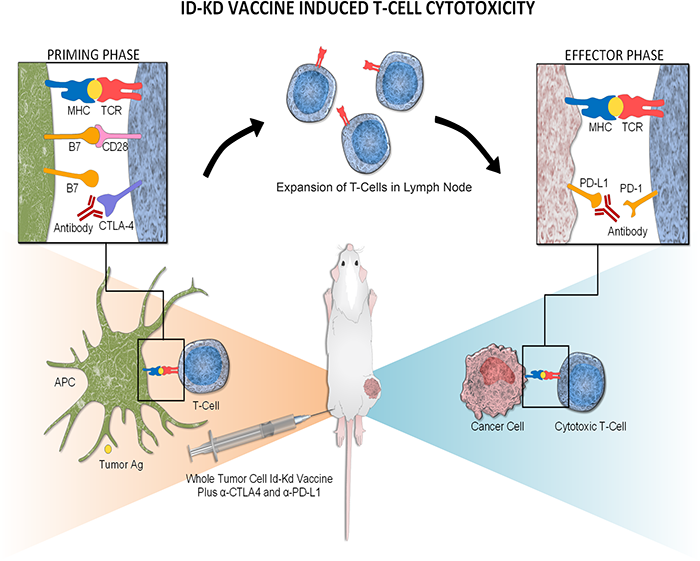
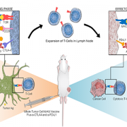
Mechanism of Id2kd Neuro2a vaccination combined with α-CTLA-4 and α-PD-L1 immunotherapy in a neuroblastoma model. During a vaccine priming phase, CTLA-4 blockade enhances activation and proliferation of T-cells that express programmed cell death 1 (PD1) and migrate to the tumor. Programmed cell death-ligand 1 (PD-L1) is up-regulated on the tumor cells, inducing adaptive resistance. Blocking PD-L1 allows for enhanced cytotoxic effector function of the CD8+ tumor-infiltrating lymphocytes. Artist: Olivia Abbate

bacteriophage

Phage therapy draws renewed interest to combat drug-resistant microbes

bacteriophage

In the face of growing antibiotic resistance and few antibiotics in the development pipeline, phages are drawing renewed research interest as a potential silver bullet.

The married professors were spending their Thanksgiving holiday in Egypt when the husband, Thomas L. Patterson, Ph.D., got very sick very quickly, experiencing fever, nausea and a racing heartbeat. By the time Patterson was accurately diagnosed with a highly multi-drug resistant bacterial infection, he was near death. His wife, Steffanie Strathdee, Ph.D., promised to “leave no stone unturned.’”

What happened next is the ultimate infectious disease feel good story: Strathdee, part of an All-Star team of infectious disease experts and epidemiologists, concocted a cocktail of viruses that killed the superbug and saved Patterson’s life.

“He was going to die,” says Roberta L. DeBiasi, M.D., MS, chief of the Division of Pediatric Infectious Diseases at Children’s National Health System. “Because of her epidemiology background – and because she loves him – Patterson became the first patient successfully treated with bacteriophages.”

Dr. DeBiasi explains that all viruses take over cells and use their machinery for their own purposes. In order to escape, viruses blow up the cell. Bacteriophages are viruses that target bacteria, taking over their machinery and ultimately killing the bacterial host.

“Infection is a race between the body’s immune response and the bacteria replicating themselves,” she adds. “Bacteria have to continually replicate. If you knock out 90 percent of them with phage therapy, that gives the immune system a fighting chance to win the race.”

She was so inspired by the team’s ingenuity that DeBiasi, program vice-chair, invited them to recount the story during IDWeek2018, held Oct. 3 to Oct. 7, 2018, in San Francisco. During the closing plenary, Patterson, a professor of psychiatry, and Strathdee, associate dean of Global Health Sciences, will be joined by Robert T. “Chip” Schooley, M.D., (all of University of California, San Diego), to discuss the clinical aspects and efficacy of phage therapy.

About 50 years ago, the U.S. military had investigated leveraging phages but ultimately placed that research portfolio on the back burner. Now, in the face of growing antibiotic resistance and few experimental antibiotics in the development pipeline, phages are drawing renewed research interest as a potential silver bullet.

“The technology has been around for 50 years. We’re going back to old things because we’re so desperate,” Dr. DeBiasi adds.

The tricky thing with phages is that each bacterium needs its own tailored phage therapy.

Children’s National is working with Adaptive Phage Therapeutics, a company based in Gaithersburg, Maryland, that developed the phage used to save Patterson, in order to help build out that library of phages, each ready to be directed to do battle against a specific pathogen.

“We have been consultants to them to think about what would be a good clinical trial, particularly in a pediatric population,” Dr. DeBiasi says.

Children’s National has been collecting and sending isolates from patients with neurogenic bladder who experience urinary tract infections to shore up the phage library in anticipation of a clinical trial. The work builds on Children’s experience as the first center to use phage therapy in a pediatric patient, a 2-year-old who had multidrug-resistant Pseudomonas aeruginosa infection complicated by bacteremia/sepsis.

Gustavo Nino

New method may facilitate childhood respiratory research

Gustavo Nino

“The use of CRC is a potentially powerful translational approach to shed light on the molecular mechanisms that control airway epithelial immune responses in infants and young children. This novel approach enables us to study the origins of respiratory disease and its chronic progression through childhood and beyond,” observes Gustavo Nino, M.D., a Children’s pulmonologist and study senior author.

A new method perfected by a team at Children’s National Health System may help expand research into pulmonary conditions experienced by infants and children, an understudied but clinically important age group. The study describing the new technique was published in the December 2017 print edition of Pediatric Allergy and Immunology.

Using conditionally reprogrammed cells (CRCs), a technique that enables indefinite proliferation of cells in the lab, the team was able to produce cell cultures that have a number of advantages over standard cultures and that may make it easier and more efficient to conduct research into pediatric respiratory immune responses.

The epithelial cells that line human airways are crucial in controlling immune responses to viruses, allergens and other environmental factors. The function and dysfunction of these airway epithelial cells (AECs) play a key role in asthma, cystic fibrosis and other pulmonary conditions, many of which begin in early life.

To generate enough of these cells for research, scientists culture AECs from primary nasal and bronchial cell samples. Cells derived from adults have fueled research leading to new therapies and the discovery of key biomarkers. But little comparable research has been conducted in infants. Airway sampling in premature infants has not been reported, likely to due to airway size limitations and underlying comorbidities. Similarly, sampling in infants is limited by the need for bronchoscopy and sedation.

“A major barrier has been the lack of a good system to culture epithelial cells, since airway sampling in infants and children is a challenge,” says co-lead author, Geovanny F. Perez, M.D., co-director of Children’s Severe Bronchopulmonary Dysplasia Program. “We needed a better way to culture cells in this age group.”

While primary AECs do not survive long in the lab, that hurdle was recently overcome by a process that generates CRCs from the primary AECs of adults, making it possible to quickly generate cell cultures from specimens.

In this study, the Children’s team adapted that approach, producing CRCs from primary AECs of neonates and infants. The CRC induction successfully enabled AEC cultures from infants born prematurely and from bronchial specimens of young children.

Geovanny Perez

“A major barrier has been the lack of a good system to culture epithelial cells, since airway sampling in infants and children is a challenge,” says co-lead author, Geovanny F. Perez, M.D., co-director of Children’s Severe Bronchopulmonary Dysplasia Program. “We needed a better way to culture cells in this age group.”

“We found that the CRCs have longer cell life and greater proliferation ability than standard cultures of epithelial cells. They preserved their original characteristics even after multiple experiments. And, they presented an innate immune response similar to that seen in primary human epithelial cells during viral respiratory responses in children,” says Dr. Perez.

“The use of CRC is a potentially powerful translational approach to shed light on the molecular mechanisms that control airway epithelial immune responses in infants and young children. This novel approach enables us to study the origins of respiratory disease and its chronic progression through childhood and beyond,” observes Gustavo Nino, M.D., a Children’s pulmonologist and study senior author.

The authors note that further studies are needed to define more precisely the differences and similarities in the immune responses of CRC and non-CRC derived from primary AEC. However, they conclude that CRC represents a new, effective method to study AEC innate immune responses in infants.

In addition to Drs. Perez and Nino, Children’s Center for Genetic Medicine Research co-authors include Co-Lead Author S. Wolf; Lana Mukharesh; Natalia Isaza Brando, M.D.; Diego Preciado, M.D., Ph.D.; Robert J. Freishtat, M.D., M.P.H.; Dinesh Pillai, M.D.; and M. C. Rose.

Financial support for this research was provided by the National Institute of Allergy and Infectious Diseases under grant number R21AI130502; Eunice Kennedy Shriver National Institute of Child Health and Human Development under grant number HD001399; National Heart, Lung and Blood Institute under grant number HL090020; and National Center for Advancing Translational Sciences under grant number UL1TR000075.