Tag Archive for: heart

Catherine Limperopoulos

Stressful pregnancies can leave fingerprint on fetal brain

Catherine Limperopoulos

“We were alarmed by the high percentage of pregnant women with a diagnosis of a major fetal heart problem who tested positive for stress, anxiety and depression,” says Catherine Limperopoulos, Ph.D., director of the Center for the Developing Brain at Children’s National and the study’s corresponding author.

When a diagnosis of fetal congenital heart disease causes pregnant moms to test positive for stress, anxiety and depression, powerful imaging can detect impaired development in key fetal brain regions, according to Children’s National Hospital research published online Jan. 13, 2020, in JAMA Pediatrics.

While additional research is needed, the Children’s National study authors say their unprecedented findings underscore the need for universal screening for psychological distress as a routine part of prenatal care and taking other steps to support stressed-out pregnant women and safeguard their newborns’ developing brains.

“We were alarmed by the high percentage of pregnant women with a diagnosis of a major fetal heart problem who tested positive for stress, anxiety and depression,” says Catherine Limperopoulos, Ph.D., director of the Center for the Developing Brain at Children’s National and the study’s corresponding author. “Equally concerning is how prevalent psychological distress is among pregnant women generally. We report for the first time that this challenging prenatal environment impairs regions of the fetal brain that play a major role in learning, memory, coordination, and social and behavioral development, making it all the more important for us to identify these women early during pregnancy to intervene,” Limperopoulos adds.

Congenital heart disease (CHD), structural problems with the heart, is the most common birth defect. Still, it remains unclear how exposure to maternal stress impacts brain development in fetuses with CHD.

The multidisciplinary study team enrolled 48 women whose unborn fetuses had been diagnosed with CHD and 92 healthy women with uncomplicated pregnancies. Using validated screening tools, they found:

  • 65% of pregnant women expecting a baby with CHD tested positive for stress
  • 27% of women with uncomplicated pregnancies tested positive for stress
  • 44% of pregnant women expecting a baby with CHD tested positive for anxiety
  • 26% of women with uncomplicated pregnancies tested positive for anxiety
  • 29% of pregnant women expecting a baby with CHD tested positive for depression and
  • 9% women with uncomplicated pregnancies tested positive for depression

All told, they performed 223 fetal magnetic resonance imaging sessions for these 140 fetuses between 21 and 40 weeks of gestation. They measured brain volume in cubic centimeters for the total brain as well as volumetric measurements for key regions such as the cerebrum, cerebellum, brainstem, and left and right hippocampus.

Maternal stress and anxiety in the second trimester were associated with smaller left hippocampi and smaller cerebellums only in pregnancies affected by fetal CHD. What’s more, specific regions — the hippocampus head and body and the left cerebellar lobe – were more susceptible to stunted growth. The hippocampus is key to memory and learning, while the cerebellum controls motor coordination and plays a role in social and behavioral development.

The hippocampus is a brain structure that is known to be very sensitive to stress. The timing of the CHD diagnosis may have occurred at a particularly vulnerable time for the developing fetal cerebellum, which grows faster than any other brain structure in the second half of gestation, particularly in the third trimester.

“None of these women had been screened for prenatal depression or anxiety. None of them were taking medications. And none of them had received mental health interventions. In the group of women contending with fetal CHD, 81% had attended college and 75% had professional educations, so this does not appear to be an issue of insufficient resources,” Limperopoulos adds. “It’s critical that we routinely to do these screenings and provide pregnant women with access to interventions to lower their stress levels. Working with our community partners, Children’s National is doing just that to help reduce toxic prenatal stress for both the health of the mother and for the future newborns. We hope this becomes standard practice elsewhere.”

Adds Yao Wu, Ph.D., a research associate working with Limperopoulos at Children’s National and the study’s lead author: “Our next goal is exploring effective prenatal cognitive behavioral interventions to reduce psychological distress felt by pregnant women and improve neurodevelopment in babies with CHD.”

In addition to Limperopoulos and Wu , Children’s National study co-authors include Kushal Kapse, MS, staff engineer; Marni Jacobs, Ph.D., biostatistician; Nickie Niforatos-Andescavage, M.D., neonatologist; Mary T. Donofrio, M.D., director, Fetal Heart Program; Anita Krishnan, M.D., associate director, echocardiography; Gilbert Vezina, M.D., director, Neuroradiology Program; David Wessel, M.D., Executive Vice President and Chief Medical Officer; and Adré  J. du Plessis, M.B.Ch.B., director, Fetal Medicine Institute. Jessica Lynn Quistorff, MPH, Catherine Lopez, MS, and Kathryn Lee Bannantine, BSN, assisted with subject recruitment and study coordination.

Financial support for the research described in this post was provided by the National Institutes of Health under grant No. R01 HL116585-01 and the Thrasher Research Fund under Early Career award No. 14764.

newborn baby

Directly measuring function in tiny hearts

newborn baby

The amount of blood the heart pumps in one minute can be directly measured safely in newborns by monitoring changes in blood velocity after injecting saline, indicates the first clinical study of direct cardiac output measurement in newborns.

The amount of blood that the heart pumps in one minute (cardiac output) can be directly measured safely in newborns by monitoring changes in blood velocity after injecting saline, indicates a paper published online Dec. 17, 2019 in the Journal of Pediatrics and Neonatal Medicine. The research, conducted by Children’s National Hospital faculty, is believed to be the first clinical study of direct cardiac output measurement in newborns.

Right now, cardiac output is measured indirectly in the nation’s neonatal intensive care units (NICU) using newborns’ blood pressure, heart rate, urine output and other indirect measures. However, these techniques can produce imprecise readings in children. And the field lacks a feasible “gold standard” to measure cardiac output in newborns.

The COstatus monitor already uses ultrasound dilution – the expected decrease in the velocity of blood when saline is injected, producing a dilution curve. A Children’s National research team used ultrasound dilution in their small pilot study to gauge the feasibility of directly measuring cardiac output in newborns.

“Infants who stand to benefit most from directly monitoring cardiac hemodynamics are often so sick they already have central venous access,” says Khodayar Rais-Bahrami, M.D., an attending neonatologist at Children’s National and the study’s senior author. “Using the COstatus monitor in these children would enable the clinical team to personalize care based on the newborn’s current hemodynamic status, while introducing minimal fluid during measurements,” Dr. Rais-Bahrami adds.

COstatus monitor

The COstatus Monitor uses an extracorporeal loop attached to arterial and venous lines to measure cardiac output using ultrasound dilution. The research team injected 1mL/kg of body temperature saline into the loop and performed up to two measurement sessions daily.

The research team recruited 12 newborns younger than 2 weeks old who already had central venous and arterial access. The venous line of the arteriovenous AV loop is connected to the umbilical venous catheter while the COstatus monitor’s arterial line is connected to the umbilical arterial catheter. During measurement sessions, two injections of solution are injected into the venous loop, allowing for two measures of cardiac output, cardiac index, active circulating volume index, central blood volume index and systemic vascular resistance index.

Infants enrolled in the pilot study underwent up to two measurement sessions per day for up to four days, for a total of 54 cardiac hemodynamic measurements. The newborns ranged from 720 to 3,740 grams in weight and 24 to 41.3 weeks in gestational age.

The infants’ mean cardiac output was 0.43L/min and increased with gestational age. By contrast, the mean cardiac index was 197mL/kg/min and changed little with infants’ increasing maturity – either by gestational age or postnatal age. Two of the study participants were undergoing therapeutic cooling for hypoxic-ischemic encephalopathy and had their measurements taken during cooling and after rewarming.

“Although this study size is small, it demonstrates that this minimally invasive technique can safely be used in newborns to directly measure cardiac hemodynamics,” says Simranjeet S. Sran, M.D., a Children’s National neonatalogist and the study’s lead author. “This technology may allow for more precise and personalized care of critically ill newborns in a range of disease states – real-world utility in NICUs that serve some of the youngest and sickest newborns,” Dr. Sran adds.

The research team notes that direct measurement by ultrasound dilution revealed a stark increase in cardiac index as infants undergoing therapeutic hypothermia were rewarmed, raising questions about whether indirect measures using other technology, such as echocardiography, underestimate hypothermia’s effect on hemodynamics.

In addition to Drs. Rais-Bahrami and Sran, Mariam Said, M.D., also a Children’s National neonatalogist, was a study co-author.

Autonomic nervous system appears to function well regardless of mode of childbirth

Late in pregnancy, the human body carefully prepares fetuses for the rigors of life outside the protection of the womb. Levels of cortisol, a stress hormone, ramp up and spike during labor. Catecholamines, another stress hormone, also rise at birth, helping to kick start the necessary functions that the baby will need to regulate breathing, heartbeat, blood pressure and energy metabolism levels at delivery. Oxytocin surges, promoting contractions for the mother during labor and stimulating milk production after the infant is born.

These processes also can play a role in preparing the fetal brain during the transition to life outside the womb by readying the autonomic nervous system and adapting its cerebral connections. The autonomic nervous system acts like the body’s autopilot, taking in information it needs to ensure that internal organs run steadily without willful action, such as ensuring the heart beats and eyelids blink at steady intervals. Its yin, the sympathetic division, stimulates body processes while its yang, the parasympathetic division, inhibits them.

Infants born preterm have reduced autonomic function compared with their full-term peers and also face possible serious neurodevelopmental impairment later in life. But is there a difference in autonomic nervous system function for full-term babies after undergoing labor compared with infants delivered via cesarean section (C-section)?

A team from the Children’s National Inova Collaborative Research Program (CNICA) – a research collaboration between Children’s National in Washington, D.C., and Inova Women’s and Children’s Hospital in Virginia – set out to answer that question in a paper published online July 30, 2019, in Scientific Reports.

They enrolled newborns who had experienced normal, full-term pregnancies and recorded their brain function and heart performance when they were about 2 days old. Infants whose conditions were fragile enough to require observation in the neonatal intensive care unit were excluded from the study. Of 167 infants recruited for the prospective cohort study, 118 newborns had sufficiently robust data to include them in the research.  Of these newborns:

  • 62 (52.5%) were born by vaginal delivery
  • 22 (18.6%) started out with vaginal delivery but ultimately switched to C-section based on failure to progress, failed labor induction or fetal intolerance to labor
  • And 34 (28.8%) were born by elective C-section.

The CNICA research team swaddled infants for comfort and slipped electrode nets over their tiny heads to simultaneously measure heart rate variability and electrocortical function through non-invasive techniques. The team hypothesized that infants who had been exposed to labor would have enhanced autonomic tone and higher cortical electroencephalogram (EEG) power than babies born via C-section.

“In a low-risk group of babies born full-term, the autonomic nervous system and cortical systems appear to function well regardless of whether infants were exposed to labor prior to birth,” says Sarah B. Mulkey, M.D., Ph.D., a fetalneonatal neurologist in the Division of Fetal and Transitional Medicine at Children’s National and the study’s lead author.

However, infants born by C-section following a period of labor had significantly increased accelerations in their heart rates. And the infants born by C-section during labor had significantly lower relative gamma frequency EEG at 25.2 hours old compared with the other two groups studied.

“Together these findings point to a possible increased stress response and arousal difference in infants who started with vaginal delivery and finished delivery with C-section,” Dr. Mulkey says. “There is so little published research about the neurologic impacts of the mode of delivery, so our work helps to provide a normal reference point for future studies looking at high-risk infants, including babies born preterm.”

Because the research team saw little differences in autonomic tone or other EEG frequencies when the infants were 1 day old, future research will explore these measures at different points in the newborns’ early life as well as the role of the sleep-wake cycle on heart rate variability.

In addition to Dr. Mulkey, study co-authors include Srinivas Kota, Ph.D., Rathinaswamy B. Govindan, Ph.D., Tareq Al-Shargabi, MSc, Christopher B. Swisher, BS, Laura Hitchings, BScM, Stephanie Russo, BS, Nicole Herrera, MPH, Robert McCarter, ScD, and Senior Author Adré  J. du Plessis, M.B.Ch.B., MPH, all of Children’s National; and Augustine Eze Jr., MS, G. Larry Maxwell, M.D., and Robin Baker, M.D., all of Inova Women’s and Children’s Hospital.

Financial support for research described in this post was provided by the National Institutes of Health National Center for Advancing Translational Sciences under award numbers UL1TR001876 and KL2TR001877.

Plastic leaching illustration

Plasticizer interaction with the heart

Calling an ambulance during an emergency, emailing a journal article before a 5 p.m. deadline and maintaining conditions during the fifth week of a 6-week lab study, without altering the light or temperature, requires electricity and translates into time, money and lives saved. During critical moments, we appreciate the tiny particles and ions in electric currents that power our phones, computers or laboratory equipment. We seldom think about the speed of these connections or potential disruptors when conditions are stable. The same applies to the electric currents, or electrophysiology, of our heart.

Arrhythmias affect millions of Americans but can be controlled with routine screenings and preventive care. In an intensive care setting, helping a patient maintain a steady heart rate, especially if they are at risk for cardiac complications, may support a faster recovery, shorter hospital stay, reduced health care costs and improved health outcomes, such as avoiding complications from heart failure or stroke.

A preclinical study, entitled “Plasticizer Interaction With the Heart,” appears in the July issue of Circulation: Arrhythmia and Electrophysiology and examines the role plastic exposure, akin to exposure in a medical setting, has on heart rhythm disruptions and arrhythmias.

changes in heart rhythm due to plastics

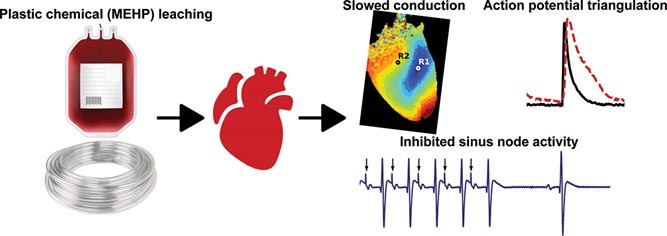
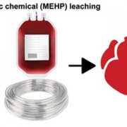
New preclinical research finds acute exposure to MEHP, a common plasticizer used in medical equipment, increases risk for alternans and arrhythmias, disruptions in heart rhythm. The images above show changes in heart rhythm, measured by slowed epicardial conduction velocity, enhanced action potential prolongation and impaired sinus node activity.

The research team, led by researchers at Children’s National Health System, discovered increased risks for irregular heart rhythms after exposing intact, in vitro heart models to 30 minutes of mono-2-ethylhexyl phthalate (MEHP), a metabolite from Di-2-ethylhexyl phthalate (DEHP). DEHP is a chemical commonly used to make plastics pliable in FDA-approved medical devices. This phthalate accounts for 40% of the weight of blood storage bags and up to 80% of the weight of tubes used in an intensive care setting, such as for assisted feeding or breathing, and for catheters used in diagnostics or to conduct minimally invasive cardiac procedures.

The team chose to study the heart’s reaction to 60 µM of MEHP, a level comparable to stored blood levels of MEHP observed in pediatric patients and in neonatal exchange transfusion procedures. They found 30-minute exposure to MEHP slowed atrioventricular conduction and increased the atrioventricular node effective refractory period. MEHP prolonged action potential duration time, enhanced action potential triangulation, increased the ventricular effective refractory period and slowed epicardial conduction velocity, which may be due to the inhibition of Nav 1.5, or sodium current.

“We chose to study the impact of MEHP exposure on cardiac electrophysiology at concentrations that are observed in an intensive care setting, since plastic medical products are known to leach these chemicals into a patient’s bloodstream,” says Nikki Gillum Posnack, Ph.D., a principal investigator with the Sheikh Zayed Institute for Pediatric Surgical Innovation at Children’s National and an assistant professor of pediatrics at the George Washington University School of Medicine and Health Sciences. “In critical conditions, a patient may have a blood transfusion, require extracorporeal membrane oxygenation, undergo cardiopulmonary bypass or require dialysis or intravenous fluid administration. All of these scenarios can lead to plastic chemical exposure. Our research team wants to investigate how these plastic chemicals can impact cardiac health.”

In this review, Dr. Posnack’s team mentions one reason for the observed changes in the preclinical heart models may be due to the structure of phthalates, which resemble hormones and can interfere with a variety of biological processes. Due to their low molecular weight, these chemicals can interact directly with ion channels, nuclear receptors and other cellular targets.

Existing epidemiological research shows associations between exposure to phthalates and adverse health outcomes, including metabolic disturbances, reproductive disorders, inflammatory conditions, neurological disorders and cardiovascular disease. This is the first study to examine the link between cardiac electrophysiology in intact hearts and exposure to MEHP, comparable to levels observed in an ICU.

Dr. Posnack’s team previously found DEHP reduced cellular electrical coupling in cardiomyocyte cell models, which slowed conduction velocity and produced an arrhythmogenic phenotype. A microarray analysis found heart cells treated with DEHP led to mRNA changes in genes responsible for contracting and calcium handling. Another preclinical study showed DEHP altered nervous system regulation of the cardiovascular system. Future studies to expand on this research may include the use of larger preclinical models or human assessments. For the latter, stem cell-derived cardiomyocytes can be used to compare the safety profile of plastic chemicals with potential alternatives.

An accompanying editorial, entitled “Shocking Aspects of Nonconductive Plastics,” authored by cardiology researchers at the University of Wisconsin-Madison, puts this novel research into perspective. Like Dr. Posnack, the team notes that while the clinical impact plasticizers have on heart health still needs to be determined, the work contributes to compelling data among multiple researchers and shows DEHP and MEHP are not inert substances.

“Toxic plasticizers in children’s toys and baby products hit public headlines 20 years ago, but exposure to these compounds is up to 25x higher in patients undergoing complex medical procedures,” write the University of Wisconsin-Madison researchers. “We readily (and unknowingly) administer these compounds, and at times in high quantity, to some of our most vulnerable patients. This work highlights the need for further investigation into short and long-term plasticizer exposure on cardiac electrophysiology.”

The Agency for Toxic Substances and Disease Registry (ATSDR), part of the Centers for Disease Control and Prevention (CDC), released a public health statement about DEHP in 2002, noting more research in humans is needed to issue formal warnings against this phthalate.

ATSDR states there is no conclusive evidence about the adverse health effects of children exposed to DEHP in a medical setting, such as procedures that require the use of flexible tubing to administer intravenous fluids or medication. However, the CDC statement includes limits of DEHP exposure, based on preclinical models, used to guide upper DEHP limits in consumer products, including food packaging, drinking water, and air quality in the workplace.

“It’s important to note that this was a preliminary study performed on an ex vivo model that is largely resilient to arrhythmias”, says Rafael Jaimes III, Ph.D., the first author of the study and a senior scientist at Children’s National. “Due to the nature of the design, it was somewhat alarming that we found such significant effects. I predict that electrophysiological disturbances will be more pronounced in models that more closely resemble humans. These types of models should absolutely be studied.”

“And, importantly, our results may incentivize the development and use of new products that are manufactured without phthalates,” Dr. Posnack adds.

These questions are powering Dr. Posnack and her team through a decade-long, multi-institution research investigation to understand how plastic chemicals and medical device biomaterials can impact cardiac health.

Additional study authors for this paper include Damon McCullough, B.S., Bryan Siegel, M.D., Luther Swift, Ph.D., Daniel McInerney, B.S., and James Hiebert, B.S., with the Sheikh Zayed Institute for Pediatric Surgical Innovation and Children’s National Heart Institute, part of Children’s National Health System in Washington, D.C.; Erick A. Perez-Alday, Ph.D., and Larisa G Tereshchenko, M.D., Ph.D., with the Knight Cardiovascular Institute at Oregon Health and Science University in Portland, Ore.; Javier Saiz, Ph.D., and Beatriz Trenor, Ph.D., with Ci2B-Universitat Politecnica de Valencia in Spain and Jiansong Sheng, Ph.D., from CiPA Lab, LLC, in Rockville, Md.

The study was supported by the National Institutes of Health (R00ES023477 and R01HL139472), Children’s Research Institute and Children’s National Heart Institute. NVIDIA corporation provided graphics processing, with partial support by the Direccion General de Politica Cientifica de la Generalitat Valenciana (PROMETEU2016/088).

heart and medical equiptment

How much do you know about congenital heart defects?


Andrea Beaton and Craig Sable

Assessing the global burden of rheumatic heart disease

Andrea Beaton and Craig Sable

A research team that included Children’s National Heart Institute experts Andrea Beaton, M.D., and Craig Sable, M.D., examined data on fatal and nonfatal Rheumatic Heart Disease for a 25 year period from 1990 through 2015 to determine the current global burden of RHD.

Rheumatic Heart Disease (RHD) is the most commonly acquired heart disease in young people under the age of 25. It’s caused by untreated streptococcal throat infections that progress into acute rheumatic fever and eventually weaken the valves of the heart. Fortunately, the devastating condition, which was endemic in the United States before 1950, is now relatively rare in the developed world due to social and economic development and the introduction of penicillin. But, as shown in a recent study published in the New England Journal of Medicine, in the developing world, RHD remains nearly as common as HIV.

As part of the 2015 Global Burden of Disease Study, a research team that included Children’s National Heart Institute experts Andrea Beaton, M.D., and Craig Sable, M.D., examined data on fatal and nonfatal RHD for a 25 year period from 1990 through 2015 to determine the current global burden of RHD. The group employed epidemiologic modeling techniques to estimate the global, regional and national prevalence of RHD, as well as death rates and disability-adjusted life years attributable to the disease.

“This study provides more detail than ever before about the global impact of RHD,” explains Dr. Sable. “It utilizes global burden of disease tools that are updated on an annual basis. These tools are considered highly reputable and allow for ongoing tracking and comparison to other diseases.”

The researchers found that overall, death rates from RHD have declined: there were 347,500 deaths from RHD in 1990 and 319,400 deaths in 2015, a decrease of 8 percent. From 1990 to 2015, the global age-standardized death rate from RHD also decreased from 9.2 to 4.8 per 100,000 — a change of 48 percent.

However, a closer look at the data shows that progress on RHD remains uneven. Although the health-related burden of RHD has declined in most countries over the 25-year period, the condition persists in some of the poorest regions in the world, with the highest estimated death rates in Central African Republic, Federated States of Micronesia, Fiji, India, Kiribati, Lesotho, Marshall Islands, Pakistan, Papua New Guinea, the Solomon Islands and Vanuatu. In several regions, mortality from RHD and the number of individuals living with RHD did not appreciably decline between 1990 and 2015. The researchers estimate that 10 out of every 1,000 people living in South Asia and central sub-Saharan Africa and 15 out of 1,000 people in Oceania were living with RHD in 2015.

“These data are critically important for increasing awareness and funding to reduce the global burden of rheumatic heart disease,” says Dr. Sable. “Dr. Beaton and I are proud to be part of a small team of global investigators leading this effort.”

Children’s National Health System was recently awarded a grant from the American Heart Association to launch a Rheumatic Heart Disease Center, with the goal of developing innovative strategies and economic incentives to improve the prevention and diagnosis of RHD in high-risk, financially disadvantaged countries and low-income communities across the United States. The program will use Children’s robust telemedicine infrastructure to connect co-collaborators around the world, as well as train the next generation of globally minded cardiovascular researchers.

Newborn pulse oximetry screening: which algorithm is best?

Gerard Martin

There’s a consensus that Pulse oximetry screening (POS) is a proven way to find critical congenital heart defects. But, screenings, specifically the algorithm used, vary. Gerard R. Martin, M.D., Medical Director of Global Health at Children’s National Health System, and Andrew K. Ewer, MD, explore which algorithm is best in their just-published article in Pediatrics. Read more.