New grant to help establish maternal mental health telehealth program
Children’s National has received a $76,000 grant from the Health Resources & Services Administration (HRSA) which will allow a cross-functional team of neonatologists and psychologists to establish a parental mental telehealth program.
Worldwide about 10% of pregnant women and 13% of women who have just given birth experience a mental health disorder, primarily depression, according to the World Health Organization.
“This is a topic that is quickly garnering attention but remains extremely underfunded,” says Lamia Soghier, M.D., F.A.A.P., C.H.S.E., medical director of the Neonatal Intensive Care Unit (NICU) at Children’s National Hospital. “We tend to focus on the babies but don’t pay enough attention to the parents.”
Dr. Soghier’s focus has been on NICU parents who experience postpartum mood and anxiety disorders (PMADs), often due to their uniquely stressful experiences.
“We have been screening on a small scale for many years and have noticed a 33-45% rate of postpartum depression symptoms in our NICU families,” she says.
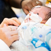
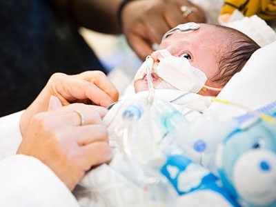
Maternal mental disorders are treatable with effective screening and interventions. Children’s National has received a $76,000 grant from the Health Resources & Services Administration (HRSA) which will allow a cross-functional team of neonatologists and psychologists to establish a parental mental telehealth program to expand screening and provide diagnosis, therapy and counseling to NICU parents who experience postpartum mood and anxiety disorders.
Dr. Soghier, along with Ololade ‘Lola’ Okito, M.D., neonatologist at Children’s National, and Erin Sadler, Psy.D., psychologist in the Division of Psychology and Behavioral Health at Children’s National, discuss the importance of this work.
Q: Tell us more about the program you’re establishing.
A: Dr. Soghier: This program will allow us to hire a licensed psychologist who will see families both in the NICU and through follow-up telehealth visits. It provides a one-stop shop for our families, which is particularly important during the COVID-19 pandemic. The grant will also allow us to develop an iPad loaner program to give loaner iPads to low income families who do not have access to a device or to reliable internet services so that they can receive therapy at home.
Dr. Sadler: We’ll be examining how the implementation of these services can increase accessibility and reduce barriers that prevent assessment and initiation of crucial mental health services for at-risk mothers. Our partnerships will be key. Mothers experiencing barriers to participating in care services in the NICU will also have access to an in-house, licensed psychologist through telehealth services within the comfort of their homes. Families experiencing problems accessing telehealth technology due to economic limits would get the loaner iPad. We’re meeting our families where they are in order to provide these critical services.
Q: Why is grant funding to important in this space?
A: Dr. Okito: Access to perinatal mental health services is limited at the local and national levels, particularly for vulnerable parents of infants admitted to the NICU. Little is known about the effect of interventions to address depression and anxiety among NICU parents, and this grant will allow us to contribute to this very important area of research.
Dr. Sadler: It is not enough to recognize the health disparities that exist amongst communities in our nation. It is imperative that we’re able to explore and examine solutions that can aid in enhancing the equity of care for children and adults alike. As Dr. Okito mentions, there is little to no research available that looks at the feasibility of the support programs we intended to put in place. We hope to create a viable model that could be used to help NICU families across the country.
Q: How is Children’s National uniquely positioned to do this work?
A: Dr. Soghier: Healthy moms and healthy dads equal happy babies. That’s why we will be taking care of the family as a whole. This is truly family-centered care and at the heart of what Children’s National is all about.
Dr. Sadler: The Children’s National NICU team has an established postpartum depression screening program. Through the piloted work, staff have identified notable barriers to universal screening, access to perinatal mental health support and the impact of PMADs on parent engagement in newborn care. As a result, Children’s National is uniquely positioned to directly address such barriers and provide specialized care.
Q: What excites you about this work?
A: Dr. Sadler: As a specialist in perinatal and infant mental health, I look forward to being able to demonstrate the lasting impact maternal mental health services can provide for not only newborns and their families, but for care providers as well. I am excited to have additional opportunities to advocate for the integration of perinatal and infant mental health in non-traditional spaces.
Dr. Okito: I am most excited about the potential to expand universal depression screening among NICU parents. Having done this work for the past three years, I know there are limitations in screening because we’ve only been able to screen parents that are at the patient’s bedside. More screening will lead to more parents getting the referrals and services that they need.