Tag Archive for: genetic

Francis Collins

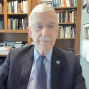
Francis S. Collins, M.D., Ph.D. from NIH: The future of genomic medicine and research funding opportunities

Kurt Newman and Francis Collins

Genomic medicine, diversity, equity and inclusion (DEI), a world post-COVID-19 and pediatric research funding were among the topics discussed during the “Special Fireside Chat” keynote lecture at the 2021 Children’s National Hospital Research, Education and Innovation Week.

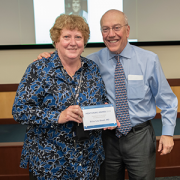
Francis S. Collins, M.D., Ph.D., director at the National Institutes of Health (NIH), is well known for his landmark discoveries of disease genes and his leadership of the international Human Genome Project, which culminated in April 2003 with the completion of a finished sequence of the human DNA instruction book.

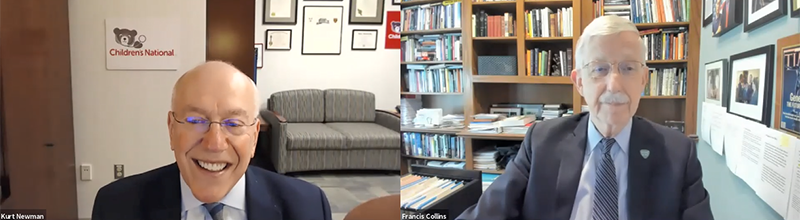
The President and CEO of Children’s National, Kurt Newman, M.D., joined Dr. Collins during the “Special Fireside Chat” keynote lecture. Dr. Newman posed several health care-related questions to Dr. Collins over the course of 30 minutes. Dr. Collins’s responses shed light on what it takes to advance various research fields focused on improving child health and develop frameworks that advocate for DEI in order to foster a more just society.

Q: You have been involved with genomic medicine since its inception. You discovered the gene causing cystic fibrosis and led the Human Genome project. What do you see as the future of genomic medicine, especially as it relates to improving child health?

A: Thank you for the question, Kurt. First, I wanted to say congratulations on your 150th anniversary. Children’s National Hospital has been such a critical component for pediatric research and care in the Washington, D.C., area, and at the national and international levels. We at the NIH consider it a great privilege to be your partner in many of the things that we can and are doing together.

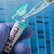
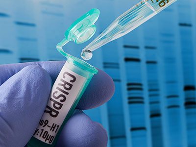
Genomic medicine has certainly come a long way. The word genomics was invented in 1980, so we have not been at this for that long. Yet, the success of the Human Genome Project and the access to cost-effective tools for rapid DNA sequencing have made many things possible. It took a lot of effort, time and money to discover the gene that causes cystic fibrosis. Kurt, if you look at what we did, while it was rewarding, it was a challenging problem that occupied the hearts of the scientific community in 1980. Now, a graduate student at Children’s National that has access to DNA samples, a thermal cycler, a DNA sequencer and the internet could do in about a week what it took us a decade and with 50 people.

We have been able to rocket forward as far as identifying the genetic causes of 6,500 diseases, where we know precisely the molecular glitch responsible for those conditions. While most of those are rare diseases, it leads to the opportunity for immediate diagnosis, which used to be a long and troubled journey.

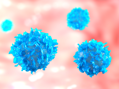
DNA sequencing has increasingly become an essential tool in newborns, especially when trying to sort out puzzling diagnosis for specific syndromes or phenotypes that are not immediately clear. Additionally, DNA sequencing significantly impacted clinical care in cancer because it made it possible to look at the mutations driving the malignancy and its genetic information that can lead to interventions. This approach is going forward in the next few years in ways that we can see now. Although I am a little reluctant to make predictions because I have to be careful about that, it may be possible to obtain complete genome sequences that can be yours for life and place them into the medical record to make predictions about future risks and choices about appropriate drugs. This path costs less than any imaging tests.

Q: The racial justice movement that was brought back to the forefront this past year has, once again, reaffirmed that this country has so much more work to do in order to end systemic racism. You have been at the forefront of promoting diversity, equity and inclusion in research and at the NIH. What do you and the NIH plan to do further DEI efforts in research and in general so that we can be a more just and equitable society?

A: I appreciate you raising this, Kurt. Diversity, equity and inclusion (DEI) is an issue where everyone should be spending a lot of time, energy and passion. You are right. 2020 will be remembered for COVID-19. I also think it will be remembered for the things that occurred around the killing of George Floyd, and the recognition of the very foundation that is still infected by this terribly difficult circumstance of structural racism. I convened a group of about 75 deep thinkers about these issues, many of them are people of color from across the NIH’s different areas of activities. I asked the group to come forward with a bold set of proposals. This effort is how the program UNITE came together to work hard on this, which is now making recommendations that I intend to follow. We are determined to close that gap and pursue additional programs that will allow us to be more successful in recruiting and retaining minority groups, for example. We need to do something with our health disparity and research portfolio as well to ensure that we are not just looking around the edges of the causes for racial inequities. We are digging deeper into what the structural racism underpinnings are and what we can do about it. I am particularly interested in supporting research projects that test intervention and not just catalog the factors involved. We have been, at times, accused and maybe rightly so of being more academic about this, and, less kindly, we have been accused of admiring the problem of health disparities as opposed to acting on it. We are ready to act.

Q: COVID has affected us all in so many ways. Could you tell us what this past year has been like for you? Also, how is the NIH preparing for a soon-to-be post-COVID pandemic?

A: This is the time to contemplate the lessons learned as everyone knows that the last worst pandemic happened over a century ago. One thing that maybe will vex us going forward, which we already started to invest in a big way, is this whole long COVID syndrome, also referred to post-acute sequelae, to understand precisely the consequences and mechanisms like Multisystem Inflammatory Syndrome in Children (MIS-C). Before moving to the next pandemic, we must think about how we will help understand those who suffer from long COVID syndrome. As far as the broader lessons learn, Kurt, we must expect that there will be other pandemics because humans are interacting more with animals, so zoonosis is likely to emerge. We need to have a clear sense of preparation for the next one. For instance, we are working on this right now, but we need to have a stronger effort to develop small molecules of anti-viral drugs aimed at the major viral classes, so we do not have to start from scratch. We also need clinical trial networks warm all the time, ready to go and to learn how valuable public partnerships can be to get things done in a hurry.

Editor’s Note: The responses in this Q+A have been modified to fit the word count.

muscle cells

Experimental model mimics early-stage myogenic deficit in boys with DMD

muscle cells

Muscle regeneration marked by incorporation of muscle stem cell nuclei (green) in the myofibers (red) in dystrophic muscles with low TGFβ level (upper image), but not with high TGFβ level (lower image). Inflammatory and other nuclei are labeled blue.

Boys with Duchenne muscular dystrophy (DMD) experience poor muscle regeneration, but the precise reasons for this remain under investigation. An experimental model of severe DMD that experiences a large spike in transforming growth factor-beta (TGFβ) activity after muscle injury shows that high TGFβ activity suppresses muscle regeneration and promotes fibroadipogenic progenitors (FAPs). This leads to replacement of the damaged muscle fibers by calcified and connective tissue, compromising muscle structure and function. While blocking FAP buildup provides a partial solution, a Children’s National Hospital study team identifies correcting the muscle micro-environment caused by high TGFβ as a ripe therapeutic target.

The team’s study was published online March 26, 2020, in JCI Insight.

DMD is a chronic muscle disease that affects 1 in 6,200 young men in the prime of their lives. The disorder, caused by genetic mutations leading to the inability to produce dystrophin protein, leads to ongoing muscle damage, chronic inflammation and poor regeneration of lost muscle tissue. The patients experience progressive muscle wasting, lose the ability to walk by the time they’re teenagers and die prematurely due to cardiorespiratory failure.

The Children’s National team finds for the first time that as early as preadolescence (3 to 4 weeks of age), their experimental model of severe DMD disease showed clear signs of the type of spontaneous muscle damage, regenerative failure and muscle fiber loss seen in preadolescent boys who have DMD.

“In boys, the challenge due to muscle loss exists from early in their lives, but had not been mimicked previously in experimental models,” says Jyoti K. Jaiswal, MSc, Ph.D., principal investigator in the Center for Genetic Medicine Research at Children’s National, and the study’s co-senior author. “TGFβ is widely associated with muscle fibrosis in DMD, when, in fact, our work shows its role in this disease process is far more significant.”

Research teams have searched for experimental models that replicate the sudden onset of symptoms in boys who have DMD as well as its complex progression.

“Our work not only offers insight into the delicate balance needed for regeneration of skeletal muscle, but it also provides quantitative information about muscle stem cell activity when this balanced is disturbed,” says Terence A. Partridge, Ph.D., principal investigator in the Center for Genetic Medicine Research at Children’s National, and the study’s co-senior author.

This schematic depicts the fate of injured myofibers in healthy or dystrophic muscle

This schematic depicts the fate of injured myofibers in healthy or dystrophic muscle (WT or mdx experimental models) that maintain low TGFβ level, compared with D2-mdx experimental models that experience a large increase in TGFβ level. As the legend shows, various cells are involved in this regenerative response.

“The D2-mdx experimental model is a relevant one to use to investigate the interplay between inflammation and muscle degeneration that is seen in humans with DMD,” adds Davi A.G. Mázala, co-lead study author.  “This model faithfully recapitulates many features of the complex disease process seen in humans.”

Between 3 to 4 weeks of age in the experimental models of severe DMD disease, the level of active TGFβ spiked up to 10-fold compared with models with milder disease. Intramuscular injections of an off-the-shelf drug that inhibits TGFβ signaling tamped down the number of FAPs, improving the muscle environment by lowering TGFβ activity.

“This work lays the foundation for studies that could lead to future therapeutic strategies to improve patients’ outcomes and lessen disease severity,” says James S. Novak, Ph.D., principal investigator in Children’s Center for Genetic Medicine Research, and co-lead study author. “Ultimately, our goal is to improve the ability of patients to continue to maintain muscle mass and regenerate muscle.”

In addition to Mázala, Novak, Jaiswal and Partridge, Children’s National study co-authors include Marshall W. Hogarth; Marie Nearing; Prabhat Adusumalli; Christopher B. Tully; Nayab F. Habib; Heather Gordish-Dressman, M.D.; and Yi-Wen Chen, Ph.D.

Financial support for the research described in this post was provided by the National Institutes of Health under award Nos. T32AR056993, R01AR055686 and U54HD090257; Foundation to Eradicate Duchenne; Muscular Dystrophy Association under award Nos. MDA295203, MDA480160 and MDA 477331; Parent Project Muscular Dystrophy; and Duchenne Parent Project – Netherlands.

Vote for STAT Madness

It’s a three-peat! Children’s National again competes in STAT Madness

Vote for STAT Madness

Children’s National Hospital collects patients’ blood, extracts T-cells and replicates them in the presence of specific proteins found on cancer cells which, in essence, teaches the T-cells to target specific tumor markers. Training the T-cells, growing them to sufficient quantities and ensuring they are safe for administration takes weeks. But when patients return to the outpatient clinic, their T-cell infusion lasts just a few minutes.

For the third consecutive year, Children’s National was selected to compete in STAT Madness, an annual bracket-style competition that chooses the year’s most impactful biomedical innovation by popular vote. Children’s entry, “Immunotherapy of relapsed and refractory solid tumors with ex vivo expanded multi-tumor associated antigen specific cytotoxic T lymphocytes,” uses the body’s own immune system to attack and eliminate cancer cells in pediatric and adult patients with solid tumor malignancies.

In 2018, Children’s first-ever STAT Madness entry advanced through five brackets in the national competition and, in the championship round, finished second. That innovation, which enables more timely diagnoses of rare diseases and common genetic disorders, helping to improve kids’ health outcomes around the world, also was among four “Editor’s Pick” finalists, entries that spanned a diverse range of scientific disciplines.

An estimated 11,000 new cases of pediatric cancer were diagnosed in children 14 and younger in the U.S. in 2019. And, when it comes to disease, cancer remains the leading cause of death among children, according to the National Institutes of Health. An enterprising research team led by Children’s National faculty leveraged T-cells – essential players in the body’s immune system – to treat pediatric and adult patients with relapsed or refractory solid tumors who had exhausted all other therapeutic options.

“We’re using the patient’s own immune system to fight their cancer, rather than more traditional chemotherapy drugs,” says Catherine M. Bollard, M.D., director of the Center for Cancer & Immunology Research at Children’s National and co-senior author of the study. “It’s more targeted and less toxic to the patient. These T-cells home in on any cancer cells that might be in the body, allowing healthy cells to continue to grow,” Dr. Bollard adds.

That means patients treated in the Phase I, first-in-human trial didn’t lose their hair and weren’t hospitalized for the treatment. After a quick clinical visit for their treatment, they returned to normal activities, like school, with good energy levels.

“With our specially trained T-cell therapy, many patients who previously had rapidly progressing disease experienced prolonged disease stabilization,” says Holly J. Meany, M.D., a Children’s National oncologist and the study’s co-senior author. “Patients treated at the highest dose level showed the best clinical outcomes, with a six-month, progression-free survival of 73% after tumor-associated antigen cytotoxic T-cell (TAA-T) infusion, compared with 38% with their immediate prior therapy.”

The multi-institutional team published their findings from the study online July 29, 2019, in the Journal of Clinical Oncology.

“Our research team and our parents are delighted that some patients treated in our study continue to do well following T-cell therapy without additional treatment. In some cases, two years after treatment, patients do not appear to have active disease and are maintaining an excellent quality of life,” says Amy B. Hont, M.D., the study’s lead author. “One of these was a patient whose parents were told his only other option was palliative care. Our innovation gives these families new hope,” Dr. Hont adds.

The 2020 STAT Madness #Core64 bracket opened March 2, and the champion will be announced April 6.

In addition to Drs. Hont, Meany and Bollard, Children’s National co-authors include C. Russell Cruz, M.D., Ph.D., Robert Ulrey, MS, Barbara O’Brien, BS, Maja Stanojevic, M.D., Anushree Datar, MS, Shuroug Albihani, MS, Devin Saunders, BA, Ryo Hanajiri, M.D., Ph.D., Karuna Panchapakesan, MS, Payal Banerjee, MS, Maria Fernanda Fortiz, BS, Fahmida Hoq, MBBS, MS, Haili Lang, M.D., Yunfei Wang, DrPH, Patrick J. Hanley, Ph.D., and Jeffrey S. Dome, M.D., Ph.D.; and Sam Darko, MS, National Institute of Allergy and Infectious Diseases.

Financial support for the research described in this post was provided by the Children’s National Hospital Heroes Gala, Alex’s Army Foundation, the Children’s National Board of Visitors and Hyundai Hope on Wheels Young Investigator Grant to Support Pediatric Cancer Research, the Children’s National Research Institute Bioinformatics Unit, the Clinical and Translational Science Institute and the National Institutes of Health under award No. UL1-TR001876.

Schistosoma

Parasitic eggs trigger upregulation in genes associated with inflammation

Schistosoma

Of the 200 million people around the globe infected with Schistosomiasis, about 100 million of them were sickened by the parasite Schistosoma haematobium.

Of the 200 million people around the globe infected with Schistosomiasis, about 100 million of them were sickened by the parasite Schistosoma haematobium. As the body reacts to millions of eggs laid by the blood flukes, people can develop fever, cough and abdominal pain, according to the Centers for Disease Control and Prevention. Schistosomiasis triggered by S. haematobium can also include hematuria, bladder calcification and bladder cancer.

Despite the prevalence of this disease, there are few experimental models specifically designed to study it, and some tried-and-true preclinical models don’t display the full array of symptoms seen in humans. It’s also unclear how S. haematobium eggs deposited in the host bladder modulate local tissue gene expression.

To better understand the interplay between the parasite and its human host, a team led by Children’s National Hospital injected 6,000 S. haematobium eggs into the bladder wall of seven-week-old experimental models.

After four days, they isolated RNA for analysis, comparing differences in gene expression in various treatment groups, including those that had received the egg injection and experimental models whose bladders were not exposed to surgical intervention.

Using the Database for Annotation, Visualization and Integrated Discovery (DAVID) – a tool that helps researchers understand the biological meaning of a long list of genes – the team identified commonalities with other pathways, including malaria, rheumatoid arthritis and the p53 signaling pathway, the team recently presented during the American Society of Tropical Medicine and Hygiene 2019 annual meeting. Some 325 genes were differentially expressed, including 34 genes in common with previous microarray data.

“Of particular importance, we found upregulation in genes associated with inflammation and fibrosis. We also now know that the body may send it strongest response on the first day it encounters a bolus of eggs,” says Michael Hsieh, M.D., Ph.D., director of transitional urology at Children’s National, and the research project’s senior author. “Next, we need to repeat these experiments and further narrow the list of candidate genes to key genes associated with immunomodulation and bladder cancer.”

In addition to Dr. Hsieh, presentation co-authors include Lead Author Kenji Ishida, Children’s National; Evaristus Mbanefo and Nirad Banskota, National Institutes of Health; James Cody, Vigene Biosciences; Loc Le, Texas Tech University; and Neil Young, University of Melbourne.

Financial support for research described in this post was provided by the National Institutes of Health under award No. R01-DK113504.

t-cells

Tailored T-cell therapies neutralize viruses that threaten kids with PID

t-cells

Tailored T-cells specially designed to combat a half dozen viruses are safe and may be effective in preventing and treating multiple viral infections, according to research led by Children’s National Hospital faculty.

Catherine Bollard, M.B.Ch.B., M.D., director of the Center for Cancer and Immunology Research at Children’s National and the study’s senior author, presented the teams’ findings Nov. 8, 2019, during a second-annual symposium jointly held by Children’s National and the National Institute of Allergy and Infectious Diseases (NIAID), part of the National Institutes of Health (NIH). Children’s National and NIAID formed a research partnership in 2017 to develop and conduct collaborative clinical research studies focused on young children with allergic, immunologic, infectious and inflammatory diseases. Each year, they co-host a symposium to exchange their latest research findings.

According to the NIH, more than 200 forms of primary immune deficiency diseases impact about 500,000 people in the U.S. These rare, genetic diseases so impair the person’s immune system that they experience repeated and sometimes rare infections that can be life threatening. After a hematopoietic stem cell transplantation, brand new stem cells can rebuild the person’s missing or impaired immune system. However, during the window in which the immune system rebuilds, patients can be vulnerable to a host of viral infections.

Because viral infections can be controlled by T-cells, the body’s infection-fighting white blood cells, the Children’s National first-in-humans Phase 1 dose escalation trial aimed to determine the safety of T-cells with antiviral activity against a half dozen opportunistic viruses: adenovirus, BK virus, cytomegalovirus (CMV), Epstein-Barr virus (EBV), Human Herpesvirus 6 and human parainfluenza-3 (HPIV3).

Eight patients received the hexa-valent, virus-specific T-cells after their stem cell transplants:

  • Three patients were treated for active CMV, and the T-cells resolved their viremia.
  • Two patients treated for active BK virus had complete symptom resolution, while one had hemorrhagic cystitis resolved but had fluctuating viral loads in their blood and urine.
  • Of two patients treated prophylactically, one developed EBV viremia that was treated with rituximab.

Two additional patients received the T-cell treatments under expanded access for emergency treatment, one for disseminated adenoviremia and the other for HPIV3 pneumonia. While these critically ill patients had partial clinical improvement, they were being treated with steroids which may have dampened their antiviral responses.

“These preliminary results show that hexaviral-specific, virus-specific T-cells are safe and may be effective in preventing and treating multiple viral infections,” says Michael Keller, M.D., a pediatric immunologist at Children’s National and the lead study author. “Of note, enzyme-linked immune absorbent spot assays showed evidence of antiviral T-cell activity by three months post infusion in three of four patients who could be evaluated and expansion was detectable in two patients.”

In addition to Drs. Bollard and Keller, additional study authors include Katherine Harris M.D.; Patrick J. Hanley Ph.D., assistant research professor in the Center for Cancer and Immunology; Allistair Abraham, M.D., a blood and marrow transplantation specialist; Blachy J. Dávila Saldaña, M.D., Division of Blood and Marrow Transplantation; Nan Zhang Ph.D.; Gelina Sani BS; Haili Lang MS; Richard Childs M.D.; and Richard Jones M.D.

###

Children’s National-NIAID 2019 symposium presentations

“Welcome and introduction”
H. Clifford Lane, M.D., director of NIAID’s Division of Clinical Research

“Lessons and benefits from collaboration between the NIH and a free-standing children’s hospital”
Marshall L. Summar, M.D., director, Rare Disease Institute, Children’s National

“The hereditary disorders of PropionylCoA and Cobalamin Metabolism – past, present and future”
Charles P. Venditti, M.D., Ph.D., National Human Genome Research Institute Collaboration

“The road(s) to genetic precision therapeutics in pediatric neuromuscular disease: opportunities and challenges”
Carsten G. Bönnemann, M.D., National Institute of Neurological Disorders and Stroke

“Genomic diagnostics in immunologic diseases”
Helen Su, M.D., Ph.D., National Institute of Allergy and Infectious Diseases

“Update on outcomes of gene therapy clinical trials for X-SCID and X-CGD and plans for future trials”
Harry Malech, M.D., National Institute of Allergy and Infectious Diseases

“Virus-specific T-cell therapies: broadening applicability for PID patients”
Catherine Bollard, M.D., Children’s National 

“Using genetic testing to guide therapeutic decisions in Primary Immune Deficiency Disease”
Vanessa Bundy, M.D., Ph.D., Children’s National 

Panel discussion moderated by Lisa M. Guay-Woodford, M.D.
Drs. Su, Malech, Bollard and Bundy
Morgan Similuk, S.C.M., NIAID
Maren Chamorro, Parent Advocate

“Underlying mechanisms of pediatric food allergy: focus on B cells
Adora Lin, M.D., Ph.D., Children’s National 

“Pediatric Lyme outcomes study – interim update”
Roberta L. DeBiasi, M.D., MS, Children’s National 

“Molecular drivers and opportunities in neuroimmune conditions of pediatric onset”
Elizabeth Wells, M.D., Children’s National 


kidneys with cysts on them

$6M gift powers new PKD clinical and research activities

kidneys with cysts on them

PKD is a genetic disorder characterized by clusters of fluid-filled sacs (cysts) multiplying and interfering with the kidneys’ ability to filter waste from the blood.

When Lisa M. Guay-Woodford, M.D., McGehee Joyce Professor of Pediatrics at Children’s National Hospital, considers a brand-new gift, she likens it to 6 million gallons of “rocket fuel” that will power new research to better understand polycystic kidney disease.

Dr. Guay-Woodford received a $5.7 million dollar gift to support PKD clinical and research activities. PKD is a genetic disorder characterized by clusters of fluid-filled sacs (cysts) multiplying and interfering with the kidneys’ ability to filter waste from the blood. The kidneys’ smooth surface transforms to a bumpy texture as the essential organs grow oversized and riddled with cysts.

The extraordinary generosity got its start in an ordinary clinical visit.

Dr. Guay-Woodford saw a young patient in her clinic at Children’s National a few times in 2015. The child’s diagnosis sparked a voyage of discovery for the patient’s extended family and, ultimately, they attended a presentation she gave during a regional meeting about PKD. That led to a telephone conversation and in-person meeting as they invited her to describe “the white space” between what was being done at the time to better understand PKD and what could be done.

“It’s the power of the art and science of medicine. They come to see people like me because of the science. If we can convey to patients and families that who they are and their unique concerns are really important to researchers, that becomes a powerful connection,” she says. “The art plus the science equals hope. That is what these families are looking for: We give people the latest insights about their disease because information is power.”

The infusion of new funding will strengthen the global initiative’s four pillars:

  • Coordinated care for children and families impacted by renal cystic disease. The Inherited and Polycystic Kidney Disease (IPKD) program, launched September 2019, includes a cadre of experts working together as a team in the medical home so that “in a single, one-stop visit, Children’s National can address the myriad concerns they have,” she explains. A multi-disciplinary team that includes nephrologists, hepatologists and endocrinology experts meets weekly to ensure the Center of Excellence provides the highest-caliber patient care. The team includes genetic counselors to empower families with knowledge about genetic risks and testing opportunities. A nurse helps families navigate the maze of who to call about which issue. Psychologists help to ease anxiety. “There is stress. There is fear. There is pain that can be associated with this set of diseases. The good news is we can control their medical issues. The bad news is some children have difficulty coping. Our psychologists help children cope so they can be a child and do the normal things that children do,” she says.
  • Strengthening global databases to capture PKD variations. The team will expand its outreach to other centers located around the world – including Australia, Europe, India and Latin America – caring for patients with both the recessive and dominant forms of polycystic kidney disease, to better understand the variety of ways the disease can manifest in children. We really don’t know a lot about kids with the dominant form of the disease. How hard should we push to control their blood pressure, knowing that could ease symptoms? What are the ramifications of experiencing acute pain compared with chronic pain? How much do these pain flareups interfere with daily life and a child’s sense of self,” she asks. Capturing the nuances of the worldwide experience offers the power of harnessing even more data. And ensuring that teams collect data in a consistent way means each group would have the potential to extract the most useful information from database queries.
  • Filling a ‘desperate need’ for biomarkers. Developing clinical trials for new therapies requires having biomarkers that indicate the disease course. Such biomarkers have been instrumental in personalizing care for patients with other chronic conditions. “We are in desperate need for such biomarkers, and this new funding will underwrite pilot studies to identify and validate these disease markers. The first bite at the apple will leverage our imaging data to identify promising biomarkers,” she says.
  • Genetic mechanisms that trigger kidney disease. About 500,000 people in the U.S. have PKD. In many cases, children inherit a genetic mutation but, often, their genetic mutation develops spontaneously. Dr. Guay-Woodford’s research about the mechanisms that make certain inherited renal disorders lethal, such as autosomal recessive polycystic kidney disease, is recognized around the world. The fourth pillar of the new project provides funding to continue her lab’s research efforts to improve the mechanistic understanding of what triggers PKD.
tube labeled "CRISPR"

$2M from NIH to extract meaningful data from CRISPR screens

tube labeled "CRISPR"

Protein-coding genes comprise a mere 1% of DNA. While the other 99% of DNA was once derided as “junk,” it has become increasingly apparent that some non-coding genes enable essential cellular functions.

Wei Li, Ph.D., a principal investigator in the Center for Genetic Medicine Research at Children’s National in Washington, D.C., proposes to develop statistical and computational methods that sidestep existing hurdles that currently complicate genome-wide CRISPR/Cas9 screening. The National Institutes of Health has granted him $2.23 million in funding over five years to facilitate the systematic study of genes, non-coding elements and genetic interactions in various biological systems and disease types.

Right now, a large volume of screening data resides in the public domain, however it is difficult to compare data that is stored in one library with data stored at a different library. Over the course of the five-year project, Li aims to:

  • Improve functional gene identification from CRISPR screens.
  • Develop new analyses algorithms for screens targeting non-coding elements.
  • Study genetic interactions from CRISPR screens targeting gene pairs.

Ultimately, Li’s work will examine a range of disease types. Take cancer.

“There is abundant information already available in the public domain, like the Project Achilles  from the Broad Institute. However, no one is looking to see what is going in inside these tumors,” Li says. “Cancer is a disease of uncontrolled cell growth that makes tumors grow faster.”

Li and colleagues are going to ask which genes control this process by looking at genes that hit the brakes on cell growth as well as genes that pump the gas.

“You knock out one gene and then look: Does the cell grow faster or does it grow more slowly? If the cell grows more slowly, you know you are knocking out a gene that has the potential to stop tumor growth. If cells are growing faster, you know that you’re hitting genes that suppress cancer cell growth.”

In a nutshell, CRISPR (clustered regularly interspaced short palindromic repeats) screens knock out different genes and monitor changes in corresponding cell populations. When CRISPR first became popular, Li decided he wanted to do something with the technology. So, as a Postdoc at Harvard, he developed comprehensive computational algorithms for functional screens using CRISPR/Cas9.

To reach as many people as possible, he offered that MAGeCK/MAGeCK-VISPR software free to as many researchers as possible, providing source code and offering internet tutorials.

“So far, I think there are quite a lot of people using this. There have been more than 40,000 software downloads,” he adds. “It’s really exciting and revolutionary technology and, eventually, we hope the outcomes also will be exciting. We hope to find something really helpful for cancer patients.”

Research reported in this publication was supported by the National Human Genome Research Institute of the National Institutes of Health under award number R01HG010753.

DNA Molecule

Decoding cellular signals linked to hypospadias

DNA Molecule

“By advancing our understanding of the genetic causes and the anatomic differences among patients, the real goal of this research is to generate knowledge that will allow us to take better care of children with hypospadias,” Daniel Casella, M.D. says.

Daniel Casella, M.D., a urologist at Children’s National, was honored with an AUA Mid-Atlantic Section William D. Steers, M.D. Award, which provides two years of dedicated research funding that he will use to better understand the genetic causes for hypospadias.

With over 7,000 new cases a year in the U.S., hypospadias is a common birth defect that occurs when the urethra, the tube that transports urine out of the body, does not form completely in males.

Dr. Casella has identified a unique subset of cells in the developing urethra that have stopped dividing but remain metabolically active and are thought to represent a novel signaling center. He likens them to doing the work of a construction foreman. “If you’re constructing a building, you need to make sure that everyone follows the blueprints.  We believe that these developmentally senescent cells are sending important signals that define how the urethra is formed,” he says.

His project also will help to standardize the characterization of hypospadias. Hypospadias is classically associated with a downward bend to the penis, a urethra that does not extend to the head of the penis and incomplete formation of the foreskin. Still, there is significant variability among patients’ anatomy and to date, no standardized method for documenting hypospadias anatomy.

“Some surgeons take measurements in the operating room, but without a standardized classification system, there is no definitive way to compare measurements among providers or standardize diagnoses from measurements that every surgeon makes,” he adds. “What one surgeon may call ‘distal’ may be called ‘midshaft’ by another.” (With distal hypospadias, the urethra opening is near the penis head; with midshaft hypospadias, the urethra opening occurs along the penis shaft.)

“By advancing our understanding of the genetic causes and the anatomic differences among patients, the real goal of this research is to generate knowledge that will allow us to take better care of children with hypospadias,” he says.

Parents worry about lingering social stigma, since some boys with hypospadias are unable to urinate while standing, and in older children the condition can be associated with difficulties having sex. Surgical correction of hypospadias traditionally is performed when children are between 6 months to 1 year old.

When reviewing treatment options with family, “discussing the surgery and postoperative care is straight forward. The hard part of our discussion is not having good answers to questions about long-term outcomes,” he says.

Dr. Casella’s study hopes to build the framework to enable that basic research to be done.

“Say we wanted to do a study to see how patients are doing 15-20 years after their surgery.  If we go to their charts now, often we can’t accurately describe their anatomy prior to surgery.  By establishing uniform measurement baselines, we can accurately track long-term outcomes since we’ll know what condition that child started with and where they ended up,” he says.

Dr. Casella’s research project will be conducted at Children’s National under the mentorship of Eric Vilain, M.D., Ph.D., an international expert in sex and genitalia development; Dolores J. Lamb, Ph.D., HCLD, an established leader in urology based at Weill Cornell Medicine; and Marius George Linguraru, DPhil, MA, MSc, an expert in image processing and artificial intelligence.

Billie Lou Short and Kurt Newman at Research and Education Week

Research and Education Week honors innovative science

Billie Lou Short and Kurt Newman at Research and Education Week

Billie Lou Short, M.D., received the Ninth Annual Mentorship Award in Clinical Science.

People joke that Billie Lou Short, M.D., chief of Children’s Division of Neonatology, invented extracorporeal membrane oxygenation, known as ECMO for short. While Dr. Short did not invent ECMO, under her leadership Children’s National was the first pediatric hospital to use it. And over decades Children’s staff have perfected its use to save the lives of tiny, vulnerable newborns by temporarily taking over for their struggling hearts and lungs. For two consecutive years, Children’s neonatal intensive care unit has been named the nation’s No. 1 for newborns by U.S. News & World Report. “Despite all of these accomplishments, Dr. Short’s best legacy is what she has done as a mentor to countless trainees, nurses and faculty she’s touched during their careers. She touches every type of clinical staff member who has come through our neonatal intensive care unit,” says An Massaro, M.D., director of residency research.

For these achievements, Dr. Short received the Ninth Annual Mentorship Award in Clinical Science.

Anna Penn, M.D., Ph.D., has provided new insights into the central role that the placental hormone allopregnanolone plays in orderly fetal brain development, and her research team has created novel experimental models that mimic some of the brain injuries often seen in very preterm babies – an essential step that informs future neuroprotective strategies. Dr. Penn, a clinical neonatologist and developmental neuroscientist, “has been a primary adviser for 40 mentees throughout their careers and embodies Children’s core values of Compassion, Commitment and Connection,” says Claire-Marie Vacher, Ph.D.

For these achievements, Dr. Penn was selected to receive the Ninth Annual Mentorship Award in Basic and Translational Science.

The mentorship awards for Drs. Short and Penn were among dozens of honors given in conjunction with “Frontiers in Innovation,” the Ninth Annual Research and Education Week (REW) at Children’s National. In addition to seven keynote lectures, more than 350 posters were submitted from researchers – from high-school students to full-time faculty – about basic and translational science, clinical research, community-based research, education, training and quality improvement; five poster presenters were showcased via Facebook Live events hosted by Children’s Hospital Foundation.

Two faculty members won twice: Vicki Freedenberg, Ph.D., APRN, for research about mindfulness-based stress reduction and Adeline (Wei Li) Koay, MBBS, MSc, for research related to HIV. So many women at every stage of their research careers took to the stage to accept honors that Naomi L.C. Luban, M.D., Vice Chair of Academic Affairs, quipped that “this day is power to women.”

Here are the 2019 REW award winners:

2019 Elda Y. Arce Teaching Scholars Award
Barbara Jantausch, M.D.
Lowell Frank, M.D.

Suzanne Feetham, Ph.D., FAA, Nursing Research Support Award
Vicki Freedenberg, Ph.D., APRN, for “Psychosocial and biological effects of mindfulness-based stress reduction intervention in adolescents with CHD/CIEDs: a randomized control trial”
Renee’ Roberts Turner for “Peak and nadir experiences of mid-level nurse leaders”

2019-2020 Global Health Initiative Exploration in Global Health Awards
Nathalie Quion, M.D., for “Latino youth and families need assessment,” conducted in Washington
Sonia Voleti for “Handheld ultrasound machine task shifting,” conducted in Micronesia
Tania Ahluwalia, M.D., for “Simulation curriculum for emergency medicine,” conducted in India
Yvonne Yui for “Designated resuscitation teams in NICUs,” conducted in Ghana
Xiaoyan Song, Ph.D., MBBS, MSc, “Prevention of hospital-onset infections in PICUs,” conducted in China

Ninth Annual Research and Education Week Poster Session Awards

Basic and Translational Science
Faculty:
Adeline (Wei Li) Koay, MBBS, MSc, for “Differences in the gut microbiome of HIV-infected versus HIV-exposed, uninfected infants”
Faculty: Hayk Barseghyan, Ph.D., for “Composite de novo Armenian human genome assembly and haplotyping via optical mapping and ultra-long read sequencing”
Staff: Damon K. McCullough, BS, for “Brain slicer: 3D-printed tissue processing tool for pediatric neuroscience research”
Staff: Antonio R. Porras, Ph.D., for “Integrated deep-learning method for genetic syndrome screening using facial photographs”
Post docs/fellows/residents: Lung Lau, M.D., for “A novel, sprayable and bio-absorbable sealant for wound dressings”
Post docs/fellows/residents:
Kelsey F. Sugrue, Ph.D., for “HECTD1 is required for growth of the myocardium secondary to placental insufficiency”
Graduate students:
Erin R. Bonner, BA, for “Comprehensive mutation profiling of pediatric diffuse midline gliomas using liquid biopsy”
High school/undergraduate students: Ali Sarhan for “Parental somato-gonadal mosaic genetic variants are a source of recurrent risk for de novo disorders and parental health concerns: a systematic review of the literature and meta-analysis”

Clinical Research
Faculty:
Amy Hont, M.D., for “Ex vivo expanded multi-tumor antigen specific T-cells for the treatment of solid tumors”
Faculty: Lauren McLaughlin, M.D., for “EBV/LMP-specific T-cells maintain remissions of T- and B-cell EBV lymphomas after allogeneic bone marrow transplantation”

Staff: Iman A. Abdikarim, BA, for “Timing of allergenic food introduction among African American and Caucasian children with food allergy in the FORWARD study”
Staff: Gelina M. Sani, BS, for “Quantifying hematopoietic stem cells towards in utero gene therapy for treatment of sickle cell disease in fetal cord blood”
Post docs/fellows/residents: Amy H. Jones, M.D., for “To trach or not trach: exploration of parental conflict, regret and impacts on quality of life in tracheostomy decision-making”
Graduate students: Alyssa Dewyer, BS, for “Telemedicine support of cardiac care in Northern Uganda: leveraging hand-held echocardiography and task-shifting”
Graduate students: Natalie Pudalov, BA, “Cortical thickness asymmetries in MRI-abnormal pediatric epilepsy patients: a potential metric for surgery outcome”
High school/undergraduate students:
Kia Yoshinaga for “Time to rhythm detection during pediatric cardiac arrest in a pediatric emergency department”

Community-Based Research
Faculty:
Adeline (Wei Li) Koay, MBBS, MSc, for “Recent trends in the prevention of mother-to-child transmission (PMTCT) of HIV in the Washington, D.C., metropolitan area”
Staff: Gia M. Badolato, MPH, for “STI screening in an urban ED based on chief complaint”
Post docs/fellows/residents:
Christina P. Ho, M.D., for “Pediatric urinary tract infection resistance patterns in the Washington, D.C., metropolitan area”
Graduate students:
Noushine Sadeghi, BS, “Racial/ethnic disparities in receipt of sexual health services among adolescent females”

Education, Training and Program Development
Faculty:
Cara Lichtenstein, M.D., MPH, for “Using a community bus trip to increase knowledge of health disparities”
Staff:
Iana Y. Clarence, MPH, for “TEACHing residents to address child poverty: an innovative multimodal curriculum”
Post docs/fellows/residents:
Johanna Kaufman, M.D., for “Inpatient consultation in pediatrics: a learning tool to improve communication”
High school/undergraduate students:
Brett E. Pearson for “Analysis of unanticipated problems in CNMC human subjects research studies and implications for process improvement”

Quality and Performance Improvement
Faculty:
Vicki Freedenberg, Ph.D., APRN, for “Implementing a mindfulness-based stress reduction curriculum in a congenital heart disease program”
Staff:
Caleb Griffith, MPH, for “Assessing the sustainability of point-of-care HIV screening of adolescents in pediatric emergency departments”
Post docs/fellows/residents:
Rebecca S. Zee, M.D., Ph.D., for “Implementation of the Accelerated Care of Torsion (ACT) pathway: a quality improvement initiative for testicular torsion”
Graduate students:
Alysia Wiener, BS, for “Latency period in image-guided needle bone biopsy in children: a single center experience”

View images from the REW2019 award ceremony.

Zhe Han

$2M NIH grant for treating disease linked to APOL1

Zhe Han

Children’s researcher Zhe Han, Ph.D., has received a $2 million award from the National Institutes of Health (NIH) to study new approaches to treat kidney disease linked to inheriting Apolipoprotein L1 (APOL1) risk alleles. These risk alleles are particularly common among persons of recent African descent, and African Americans are disproportionately affected by the increased risk in kidney disease associated with these risk alleles.

Han, an associate professor in Children’s Center for Genetic Medicine Research, has established a leading research program that uses the fruit fly Drosophila as a model system to study how genetic mutations lead to disease.

Drosophila is a very basic model, but studies in the fly have led to major breakthroughs in understanding fundamental biological processes that underlie health and disease in humans,” Han says. “Since coming to Children’s National five years ago, I have focused a significant part of my research studying particular fly cells called nephrocytes that carry out many of the important roles of human kidney glomeruli, units within the kidney where blood is cleaned. Working together with clinician colleagues here, we have demonstrated that these Drosophila cells can be used to very efficiently study different types of renal disease caused by genetic mutations.”

The APOL1 risk alleles are genetic variants, termed G1 and G2, found almost exclusively in people of African ancestry and can lead to a four-fold higher risk of end-stage kidney disease, the last of five stages of chronic kidney disease. Exactly how inheriting these risk alleles increases the risk of kidney disease remains an unanswered question and the focus of considerable research activity. Han’s laboratory has developed a Drosophila model of APOL1-linked renal disease by producing the G1 and G2 forms of APOL1 specifically in nephrocytes. This led to defects in fly renal cells that strikingly overlap with disease-associated changes in experimental model and human kidney cells expressing APOL1 risk alleles.

The new NIH award will fund large-scale screening and functional testing to identify new treatment targets and new drugs to treat kidney disease linked to APOL1. Using a genetic screening approach, Han’s lab will identify nephrocyte “modifier” genes that interact with APOL1 proteins and counter the toxic effects of risk-associated G1 and G2 variants.

The team also will identify nephrocyte genes that are turned on or off in the presence of APOL1 risk alleles, and confirm that such “downstream” APOL1-regulated genes are similarly affected in experimental model and human kidney cells. The potential of the newly identified “modifier” and “downstream” genes to serve as targets of novel therapeutic interventions will be experimentally tested in fly nephrocytes in vivo and in cultured mammalian kidney cells.

Finally, the Drosophila model will be used as a drug screening platform for in vivo evaluation of positive “hits” from a cell-based APOL1 drug screening study in order to identify compounds that are most effective with the fewest side effects.

“These types of studies can be most efficiently performed in Drosophila,” Han adds.  “They take advantage of the speed and low cost of the fly model system and the amazing array of well-established, sophisticated genetic tools available for the fly. Using this model to elucidate human disease mechanisms and to identify new effective therapies has truly become my research passion.”