As a provider with international experience, Suvankar Majumdar, M.D., joined Children’s National in August 2017 as chief of Children’s Division of Hematology within the Center for Cancer and Blood Disorders. Dr. Majumdar is excited to be at Children’s National because of the opportunities for growth, cutting-edge research and continuing education that our diverse population of patients can provide clinicians.
Born in Zambia, in southern Africa, and educated in the United Kingdom, Dr. Majumdar moved to Zimbabwe to study medicine, which he considers the turning point of his career. While in medical school, Dr. Majumdar oversaw and managed the treatment of patients with HIV and other chronic illnesses and determined that blood disorders, particularly sickle cell, was where he wanted to place his focus. Since then, he has served as the Director of the Comprehensive Pediatric Sickle Cell Program as well as Director of the Hemophilia Treatment Center at the University of Mississippi and is a recognized leader in hematology and sickle cell disease. It is this expertise, as well as his dedication to research studies, that have already made him an asset to Children’s National.
Within the Division of Hematology, Children’s providers focus on treating patients with blood disorders, bleeding and clotting disorders, red blood cell disorders (such as sickle cell) and more. Since coming to Children’s National, Dr. Majumdar has experienced a tremendous amount of dedication and enthusiasm from his colleagues. “I’m excited to build on what our faculty has accomplished so far. We’re already well poised to become a national leader in hematology,” he says. “I have no doubt that we will continue to accomplish our goals through collaboration and working toward a common life-saving cause.”
One of his immediate goals for the division is to focus on bringing improved patient care and accessibility in the surrounding Washington area. Additionally, Dr. Majumdar is currently conducting two research studies for sickle cell disease. As one of his studies enters the second phase, he’s focused on seeing the impact of an intravenous citrulline, a nitric oxide booster, on patients with sickle cell disease. Another study has begun to determine if specific genetic mutations that cause prolonged QT, or irregular heartbeats in patients, cause mortality, as sickle cell patients are predisposed to cardiac episodes.
It is Dr. Majumdar’s hope that the hematology team at Children’s National will also continue training the next generation of providers to advance research, education and clinical aspects of the field. To those looking to join the specialty, Dr. Majumdar suggests keeping an open mind when it comes to collaborating with colleagues. “My dad always said to my siblings and I that ‘to break one stick is easy, but to break three sticks is harder’ and really impressed upon us that we’re stronger together,” he says. “By working together, we’re more likely to produce the results that we’re looking for.”
Being located in the nation’s capital, providers at Children’s National are accustomed to seeing a diverse array of patients. For Dr. Majumdar, this presents a unique opportunity. “Meeting and interacting with different patients and families was really appealing when I decided to come to Children’s National. The variety of cases we see in the Division of Hematology can definitely present new challenges, but it’s also more rewarding,” he says.
Working with the pediatric population is also a passion of his. “Children are resilient and tend to bounce back quickly,” Dr. Majumdar says. “As a parent, I try to empathize with treatment concerns and always treat every child as if they were my own. I’m always going to make sure it’s the best level of care possible.”
https://innovationdistrict.childrensnational.org/wp-content/uploads/2019/06/Suvankar-Majumdar.jpg300400Innovation Districthttps://innovationdistrict.childrensnational.org/wp-content/uploads/2023/12/innovationdistrict_logo-1-1030x165.pngInnovation District2019-06-04 16:19:322019-06-12 15:15:31Spotlight on Suvankar Majumdar, M.D.
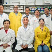
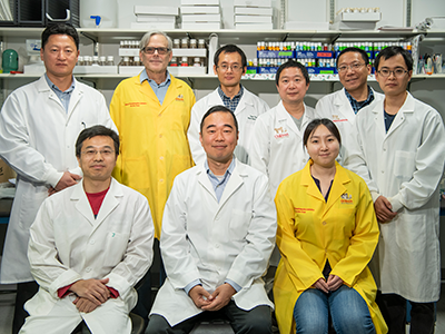
A Children’s researcher has received a $2 million grant from the National Institutes of Health (NIH) to study nephrotic syndrome in Drosophila, a basic model system that has revealed groundbreaking insights into human health. The award for Zhe Han, Ph.D., an associate professor in Children’s Center for Genetic Medicine Research, is believed to be the first ever NIH Research Project grant (R01) to investigate glomerular kidney disease using Drosophila. Nephrotic syndrome is mostly caused by damage of glomeruli, so it is equivalent to glomerular kidney disease.
“Children’s National leads the world in using Drosophila to model human kidney diseases,” Han says.
In order to qualify for the five-year funding renewal, Han’s lab needed to successfully accomplish the aims of its first five years of NIH funding. During the first phase of funding, Han established that nephrocytes in Drosophila serve the same functions as glomeruli in humans, and his lab created a series of fly models that are relevant for human glomerular disease.
“Some 85 percent of the genes known to be involved in nephrotic syndrome are conserved from the fly to humans. They play similar roles in the nephrocyte as they play in the podocytes in human kidneys,” he adds.
Pediatric nephrotic syndrome is a constellation of symptoms that indicate when children’s kidneys are damaged, especially the glomeruli, units within the kidney that filter blood. Babies as young as 1 year old can suffer proteinuria, which is characterized by too much protein being released from the blood into the urine.
“It’s a serious disease and can be triggered by environmental factors, taking certain prescription medicines or inflammation, among other factors. Right now, that type of nephrotic syndrome is mainly treated by steroids, and the steroid treatment works in many cases,” he says.
However, steroid-resistant nephrotic syndrome occurs primarily due to genetic mutations that affect the kidney’s filtration system: These filters are either broken or the protein reabsorption mechanism is disrupted.
“When genetics is to blame, we cannot turn to steroids. Right now there is no treatment. And many of these children are too young to be considered for a kidney transplant,” he adds. “We have to understand exactly which genetic mutation caused the disease in order to develop a targeted treatment.”
With the new funding, Han will examine a large array of genetic mutations that cause nephrotic syndrome. He’s focusing his efforts on genes involved in the cytoskeleton, a network of filaments and tubules in the cytoplasm of living cells that help them to maintain shape and carry out important functions.
“Right now, we don’t really understand the cytoskeleton of podocytes – highly specialized cells that wrap around the capillaries of the glomerulus – because podocytes are difficult to access. To change a gene requires time and considerable effort in other experimental models. However, changing genes in Drosophila is very easy, quick and inexpensive. We can examine hundreds of genes involving the cytoskeleton and see how changing those genes affect kidney cell function,” he says.
Han’s lab already found that Coenzyme Q10, one of the best-selling nutrient supplements to support heart health also could be beneficial for kidney health. For the cytoskeleton, he has a different targeted medicine in mind to determine whether Rho inhibitors also could be beneficial for kidney health for patients with certain genetic mutations affecting their podocyte cytoskeleton.
“One particular aim of our research is to use the same strategy as we employed for the Coq2 gene to generate a personalized fly model for patients with cytoskeleton gene mutations and test potential target drugs, such as Rho inhibitors.” Han added. “As far as I understand, this is where the future of medicine is headed.”
https://innovationdistrict.childrensnational.org/wp-content/uploads/2019/03/Zhe-Han-lab_2018.png300400Innovation Districthttps://innovationdistrict.childrensnational.org/wp-content/uploads/2023/12/innovationdistrict_logo-1-1030x165.pngInnovation District2019-03-11 13:54:382019-11-18 14:31:31$2 million NIH grant to study nephrotic syndrome
Research by an international team that includes Children’s National faculty, published online Jan. 25, 2019 in Human Molecular Genetics, suggests that genetic mutations that cause cleft lip and palate also may contribute to neural tube defects, such as spina bifida.
Oral clefts are some of the most common birth defects worldwide, affecting about one in every 700 births. In the U.S., more than 4,000 babies are born each year with cleft lip, with or without cleft palate.
This defect isn’t simply a cosmetic manner: Oral clefts can severely affect feeding, speech and hearing, and they cause about 3,300 deaths annually worldwide.
To better understand these conditions, researchers have isolated a number of genetic mutations that appear to play contributing roles. These include those in a gene known as Interferon Regulatory Factor 6. New research by an international team that includes Children’s National faculty, published online Jan. 25, 2019 in Human Molecular Genetics, suggests that these mutations also may contribute to neural tube defects such as spina bifida.
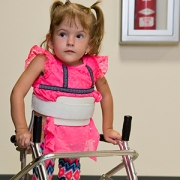
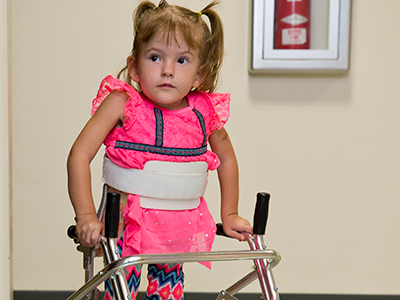
In the first weeks of fetal development, the neural plate curves, creating a neural tube that, once fused shut, becomes the fetal brain and fetal spinal cord. Neural tube defects, which can range from mild to severe, are characterized by incomplete development of the brain, spinal cord or meninges. These defects can potentially result in paralysis or even fetal or neonatal demise. According to the National Institutes of Health, spina bifida, which affects the spinal cord, is the most common neural tube defect in the U.S., affecting up to 2,000 infants each year.
“Despite its high frequency, spina bifida remains among the least understood structural birth defects,” says Brian C. Schutte, an associate professor of Microbiology and Molecular Genetics, Pediatrics and Human Development at Michigan State University and the study’s senior author. “There is strong evidence that genetic factors are a leading cause of such structural birth defects, but in most cases, the cause is unknown. Our team’s study is the first published research to demonstrate that DNA variants in the gene IRF6 can cause spina bifida,” Schutte says.
What’s more, the research team identified a mechanism to explain how altering IRF6 leads to neural tube defects. This mechanism links IRF6 function to two other genes – known as transcription Factor AP2A (TFAP2A) and Grainyhead Like 3 (GRHL3) – that are also known to be required for the development of the neural tube, lip and palate.
“We’re all on the hunt for the reasons when, how and why birth defects happen,” adds Youssef A. Kousa, MS, D.O., Ph.D., a clinical fellow in the Division of Child Neurology at Children’s National Health System and the study’s lead author. “Our main goal is prevention. This paper is a significant development because our team has identified a group of genes that can potentially contribute to very common types of birth defects: craniofacial as well as neural tube defects.”
The scientific odyssey is a wonderful example of serendipity. Kousa, then working in Schutte’s lab, was studying the effects of a new mutant experimental model strain on development of the palate. But one day, he walked into Schutte’s office holding a deformed preclinical embryo and said: “Brian, look at this!”
“Weird things happen in biology,” Schutte replied and counseled him to return if it happened again. Less than two weeks later, Kousa was back with several more of the deformed preclinical embryos, saying: “OK, Brian. It happened again.”
Within hours Kousa had unearthed recently published research that included an image of a similarly affected preclinical embryo. The pair then sketched out possible intersecting genetic pathways, as they brainstormed the myriad ways to end up with that specific phenotype. Initially, they tested their hypotheses in experimental models and eventually corroborated findings through human genetic studies.
The human studies could only be performed by collaborations. Schutte shared their initial observations with human genetics researchers scattered across the country. Those labs then generously agreed to test whether DNA variants in IRF6 were associated with neural tube defects in samples from patients that they had collected over decades of research.
The team found that Tfap2a, Irf6 and Grhl3 are components of a gene regulatory network required for neurulation, a folding process that results in the neural tube bending and then fusing to become the basis of the embryo’s nervous system, from brain to spinal cord.
“Since this network is also required for formation of the lip, palate, limbs and epidermis, which develop at different times and places during embryogenesis, we suggest that the Tfap2a–Irf6–Grhl3 network is a fundamental pathway for multiple morphogenetic processes,” the researchers write.
Interferon Regulatory Factor 6 functions best when there is neither too much expression nor too little. Overexpression of Irf6 suppresses Transcription Factor Activation Protein 2A and Grainyhead Like 3, causing exencephaly, a neural tube defect characterized by the brain being located outside of the skull. Counterintuitively, experimental models that had too little Irf6 also ended up with reduced levels of Tfap2a and Grhl3 that led to a structural birth defect, but at the opposite end of the neural tube.
To test whether the experimental model findings held true in humans, they sequenced samples from people who had spina bifida and anencephaly – the rare birth defect that Kousa spotted in the experimental models – and found IRF6 function was conserved in people. Because of the genetic complexity of these birth defects, and the challenges inherent in collecting samples from cases of severe birth defects, many research teams were invited to participate in the study.
As testament to their collegiality, researchers from Stanford University, University of Texas at Austin, University of Iowa, University of Texas at Houston and Duke University agreed to share precious samples from the California Birth Defects Monitoring Program, from the Hereditary Basis of Neural Tube Defects study and from their own institutional sample collections.
“As we get better at personalized medicine, we could use this information to one day help to counsel families about their own risk and protective factors,” Kousa adds. “If we can identify the genetic pathway, we might also be able to modify it to prevent a birth defect. For example, prenatal supplementation with folic acid has led to a decrease in babies born with neural tube defects, but not all neural tube defects are sensitive to folic acid. This knowledge will help us develop individual-based interventions.”
Financial support for the research covered in this post was provided by the National Institutes of Health under grants DE13513, F31DE022696, DE025060, P01HD067244 and GM072859; startup funding from Michigan State University and the UT-Health School of Dentistry in Houston; and the Centers for Disease Control and Prevention under award number 5U01DD001033.
In addition to Kousa and Schutte, study co-authors include Huiping Zhu, Yunping Lei and Richard H. Finnell, University of Texas at Austin; Walid D. Fakhouri, University of Texas Health Science Center at Houston; Akira Kinoshita, Nagasaki University; Raeuf R. Roushangar, Nicole K. Patel, Tamer Mansour, Arianna L. Smith, and Dhruv B. Sharma, Michigan State University; A.J. Agopian and Laura E. Mitchell, University of Texas School of Public Health; Wei Yang and Gary M. Shaw, Stanford University School of Medicine; Elizabeth J. Leslie, Emory University; Xiao Li, Tamara D. Busch, Alexander G. Bassuk and Brad A. Amendt, University of Iowa; Edward B. Li and Eric C. Liao, Massachusetts General Hospital; Trevor J. Williams, University of Colorado Denver at Anschutz Medical Campus; Yang Chai, University of Southern California; and Simon Gregory and Allison Ashley-Koch, Duke University Medical Center.
https://innovationdistrict.childrensnational.org/wp-content/uploads/2019/01/little-girl-with-spina-bifida.jpg300400Innovation Districthttps://innovationdistrict.childrensnational.org/wp-content/uploads/2023/12/innovationdistrict_logo-1-1030x165.pngInnovation District2019-01-25 12:58:112024-12-30 12:46:57Oral clefts may stem from a shared genetic cause as neural tube defects