Tag Archive for: GABA

Representative OFF and DIFF spectra

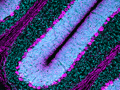
GABA and glutamate in the preterm neonatal brain

Preterm and sick newborns are at high risk of brain injury that can lead to cognitive delays and behavioral disorders including autism and ADHD. Gamma-aminobutyric acid (GABA) and glutamate system disruptions may underlie these neonatal brain injuries and hence it is important to describe their normative profile in the developing neonatal brain.

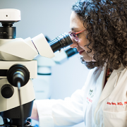
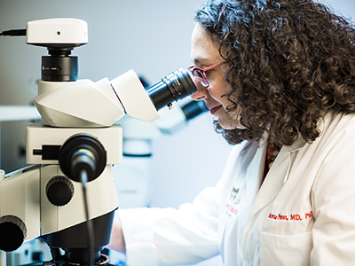
In a study led by Sudeepta Basu, M.D., neonatologist at Children’s National Hospital and Assistant Professor of Pediatrics at George Washington University School of Medicine and Health Sciences, specialized GABA editing spectroscopy (MEGA-PRESS) was acquired on a 3Tesla MRI scanner. Although MEGA-PRESS has been used in older subjects, there are challenges in the newborn population that have limited investigations with only a few institutions worldwide. Under the leadership of Catherine Limperopoulos, Ph.D., in the Developing Brain Institute (DBI) at Children’s National, a team of scientists (in particular, Dr. Subechhya Pradhan) have diligently overcome the technical challenges to enable use of this cutting-edge technology for research at the institute.

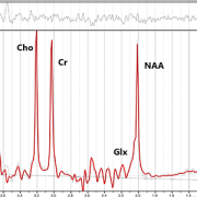
With this unique capability, Dr. Basu’s team prospectively enrolled 58 healthy newborns to describe the normal GABA and glutamate concentrations in different regions of the developing brain. In a recent article published in the American Journal of Neuroradiology, Dr. Basu reports that GABA and glutamate concentrations were highest in the cerebellum, slightly lower in the basal ganglia, but significantly lower in the frontal lobe.

“Our ability to reliably describe the normal metabolic-neurotransmitter milieu of the developing newborn brain is the first step in filling a critical gap in knowledge,” says Dr. Basu. “We hope to identify early bio-markers of brain injury of cognitive delays and autism and ADHD risk which remains a major challenge until clinical symptoms manifest later in childhood.”

Under the direction of Dr. Limperopoulos, advanced multi-modal high precision MRI protocols have been developed for use in research studies at Children’s National that allows the scientists to identify subtle signs of delayed growth and development of the newborn brain. With the optimization of MEGA-PRESS for newborns, Children’s National is one of a few institutions worldwide capable of investigating the newborn brain neurotransmitters in future research studies.

Read the full article in American Journal of Neuroradiology.

Representative OFF and DIFF spectra

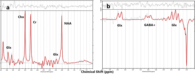
Representative OFF and DIFF spectra.

Vittorio Gallo

Special issue of “Neurochemical Research” honors Vittorio Gallo, Ph.D.

Vittorio Gallo

Investigators from around the world penned manuscripts that were assembled in a special issue of “Neurochemical Research” that honors Vittorio Gallo, Ph.D., for his leadership in the field of neural development and regeneration.

At a pivotal moment early in his career, Vittorio Gallo, Ph.D., was accepted to work with Professor Giulio Levi at the Institute for Cell Biology in Rome, a position that leveraged courses Gallo had taken in neurobiology and neurochemistry, and allowed him to work in the top research institute in Italy directed by the Nobel laureate, Professor Rita Levi-Montalcini.

For four years as a student and later as Levi’s collaborator, Gallo focused on amino acid neurotransmitters in the brain and mechanisms of glutamate and GABA release from nerve terminals. Those early years cemented a research focus on glutamate neurotransmission that would lead to a number of pivotal publications and research collaborations that have spanned decades.

Now, investigators from around the world who have worked most closely with Gallo penned tributes in the form of manuscripts that were assembled in a special issue of “Neurochemical Research” that honors Gallo “for his contributions to our understanding of glutamatergic and GABAergic transmission during brain development and to his leadership in the field of neural development and regeneration,” writes guest editor Arne Schousboe, of the University of Copenhagen in Denmark.

Dr. Gallo as a grad student

Vittorio Gallo, Ph.D. as a 21-year-old mustachioed graduate student.

“In spite of news headlines about competition in research and many of the negative things we hear about the research world, this shows that research is also able to create a community around us,” says Gallo, chief research officer at Children’s National Hospital and scientific director for the Children’s National Research Institute.

As just one example, he first met Schousboe 44 years ago when Gallo was a 21-year-old mustachioed graduate student.

“Research can really create a sense of community that we carry on from the time we are in training, nurture as we meet our colleagues at periodic conferences, and continue up to the present. Creating community is bi-directional: influencing people and being influenced by people. People were willing to contribute these 17 articles because they value me,” Gallo says. “This is a lot of work for the editor and the people who prepared papers for this special issue.”

In addition to Gallo publishing more than 140 peer-reviewed papers, 30 review articles and book chapters, Schousboe notes a number of Gallo’s accomplishments, including:

  • He helped to develop the cerebellar granule cell cultures as a model system to study how electrical activity and voltage-dependent calcium channels modulate granule neuron development and glutamate release.
  • He developed a biochemical/neuropharmacological assay to monitor the effects of GABA receptor modulators on the activity of GABA chloride channels in living neurons.
  • He and Maria Usowicz used patch-clamp recording and single channel analysis to demonstrate for the first time that astrocytes express glutamate-activated channels that display functional properties similar to neuronal counterparts.
  • He characterized one of the spliced isoforms of the AMPA receptor subunit gene Gria4 and demonstrated that this isoform was highly expressed in the cerebellum.
  • He and his Children’s National colleagues demonstrated that glutamate and GABA regulate oligodendrocyte progenitor cell proliferation and differentiation.
Purkinje cells

Purkinje cells are large neurons located in the cerebellum that are elaborately branched like interlocking tree limbs and represent the only source of output for the entire cerebellar cortex.

Even the image selected to grace the special issue’s cover continues the theme of continuity and leaving behind a legacy. That image of Purkinje cells was created by a young scientist who works in Gallo’s lab, Aaron Sathyanesan, Ph.D. Gallo began his career working on the cerebellum – a region of the brain important for motor control – and now studies with a team of scientists and clinician-scientists Purkinje cells’ role in locomotor adaptive behavior and how that is disrupted after neonatal brain injury.

“These cells are the main players in cerebellar circuitry,” Gallo says. “It’s a meaningful image because goes back to my roots as a graduate student and is also an image that someone produced in my lab early in his career. It’s very meaningful to me that Aaron agreed to provide this image for the cover of the special issue.”

Dr. Anna Penn uses a microscope

New model mimics persistent interneuron loss seen in prematurity

Dr. Anna Penn uses a microscope

Children’s research-clinicians created a novel preclinical model that mimics the persistent interneuron loss seen in preterm human infants, identifying interneuron subtypes that could become future therapeutic targets to prevent or lessen neurodevelopmental risks.

Research-clinicians at Children’s National Health System have created a novel preclinical model that mimics the persistent interneuron loss seen in preterm human infants, identifying interneuron subtypes that could become future therapeutic targets to prevent or lessen neurodevelopmental risks, the team reports Jan. 31, 2019, in eNeuro. The open access journal for Society for Neuroscience recognized the team’s paper as its “featured” article.

In the prefrontal cortex (PFC) of infants born preterm, there are decreased somatostatin and calbindin interneurons seen in upper cortical layers in infants who survived for a few months after preterm birth. This neuronal damage was mimicked in an experimental model of preterm brain injury in the PFC, but only when the newborn experimental models had first experienced a combination of prenatal maternal immune activation and postnatal chronic sublethal hypoxia. Neither neuronal insult on its own produced the pattern of interneuron loss in the upper cortical layers observed in humans, the research team finds.

“These combined insults lead to long-term neurobehavioral deficits that mimic what we see in human infants who are born extremely preterm,” says Anna Penn, M.D., Ph.D., a neonatologist in the Division of Neonatology and the Fetal Medicine Institute and a developmental neuroscientist at Children’s National Health System, and senior study author. “Future success in preventing neuronal damage in newborns relies on having accurate experimental models of preterm brain injury and well-defined outcome measures that can be examined in young infants and experimental models of the same developmental stage.”

According to the Centers for Disease Control and Prevention 1 in 10 infants is born preterm, before the 37th week of pregnancy. Many of these preterm births result from infection or inflammation in utero. After delivery, many infants experience other health challenges, like respiratory failure. These multi-hits can exacerbate brain damage.

Prematurity is associated with significantly increased risk of neurobehavioral pathologies, including autism spectrum disorder and schizophrenia. In both psychiatric disorders, the prefrontal cortex inhibitory circuit is disrupted due to alterations of gamma-aminobutyric acid (GABA) interneurons in a brain region involved in working memory and social cognition.

Cortical interneurons are created and migrate late in pregnancy and early infancy. That timing leaves them particularly vulnerable to insults, such as preterm birth.

In order to investigate the effects of perinatal insults on GABAergic interneuron development, the Children’s research team, led by Helene Lacaille, Ph.D., in Dr. Penn’s laboratory, subjected the new preterm encephalopathy experimental model to a battery of neurobehavioral tests, including working memory, cognitive flexibility and social cognition.

“This translational study, which examined the prefrontal cortex in age-matched term and preterm babies supports our hypothesis that specific cellular alterations seen in preterm encephalopathy can be linked with a heightened risk of children experiencing neuropsychiatric disorders later in life,” Dr. Penn adds. “Specific interneuron subtypes may provide specific therapeutic targets for medicines that hold the promise of preventing or lessening these neurodevelopmental risks.”

In addition to Dr. Penn and Lead Author Lacaille, Children’s co-authors include Claire-Marie Vacher; Dana Bakalar, Jiaqi J. O’Reilly and Jacquelyn Salzbank, all of Children’s Center for Neuroscience Research.

Financial support for research described in this post was provided by the National Institutes of Health under award R01HD092593, District of Columbia Intellectual Developmental Disabilities Research Center under award U54HD090257, Cerebral Palsy Alliance Research Foundation, Children’s National Board of Visitors, Children’s Research Institute and Fetal Medicine Institute.

Sudeepta Basu

GABA concentration in pre-term brain increases with gestational age

Sudeepta Basu

“A more complete understanding of the diagnostic and prognostic importance of GABA and glutamate in the preterm brain will help us to direct treatment strategies for the most vulnerable preterm infants at risk of brain injury,” says Sudeepta K. Basu, M.D.

The major neurotransmitters gamma-aminobutyric acid (GABA) and glutamate are pivotal to fetal and newborn brain development and influence evolution of brain injury and repair following preterm birth. Magnetic resonance spectroscopy (MRS) enables in vivo measurement of brain metabolites. However, GABA and glutamate are found in the developing brain in low concentrations, and their weak signal can be swamped by the stronger signal of more dominant metabolites.

A Children’s research team reports findings from a pilot study utilizing an innovative technique of MRS to reliably measure in vivo GABA in the developing preterm brain. The groundbreaking research done by the team that includes Principal Investigator Sudeepta K. Basu, M.D., neonatology attending at Children’s National Health System, is very unique and original since there are no existing data of in vivo GABA concentrations in the developing cerebellum. Under the mentorship of Catherine Limperopoulos, Ph.D., director of Children’s Developing Brain Research Laboratory, the team of multi-disciplinary specialists is pursuing cutting-edge technologies in advanced MRI neuroimaging to explore brain development and injury in preterm infants.

The research, presented at the Eastern Society for Pediatric Research (ESPR) annual meeting by Dr. Basu, was honored with the “2018 Meritorious Poster Award.” The research titled “Distinct temporal trends of GABA and glutamate in the cerebellum and frontal cortex of preterm infants” reports, for the first time, positive temporal trends in the specific regions of the developing brain intricately involved in cognitive and motor functions. This work lays the foundation for developing novel ways to diagnose, monitor and investigative brain protective therapies for vulnerable prematurely born infants.

The Children’s team performed non-sedated MRS in 44 preterm infants whose mean gestational age at birth was 26.5 weeks, placing voxels at the middle of the cerebellum and the right frontal cortex. GABA and GIx (glutamate combined with glutamine) were positively correlated with post-menstrual age in the frontal cortex, but not the cerebellum.  At the ESPR meeting, the team also presented for the first time that caffeine, a neuroprotective agent in preemies, leads to increased in vivo GABA concentration in the developing frontal cortex.

“Open questions include whether these findings reflect varying paces of maturation and vulnerability to injury among specific regions of the brain. Also, the relationship between clinical factors and medication exposure and changes in the concentration of these neurotransmitters may guide brain protective therapies in future,” Dr. Basu says. “A more complete understanding of the diagnostic and prognostic importance of GABA and glutamate in the preterm brain will help us to direct treatment strategies for the most vulnerable preterm infants at risk of brain injury.”

Children’s senior fellows from Division of Neonatology made four platform presentations during the ESPR conference:

  • “Caffeine increases GABA/Cr ratio in frontal cortex of preterm infants on spectroscopy.” Aditi Gupta; Sudeepta K. Basu, M.D.; Mariam Said, M.D.; Subechhya Pradhan, Linda White; Kushal Kapse; Jonathan Murnick, M.D., Ph.D.; Taeun Chang, M.D.; and Catherine Limperopoulos, Ph.D.
  • “Impact of early nutrition on microstructural brain development in VLBW Infants.” Katherine M. Ottolini, Nickie Andescavage, M.D.; Kushal Kapse; and Catherine Limperopoulos, Ph.D.
  • “Direct measurement of neonatal cardiac output utilizing the CO status monitor.” Simranjeet S. Sran, Mariam Said, M.D.; and Khodayar Rais-Bahrami, M.D.
  • “Cerebro-cerebellar diaschisis in preterm infants following unilateral cerebral parenchymal injury.” Huma Mirza, Yao Wu, Kushal Kapse, Jonathan Murnick, M.D., Ph.D.; Taeun Chang, M.D.; and Catherine Limperopoulos, Ph.D.