Tag Archive for: experimental model

mitochondria

Molecular gatekeepers that regulate calcium ions key to muscle function

mitochondria

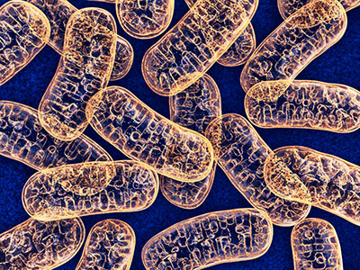
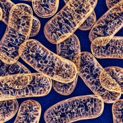
Controlled entry of calcium ions into the mitochondria, the cell’s energy powerhouses, makes the difference between whether muscles grow strong or easily tire and perish from injury, according to research published in Cell Reports.

Calcium ions are essential to how muscles work effectively, playing a starring role in how and when muscles contract, tap energy stores to keep working and self-repair damage. Not only are calcium ions vital for the repair of injured muscle fibers, their controlled entry into the mitochondria, the cell’s energy powerhouses, spells the difference between whether muscles will be healthy or if they will easily tire and perish following an injury, according to research published Oct. 29, 2019, in Cell Reports.

“Lack of the protein mitochondrial calcium uptake1 (MICU1) lowers the activation threshold for calcium uptake mediated by the mitochondrial calcium uniporter in both, muscle fibers from an experimental model and fibroblast of  a patient lacking MICU1,” says Jyoti K. Jaiswal, MSc, Ph.D., a principal investigator in the Center for Genetic Medicine Research at Children’s National Hospital and one of the paper’s corresponding authors. “Missing MICU1 also tips the calcium ion balance in the mitochondria when muscles contract or are injured, leading to more pronounced muscle weakness and myofiber death.”

Five years ago, patients with a very rare disease linked to mutations in the mitochondrial gene MICU1 were described to suffer from a neuromuscular disease with signs of muscle weakness and damage that could not be fully explained.

To determine what was going awry, the multi-institutional research team used a comprehensive approach that included fibroblasts donated by a patient lacking MICU1 and an experimental model whose MICU1 gene was deleted in the muscles.

Loss of MICU1 in skeletal muscle fibers leads to less contractile force, increased fatigue and diminished capacity to repair damage to their cell membrane, called the sarcolemma. Just like human patients, the experimental model suffers more pronounced muscle weakness, increased numbers of dead myofibers, with greater loss of muscle mass in certain muscles, like the quadriceps and triceps, the research team writes.

“What was happening to the patient’s muscles was a big riddle that our research addressed,” Jaiswal adds. “Lacking this protein is not supposed to make the muscle fiber die, like we see in patients with this rare disease. The missing protein is just supposed to cause atrophy and weakness.”

Patients with this rare disease show early muscle weakness, fluctuating levels of fatigue and lethargy, muscle aches after exercise, and elevated creatine kinase in their bloodstream, an indication of cell damage due to physical stress.

“One by one, we investigated these specific features in experimental models that look normal and have normal body weight, but also show lost muscle mass in the quadriceps and triceps,” explains Adam Horn, Ph.D., the lead researcher in Jaiswal’s lab who conducted this study. “Our experimental model lacking MICU1 only in skeletal muscles responded to muscle deficits so similar to humans that it suggests that some of the symptoms we see in patients can be attributed to MICU1 loss in skeletal muscles.”

Future research will aim to explore the details of how the impact of MICU1 deficit in muscles may be addressed therapeutically and possible implications of lacking MICU1 or its paralog in other organs.

In addition to Jaiswal and Horn, Children’s National Hospital Center for Genetic Medicine Research co-authors include Marshall W. Hogarth and Davi A. Mazala. Additional co-authors include Lead Author Valentina Debattisti, Raghavendra Singh, Erin L. Seifert, Kai Ting Huang, and Senior Author György Hajnóczky, all from Thomas Jefferson University; and Rita Horvath, from Newcastle University.

Financial support for research described in this post was provided by the National Institutes of Health under award numbers R01AR55686, U54HD090257 and RO1 GM102724; National Institute of Arthritis and Musculoskeletal and Skin Diseases under award number T32AR056993; and Foundation Leducq.

Claire Marie Vacher

Placental function linked to brain injuries associated with autism

Claire Marie Vacher

“We saw long-term cerebellar white matter alterations in male experimental models, and behavioral testing revealed social impairments and increased repetitive behaviors, two hallmark features of ASD,” says Claire-Marie Vacher, Ph.D., lead study author.

Allopregnanolone (ALLO), a hormone made by the placenta late in pregnancy, is such a potent neurosteroid that disrupting its steady supply to the developing fetus can leave it vulnerable to brain injuries associated with autism spectrum disorder (ASD), according to Children’s research presented during the Pediatric Academic Societies 2019 Annual Meeting.

In order to more effectively treat vulnerable babies, the Children’s research team first had to tease out what goes wrong in the careful choreography that is pregnancy. According to the Centers for Disease Control and Prevention, about 1 in 10 babies is born preterm, before 37 weeks of gestation. Premature birth is a major risk factor for ASD.

The placenta is an essential and understudied organ that is shared by the developing fetus and the pregnant mother, delivering oxygen, glucose and nutrients and ferrying out waste products. The placenta also delivers ALLO, a progesterone derivative, needed to ready the developing fetal brain for life outside the womb.

ALLO ramps up late in gestation. When babies are born prematurely, their supply of ALLO stops abruptly. That occurs at the same time the cerebellum – a brain region essential for motor coordination, posture, balance and social cognition– typically undergoes a dramatic growth spurt.

“Our experimental model demonstrates that losing placental ALLO alters cerebellar development, including white matter development,” says Anna Penn, M.D., Ph.D., a neonatologist in the divisions of Neonatology and Fetal Medicine, and a developmental neuroscientist at Children’s National. “Cerebellar white matter development occurs primarily after babies are born, so connecting a change in placental function during pregnancy with lingering impacts on later brain development is a particularly striking result.”

The research team created a novel experimental model in which the gene encoding the enzyme responsible for producing ALLO is deleted in the placenta. They compared these preclinical models with a control group and performed whole brain imaging and RNAseq gene expression analyses for both groups.

“We saw long-term cerebellar white matter alterations in male experimental models, and behavioral testing revealed social impairments and increased repetitive behaviors, two hallmark features of ASD,” says Claire-Marie Vacher, Ph.D., lead study author. “These male-specific outcomes parallel the increased risk of brain injury and ASD we see in human babies born prematurely.”

ALLO binds to specific GABA receptors, which control most inhibitory signaling in the nervous system.

“Our findings provide a new way to frame poor placental function: Subtle but significant changes in utero may set in motion neurodevelopmental disorders that children experience later in life,” adds Dr. Penn, the study’s senior author. “Future directions for our research could include identifying new targets in the placenta or brain that could be amenable to hormone supplementation, opening the potential for earlier treatment for high-risk fetuses.”

Pediatric Academic Societies 2019 Annual Meeting presentation

  • “Placental allopregnanolone loss alters postnatal cerebellar development and function.”
    • Sunday, April 28, 2019, 5:15 p.m. to 5:30 p.m. (EST)

Claire-Marie Vacher, Ph.D., lead author; Jackie Salzbank, co-author; Helene Lacaille, co-author; Dana Bakalar, co-author; Jiaqi O’Reilly, co-author; and Anna Penn, M.D., Ph.D., a neonatologist in the divisions of Neonatology and Fetal Medicine, developmental neuroscientist and senior study author.

Zhe Han lab 2018

$2 million NIH grant to study nephrotic syndrome

Zhe Han lab 2018

A Children’s researcher has received a $2 million grant from the National Institutes of Health (NIH) to study nephrotic syndrome in Drosophila, a basic model system that has revealed groundbreaking insights into human health. The award for Zhe Han, Ph.D., an associate professor in Children’s Center for Genetic Medicine Research, is believed to be the first ever NIH Research Project grant (R01)  to investigate glomerular kidney disease using Drosophila. Nephrotic syndrome is mostly caused by damage of glomeruli, so it is equivalent to glomerular kidney disease.

“Children’s National leads the world in using Drosophila to model human kidney diseases,” Han says.

In order to qualify for the five-year funding renewal, Han’s lab needed to successfully accomplish the aims of its first five years of NIH funding.  During the first phase of funding, Han established that nephrocytes in Drosophila serve the same functions as glomeruli in humans, and his lab created a series of fly models that are relevant for human glomerular disease.

“Some 85 percent of the genes known to be involved in nephrotic syndrome are conserved from the fly to humans. They play similar roles in the nephrocyte as they play in the podocytes in human kidneys,” he adds.

Pediatric nephrotic syndrome is a constellation of symptoms that indicate when children’s kidneys are damaged, especially the glomeruli, units within the kidney that filter blood. Babies as young as 1 year old can suffer proteinuria, which is characterized by too much protein being released from the blood into the urine.

“It’s a serious disease and can be triggered by environmental factors, taking certain prescription medicines or inflammation, among other factors.  Right now, that type of nephrotic syndrome is mainly treated by steroids, and the steroid treatment works in many cases,” he says.

However, steroid-resistant nephrotic syndrome occurs primarily due to genetic mutations that affect the kidney’s filtration system: These filters are either broken or the protein reabsorption mechanism is disrupted.

“When genetics is to blame, we cannot turn to steroids. Right now there is no treatment. And many of these children are too young to be considered for a kidney transplant,” he adds. “We have to understand exactly which genetic mutation caused the disease in order to develop a targeted treatment.”

With the new funding, Han will examine a large array of genetic mutations that cause nephrotic syndrome. He’s focusing his efforts on genes involved in the cytoskeleton, a network of filaments and tubules in the cytoplasm of living cells that help them to maintain shape and carry out important functions.

“Right now, we don’t really understand the cytoskeleton of podocytes – highly specialized cells that wrap around the capillaries of the glomerulus – because podocytes are difficult to access. To change a gene requires time and considerable effort in other experimental models. However, changing genes in Drosophila is very easy, quick and inexpensive. We can examine hundreds of genes involving the cytoskeleton and see how changing those genes affect kidney cell function,” he says.

Han’s lab already found that Coenzyme Q10, one of the best-selling nutrient supplements to support heart health also could be beneficial for kidney health. For the cytoskeleton, he has a different targeted medicine in mind to determine whether Rho inhibitors also could be beneficial for kidney health for patients with certain genetic mutations affecting their podocyte cytoskeleton.

“One particular aim of our research is to use the same strategy as we employed for the Coq2 gene to generate a personalized fly model for patients with cytoskeleton gene mutations and test potential target drugs, such as Rho inhibitors.” Han added. “As far as I understand, this is where the future of medicine is headed.”

Fetal Brain Cells

Tracking environmental stress damage in the brain

Fluorescence Reporter

A team led by Children’s National developed a fluorescence reporter system in an experimental model that can single out neurons that have survived prenatal damage but remain vulnerable after birth.

PDF Version

What’s known

When fetuses are exposed to environmental stressors, such as maternal smoking or alcohol consumption, radiation or too little oxygen, some of these cells can die. A portion of those that survive often have lingering damage and remain more susceptible to further environmental insults than healthy cells; however, researchers haven’t had a way to identify these weakened cells. This lack of knowledge has made it difficult to discover the mechanisms behind pathological brain development thought to arise from these very early environmental exposures, as well as ways to prevent or treat it.

What’s new

A team led by Kazue Hashimoto-Torii, Ph.D., a principal investigator in the Center for Neuroscience Research at Children’s National Health System, developed a marker that makes a protein known as Heat Shock Factor 1 glow red. This protein is produced in cells that become stressed through exposure to a variety of environmental insults. Gestation is a particularly vulnerable time for rapidly dividing nerve cells in the fetal brain. Tests showed that this marker worked not just on cells in petri dishes but also in an experimental model to detect brain cells that were damaged and remained vulnerable after exposure to a variety of different stressors. Tweaks to the system allowed the researchers to follow the progeny of cells that were affected by the initial stressor and track them as they divided and spread throughout the brain. By identifying which neurons are vulnerable, the study authors say, researchers eventually might be able to develop interventions that could slow or stop damage before symptoms arise.

Questions for future research

Q: How do different environmental insults damage brain cells during gestation?
Q: How does this damage translate into pathology in organisms as they mature?
Q: Do the progeny of damaged brain cells retain the same degree of damage as they divide and spread?
Q: Can this new detection system be used to find and track damage in other organs, such as the heart, eye and liver?

Source: Torii, M., S. Masanori, Y.W. Chang, S. Ishii, S.G. Waxman, J.D. Kocsis, P. Rakic and K. Hashimoto-Torii. “Detection of vulnerable neurons damaged by environmental insults in utero.” Published Dec. 22, 2016 by Proceedings of the National Academy of Sciences. doi: 10.1073/pnas.1620641114