Tag Archive for: development

Vittorio Gallo

Special issue of “Neurochemical Research” honors Vittorio Gallo, Ph.D.

Vittorio Gallo

Investigators from around the world penned manuscripts that were assembled in a special issue of “Neurochemical Research” that honors Vittorio Gallo, Ph.D., for his leadership in the field of neural development and regeneration.

At a pivotal moment early in his career, Vittorio Gallo, Ph.D., was accepted to work with Professor Giulio Levi at the Institute for Cell Biology in Rome, a position that leveraged courses Gallo had taken in neurobiology and neurochemistry, and allowed him to work in the top research institute in Italy directed by the Nobel laureate, Professor Rita Levi-Montalcini.

For four years as a student and later as Levi’s collaborator, Gallo focused on amino acid neurotransmitters in the brain and mechanisms of glutamate and GABA release from nerve terminals. Those early years cemented a research focus on glutamate neurotransmission that would lead to a number of pivotal publications and research collaborations that have spanned decades.

Now, investigators from around the world who have worked most closely with Gallo penned tributes in the form of manuscripts that were assembled in a special issue of “Neurochemical Research” that honors Gallo “for his contributions to our understanding of glutamatergic and GABAergic transmission during brain development and to his leadership in the field of neural development and regeneration,” writes guest editor Arne Schousboe, of the University of Copenhagen in Denmark.

Dr. Gallo as a grad student

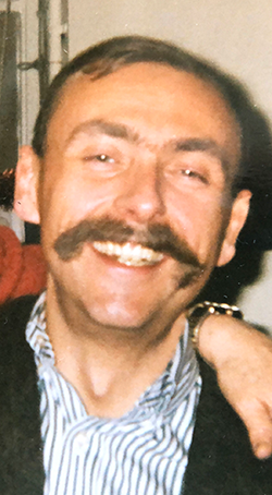
Vittorio Gallo, Ph.D. as a 21-year-old mustachioed graduate student.

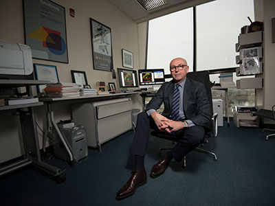
“In spite of news headlines about competition in research and many of the negative things we hear about the research world, this shows that research is also able to create a community around us,” says Gallo, chief research officer at Children’s National Hospital and scientific director for the Children’s National Research Institute.

As just one example, he first met Schousboe 44 years ago when Gallo was a 21-year-old mustachioed graduate student.

“Research can really create a sense of community that we carry on from the time we are in training, nurture as we meet our colleagues at periodic conferences, and continue up to the present. Creating community is bi-directional: influencing people and being influenced by people. People were willing to contribute these 17 articles because they value me,” Gallo says. “This is a lot of work for the editor and the people who prepared papers for this special issue.”

In addition to Gallo publishing more than 140 peer-reviewed papers, 30 review articles and book chapters, Schousboe notes a number of Gallo’s accomplishments, including:

  • He helped to develop the cerebellar granule cell cultures as a model system to study how electrical activity and voltage-dependent calcium channels modulate granule neuron development and glutamate release.
  • He developed a biochemical/neuropharmacological assay to monitor the effects of GABA receptor modulators on the activity of GABA chloride channels in living neurons.
  • He and Maria Usowicz used patch-clamp recording and single channel analysis to demonstrate for the first time that astrocytes express glutamate-activated channels that display functional properties similar to neuronal counterparts.
  • He characterized one of the spliced isoforms of the AMPA receptor subunit gene Gria4 and demonstrated that this isoform was highly expressed in the cerebellum.
  • He and his Children’s National colleagues demonstrated that glutamate and GABA regulate oligodendrocyte progenitor cell proliferation and differentiation.
Purkinje cells

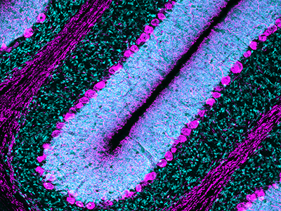
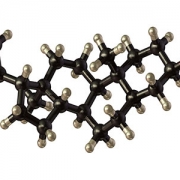
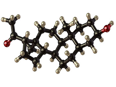
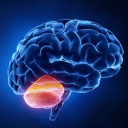
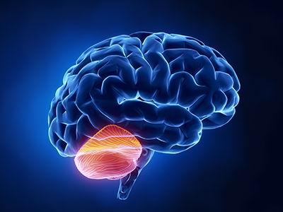
Purkinje cells are large neurons located in the cerebellum that are elaborately branched like interlocking tree limbs and represent the only source of output for the entire cerebellar cortex.

Even the image selected to grace the special issue’s cover continues the theme of continuity and leaving behind a legacy. That image of Purkinje cells was created by a young scientist who works in Gallo’s lab, Aaron Sathyanesan, Ph.D. Gallo began his career working on the cerebellum – a region of the brain important for motor control – and now studies with a team of scientists and clinician-scientists Purkinje cells’ role in locomotor adaptive behavior and how that is disrupted after neonatal brain injury.

“These cells are the main players in cerebellar circuitry,” Gallo says. “It’s a meaningful image because goes back to my roots as a graduate student and is also an image that someone produced in my lab early in his career. It’s very meaningful to me that Aaron agreed to provide this image for the cover of the special issue.”

preterm baby

Validating a better way to stratify BPD risk in vulnerable newborns

preterm baby

Factoring in the total number of days that extremely preterm infants require supplemental oxygen and tracking this metric for weeks longer than usual improves clinicians’ ability to predict respiratory outcomes according to bronchopulmonary dysplasia severity.

Factoring in the total number of days that extremely preterm infants require supplemental oxygen and tracking this metric for weeks longer than usual improves clinicians’ ability to predict respiratory outcomes according to bronchopulmonary dysplasia (BPD) severity, a research team led by Children’s National Hospital writes in Scientific Reports. What’s more, the researchers defined a brand-new category (level IV) for newborns who receive supplemental oxygen more than 120 days as a reliable way to predict which infants are at the highest risk of returning to the hospital due to respiratory distress after discharge.

About 1 in 10 U.S. infants is born preterm, before 37 weeks gestation, according to the Centers for Disease Control and Prevention. That includes extremely preterm infants who weigh about 1 lb. at birth. These very low birthweight newborns have paper thin skin, frail hearts and lungs that are not yet mature enough to deliver oxygen throughout the body as needed. Thanks to advances in neocritical care, an increasing number of them survive prematurity, and many develop BPD, a chronic lung disease characterized by abnormal development of the lungs and pulmonary vasculature.

“About half of the babies born prematurely will come back to the hospital within the first year of life with a respiratory infection. The key is identifying them and, potentially, preventing complications in this high-risk population,” says Gustavo Nino, M.D., a Children’s National pulmonologist and the study’s lead author.

For decades, the most common way to stratify BPD risk in these vulnerable newborns has been to see if they require supplemental oxygen at 36 weeks corrected gestational age.

“The problem with this classification is it doesn’t take into account the very premature babies who are on oxygen for much longer than other babies. So, we asked the question: Can we continue risk stratification beyond 36 weeks in order to identify a subset of babies who are at much higher risk of complications,” Dr. Nino says.

The longitudinal cohort study enrolled 188 infants born extremely preterm who were admitted to the neonatal intensive care unit (NICU) at Children’s National and tracked their data for at least 12 months after discharge. The team used a multidimensional approach that tracked duration of supplemental oxygen during the newborns’ NICU stay as well as scoring lung imaging as an independent marker of BPD severity. To validate the findings, these U.S.-born newborns were matched with 130 infants who were born preterm and hospitalized at two NICUs located in Bogotá, Colombia.

“Babies who are born very preterm and require oxygen beyond 120 days should have expanded ventilation of the lungs and cardiovascular pulmonary system before going home,” he notes. “We need to identify these newborns and optimize their management before they are discharged.”

And, the babies with level IV BPD risk need a different type of evaluation because the complications they experience – including pulmonary hypertension – place them at the highest risk of developing sleep apnea and severe respiratory infection, especially during the first year of life.

“The earlier we identify them, the better their outcome is likely to be,” Dr. Nino says. “We really need to change the risk stratification so we don’t call them all ‘severe’ and treat them the same when there is a subset of newborns who clearly are at a much higher risk for experiencing respiratory complications after hospital discharge.”

In addition to Dr. Nino, Children’s National study co-authors include Awais Mansoor, Ph.D., staff scientist at the Sheikh Zayed Institute for Pediatric Surgical Innovation (SZI); Geovanny F. Perez, M.D., pediatric pulmonologist; Maria Arroyo, M.D., pulmonologist; Xilei Xu Chen, M.D., postdoctoral fellow; Jered Weinstock, pediatric pulmonary fellow; Kyle Salka, MS, research technician; Mariam Said, M.D., neonatologist, and Marius George Linguraru, DPhil, MA, MSc, SZI principal investigator and senior author. Additional co-authors include Ranniery Acuña-Cordero, Universidad Militar Nueva Granada, Bogotá, Colombia; and Monica P. Sossa-Briceño and Carlos E. Rodríguez-Martínez, both of Universidad Nacional de Colombia.

Funding for research described in this post was provided by the National Institutes of Health (NIH) under award Nos. HL145669, AI130502 and HL141237. In addition, the NIH has awarded Dr. Nino an RO1 grant to continue this research.

allopregnanolone molecule

Autism spectrum disorder risk linked to insufficient placental steroid

allopregnanolone molecule

A study led by Children’s National Hospital and presented during Neuroscience 2019 finds that loss of allopregnanolone, a key hormone supplied by the placenta, leads to long-term structural alterations of the cerebellum – a brain region essential for smooth motor coordination, balance and social cognition – and increases the risk of developing autism.

An experimental model study suggests that allopregnanolone, one of many hormones produced by the placenta during pregnancy, is so essential to normal fetal brain development that when provision of that hormone decreases – as occurs with premature birth – offspring are more likely to develop autism-like behaviors, a Children’s National Hospital research team reports at the Neuroscience 2019 annual meeting.

“To our knowledge, no other research team has studied how placental allopregnanolone (ALLO) contributes to brain development and long-term behaviors,” says Claire-Marie Vacher, Ph.D., lead author. “Our study finds that targeted loss of ALLO in the womb leads to long-term structural alterations of the cerebellum – a brain region that is essential for motor coordination, balance and social cognition ­– and increases the risk of developing autism,” Vacher says.

According to the Centers for Disease Control and Prevention, about 1 in 10 infants is born preterm, before 37 weeks gestation; and 1 in 59 children has autism spectrum disorder.

In addition to presenting the abstract, on Monday, Oct. 21, Anna Penn, M.D., Ph.D., the abstract’s senior author, will discuss the research with reporters during a Neuroscience 2019 news conference. This Children’s National abstract is among 14,000 abstracts submitted for the meeting, the world’s largest source of emerging news about brain science and health.

ALLO production by the placenta rises in the second trimester of pregnancy, and levels of the neurosteroid peak as fetuses approach full term.

To investigate what happens when ALLO supplies are disrupted, a research team led by Children’s National created a novel transgenic preclinical model in which they deleted a gene essential in ALLO synthesis. When production of ALLO in the placentas of these experimental models declines, offspring had permanent neurodevelopmental changes in a sex- and region-specific manner.

“From a structural perspective, the most pronounced cerebellar abnormalities appeared in the cerebellum’s white matter,” Vacher adds. “We found increased thickness of the myelin, a lipid-rich insulating layer that protects nerve fibers. From a behavioral perspective, male offspring whose ALLO supply was abruptly reduced exhibited increased repetitive behavior and sociability deficits – two hallmarks in humans of autism spectrum disorder.”

On a positive note, providing a single ALLO injection during pregnancy was enough to avert both the cerebellar abnormalities and the aberrant social behaviors.

The research team is now launching a new area of research focus they call “neuroplacentology” to better understand the role of placenta function on fetal and newborn brain development.

“Our team’s data provide exciting new evidence that underscores the importance of placental hormones on shaping and programming the developing fetal brain,” Vacher notes.

  • Neuroscience 2019 presentation
    Sunday, Oct. 20, 9:30 a.m. (CDT)
    “Preterm ASD risk linked to cerebellar white matter changes”
    Claire-Marie Vacher, lead author; Sonia Sebaoui, co-author; Helene Lacaille, co-author; Jackie Salzbank, co-author; Jiaqi O’Reilly, co-author; Diana Bakalar, co-author; Panagiotis Kratimenos, M.D., neonatologist and co-author; and Anna Penn, M.D., clinical neonatologist and developmental neuroscientist and senior author.

Paradoxical outcomes for Zika-exposed tots

In the midst of an unprecedented Zika crisis in Brazil, there were a few flickers of hope: Some babies appeared to be normal at birth, free of devastating birth defects that affected other Brazilian children exposed to the virus in utero.

In the midst of an unprecedented Zika crisis in Brazil, there were a few flickers of hope: Some babies appeared to be normal at birth, free of devastating birth defects that affected other Brazilian children exposed to the virus in utero. But according to a study published online July 8, 2019, in Nature Medicine and an accompanying commentary co-written by a Children’s National clinician-researcher, the reality for Zika-exposed infants is much more complicated.

Study authors led by Karin Nielsen-Saines at David Geffen UCLA School of Medicine followed 216 infants in Rio de Janeiro who had been exposed to the Zika virus during pregnancy, performing neurodevelopmental testing when the babies ranged in age from 7 to 32 months. These infants’ mothers had had Zika-related symptoms themselves, including rash.

Although many children had normal assessments, 29% scored below average in at least one domain of neurological development, including cognitive performance, fine and gross motor skills and expressive language, Sarah B. Mulkey, M.D., Ph.D., and a colleague write in a companion commentary published online by Nature Medicine July 29, 2019.

The study authors found progressively higher risks for developmental, hearing and eye abnormality depending on how early the pregnancy was at the time the infants were exposed. Because Zika virus has an affinity for immature neurons, even babies who were not born with microcephaly remained at continued risk for suffering abnormalities.

Of note, 24 of 49 (49%) infants who had abnormalities at birth went on to have normal test results in the second or third year of life. By contrast, 17 of 68 infants (25%) who had normal assessments at birth had below-average developmental testing or had abnormalities in hearing or vision by age 32 months.

“This work follows babies who were born in 2015 and 2016. It’s heartening that some babies born with abnormalities tested in the normal range later in life, though it’s unclear whether any specific interventions help to deliver these positive findings,” says Dr. Mulkey, a fetalneonatal neurologist in the Division of Fetal and Transitional Medicine at Children’s National in Washington, D.C. “And it’s quite sobering that babies who appeared normal at birth went on to develop abnormalities due to that early Zika exposure.”

It’s unclear how closely the findings apply to the vast majority of U.S. women whose Zika infections were asymptomatic.

“This study adds to the growing body of research that argues in favor of ongoing follow-up for Zika-exposed children, even if their neurologic exams were reassuring at birth,” Dr. Mulkey adds. “As Zika-exposed children approach school age, it’s critical to better characterize the potential implications for the education system and public health.”

In addition to Dr. Mulkey, the perspective’s senior author, William J. Muller, Northwestern University, was the commentary’s lead author.

illustration of brain showing cerebellum

Focusing on the “little brain” to rescue cognition

illustration of brain showing cerebellum

Research faculty at Children’s National in Washington, D.C., with colleagues recently published a review article in Nature Reviews Neuroscience that covers the latest research about how abnormal development of the cerebellum leads to a variety of neurodevelopmental disorders.

Cerebellum translates as “little brain” in Latin. This piece of anatomy – that appears almost separate from the rest of the brain, tucked under the two cerebral hemispheres – long has been known to play a pivotal role in voluntary motor functions, such as walking or reaching for objects, as well as involuntary ones, such as maintaining posture.

But more recently, says Aaron Sathyanesan, Ph.D., a postdoctoral research fellow at the Children’s Research Institute, the research arm of Children’s National  in Washington, D.C., researchers have discovered that the cerebellum is also critically important for a variety of non-motor functions, including cognition and emotion.

Sathyanesan, who studies this brain region in the laboratory of Vittorio Gallo, Ph.D., Chief Research Officer at Children’s National and scientific director of the Children’s Research Institute, recently published a review article with colleagues in Nature Reviews Neuroscience covering the latest research about how altered development of the cerebellum contributes to a variety of neurodevelopmental disorders.

These disorders, he explains, are marked by problems in the nervous system that arise while it’s maturing, leading to effects on emotion, learning ability, self-control, or memory, or any combination of these. They include diagnoses as diverse as intellectual disability, autism spectrum disorder (ASD), attention-deficit/hyperactivity disorder and Down syndrome.

“One reason why the cerebellum might be critically involved in each of these disorders,” Sathyanesan says, “is because its developmental trajectory takes so long.”

Unlike other brain structures, which have relatively short windows of development spanning weeks or months, the principal cells of the cerebellum – known as Purkinje cells – start to differentiate from stem cell precursors at the beginning of the seventh gestational week, with new cells continuing to appear until babies are nearly one year old.  In contrast, cells in the neocortex, a part of the brain involved in higher-order brain functions such as cognition, sensory perception and language is mostly finished forming while fetuses are still gestating in the womb.

This long window for maturation allows the cerebellum to make connections with other regions throughout the brain, such as extensive connections with the cerebral cortex, the outer layer of the cerebrum that plays a key role in perception, attention, awareness, thought, memory, language and consciousness. It also allows ample time for things to go wrong.

“Together,” Sathyanesan says, “these two characteristics are at the root of the cerebellum’s involvement in a host of neurodevelopmental disorders.”

For example, the review article notes, researchers have discovered both structural and functional abnormalities in the cerebellums of patients with ASD. Functional magnetic resonance imaging (MRI), an imaging technique that measures activity in different parts of the brain, suggests that significant differences exist between connectivity between the cerebellum and cortex in people with ASD compared with neurotypical individuals. Differences in cerebellar connectivity are also evident in resting-state functional connectivity MRI, an imaging technique that measures brain activity in subjects when they are not performing a specific task. Some of these differences appear to involve patterns of overconnectivity to different brain regions, explains Sathyanesan; other differences suggest that the cerebellums of patients with ASD don’t have enough connections to other brain regions.

These findings could clarify research from Children’s National and elsewhere that has shown that babies born prematurely often sustain cerebellar injuries due to multiple hits, including a lack of oxygen supplied by infants’ immature lungs, he adds. Besides having a sibling with ASD, premature birth is the most prevalent risk factor for an ASD diagnosis.

The review also notes that researchers have discovered structural changes in the cerebellums of patients with Down syndrome, who tend to have smaller cerebellar volumes than neurotypical individuals. Experimental models of this trisomy recapitulate this difference, along with abnormal connectivity to the cerebral cortex and other brain regions.

Although the cerebellum is a pivotal contributor toward these conditions, Sathyanesan says, learning more about this brain region helps make it an important target for treating these neurodevelopmental disorders. For example, he says, researchers are investigating whether problems with the cerebellum and abnormal connectivity could be lessened through a non-invasive form of brain stimulation called transcranial direct current stimulation or an invasive one known as deep brain stimulation. Similarly, a variety of existing pharmaceuticals or new ones in development could modify the cerebellum’s biochemistry and, consequently, its function.

“If we can rescue the cerebellum’s normal activity in these disorders, we may be able to alleviate the problems with cognition that pervade them all,” he says.

In addition to Sathyanesan and Senior Author Gallo, Children’s National study co-authors include Joseph Scafidi, D.O., neonatal neurologist; Joy Zhou and Roy V. Sillitoe, Baylor College of Medicine; and Detlef H. Heck, of University of Tennessee Health Science Center.

Financial support for research described in this post was provided by the National Institute of Neurological Disorders and Stroke under grant numbers 5R01NS099461, R01NS089664, R01NS100874, R01NS105138 and R37NS109478; the Hamill Foundation; the Baylor College of Medicine Intellectual and Developmental Disabilities Research Center under grant number U54HD083092; the University of Tennessee Health Science Center (UTHSC) Neuroscience Institute; the UTHSC Cornet Award; the National Institute of Mental Health under grant number R01MH112143; and the District of Columbia Intellectual and Developmental Disabilities Research Center under grant number U54 HD090257.